
Autoimmunity

Abstract
In rheumatoid arthritis (RA), CD4+ T cells specific for citrullinated antigens (cit-antigens) are key drivers of disease, but knowledge about epitopes and phenotypes remains limited. We characterized the frequency and phenotype of cit-specific CD4+ T cells in peripheral blood using HLA class II tetramers combined with computational analysis of phenotypic clusters to simultaneously detect peptides derived from 5 cit-antigens (aggrecan, vimentin, fibrinogen, cartilage intermediate layer protein, and α-enolase) previously implicated in RA pathogenesis. In a cross-sectional cohort, cit-aggrecan–, cit-vimentin–, and cit-fibrinogen–specific T cells were more frequent in participants with RA than healthy volunteers, associated with active disease, and had Th1-like and stem-like lineages in RA. In a longitudinal cohort investigating response to therapy, the frequency of cit-aggrecan–, cit-vimentin–, and cit-fibrinogen–specific CD4+ T cells was significantly higher at baseline and further elevated in responders. Furthermore, the frequency of cit-specific Th1-like cells in responders decreased over time. In contrast, the frequency of Th1-like cells in non-responders increased over time. Collectively, these findings demonstrate that cit-specific CD4+ T cells are expanded in RA and target a broad number of antigens across a breadth of phenotypes. Furthermore, the predominant antigen specificities associate with disease activity and exhibit dynamic changes in phenotype that reflect response to therapy.
Authors
Cliff Rims, Hannah A. DeBerg, Sylvia E. Posso, Virginia S. Muir, Hannes Uchtenhagen, Anne M. Hocking, Heather Bukiri, Jeffrey Carlin, Bernard Ng, Peter S. Linsley, Eddie A. James, Jane H. Buckner

Abstract
Foxp3 deficiency causes a profound loss of immune tolerance, unleashing autoreactive T and B cells, lymphoproliferation, cytokine-driven inflammation, and autoantibody production. This autoimmune pathology is fueled by increased glutamine usage, but it remains unresolved whether glutamine is necessary to produce energy or for intermediate metabolite biosynthesis responsible for immunomodulation. Here, we demonstrate that glutamine utilization for biosynthetic pathways supported autoimmune inflammation in the settings of Foxp3 deficiency and dextran sodium sulfate–induced colitis. By employing a model of autoimmunity driven by Treg-specific loss of Foxp3, we showed that this effect is independent of pathogenic Foxp3-deficient Treg reprogramming. Mechanistically, glutamine biosynthetic pathways sustained conventional T cell activation and proinflammatory cytokine production by preventing inosine accumulation and signaling, thus implicating adenosine pathway modulation in autoreactive T cell dysregulation. Conversely, autoreactive B cell activation and autoantibody production relied on glutamine-dependent asparagine availability, which we identified as a targetable vulnerability for autoantibody formation. These findings highlighted glutamine-driven biosynthetic processes as critical drivers of autoimmunity and revealed distinct metabolic vulnerabilities in autoreactive T and B cells that could be targeted for therapeutic intervention.
Authors
Mohammad Adeel Zafar, Charlotte N. Hill Machado, Jyotirmaya Behera, Yuelin Zhong, Xiao Li, Shakchhi Joshi, Yassine El Fazaa, Virginia Camacho, Peter Georgiev, Kiran Kurmi, Marcia Haigis, Louis-Marie Charbonnier
Abstract
Inflammatory cytokines reprogram keratinocyte metabolism, but the metabolic pathways that couple immune signals to pathological epidermal growth remain incompletely defined. Here, we identify GLS1-mediated glutaminolysis as a metabolic program preferentially induced in keratinocytes under type 3 inflammatory conditions. Integrated transcriptomic, metabolomic, genetic, and functional analyses showed that IL-17A induced GLS1 expression and glutaminolysis in keratinocytes. Keratinocyte-specific Gls1 deletion reduced the intracellular availability of arginine, proline, and methionine, impaired amino acid-dependent mTORC1 activation, disrupted redox homeostasis, and limited keratinocyte proliferation. Amino acid or antioxidant supplementation partially rescued these defects, whereas rapamycin blocked the amino acid-mediated proliferative rescue. Gls1 deletion did not impair steady-state skin development or homeostasis and did not alter MC903-induced type 2 dermatitis, but it delayed wound re-epithelialization and attenuated IMQ-induced psoriasiform inflammation. Loss of keratinocyte GLS1 also reduced epidermal chemokine expression and the accumulation of neutrophils and IL-17A-producing γδ T cells, revealing a role for glutaminolysis in amplifying epithelial-immune crosstalk. These findings define GLS1-mediated glutaminolysis as a context-specific metabolic checkpoint linking type 3 inflammation to keratinocyte proliferation and cutaneous immune amplification, and support locally or temporally controlled GLS1 inhibition as a potential therapeutic strategy for psoriasis.
Authors
Yongfei Hu, Hai Yu, Kaiyu Liang, Liyan Yuan, Wenjun Zhang, Zhe Zhuang, Suyun Ji, Xichun Xia
Abstract
Identifying and monitoring autoreactive T cells that drive beta cell destruction remains a major obstacle to developing effective immunotherapies for type 1 diabetes (T1D). These cells are extremely rare in peripheral blood and cannot be accessed directly from the pancreas. We used intradermal injection of Glutamic Acid Decarboxylase (GAD)-Alum to recruit GAD-specific T cells to accessible sites in the skin and skin-draining lymph nodes (LNs), sampled by skin suction blisters and ultrasound-guided LN aspiration. Peripheral blood samples obtained before GAD injection were restimulated with GAD in vitro to detect reactive CD4+ T cells. Single-cell RNA sequencing (scRNAseq) followed by re-expression of selected T cell receptors (TCRs) confirmed antigen specificity. Up to 70% of T cells at the skin injection site were clonally-expanded and 4 of 14 (28%) re-expressed TCRs were GAD-reactive. In LNs 1 of 14 (4%) clonally-expanded TCRs was GAD-reactive, representing ~0.08% of all T-cells. GAD-reactive cells across compartments displayed Th1 and Th17-associated transcription signatures. These results demonstrate the intradermal autoantigen challenge and scRNAseq, enable direct identification and molecular profiling of autoreactive T cells in vivo. This minimally invasive approach provides a powerful platform for tracking antigen-specific T cells to monitor disease activity and evaluate immune interventions in T1D.
Authors
Stephanie J. Hanna, Emma J.S. Robinson, Terri C. Thayer, Maki Nakayama, Laurie Landry, Robert Andrews, Garry Dolton, Joanne Davies, Evangelia Williams, James A. Pearson, Andrew K. Sewell, Parth Narendran, David Wraith, Alexandra Howell, Philippa Young, Mary Hart, Anton Lindqvist, F. Susan Wong, Tim I.M. Tree, Colin M. Dayan, Danijela Tatovic
Abstract
Systemic lupus erythematosus (SLE) is a progressive autoimmune disease that affects multiple organs and tissues, with lupus nephritis (LN) as one of its most severe complications. While LN progression is associated with compromised permeability of human renal glomerular endothelial cells (HRGECs), the underlying mechanisms are not fully defined. Herein, we demonstrate that aberrant glycolysis drives this glomerular endothelial barrier defect by suppressing the transcription of tight junction (TJ) genes. Mechanistically, circulating self-DNA in SLE plasma acts as a ligand that activates the cyclic GMP-AMP synthase (cGAS)-stimulator of interferon genes (STING) pathway in HRGECs, driving aberrant glycolytic adaption. The resulting glycolytic product, lactate, serves as a substrate for protein lactylation, leading to extensive lactylation and subsequent ubiquitination of the enhancer of zeste homolog 2 (EZH2). In consequence, EZH2 deficiency results in reduced H3K27me3 levels, thereby suppressing the transcription of TJ genes. In a self-DNA-induced SLE model, inhibition of cGAS-STING signaling or lactate production effectively restored the integrity of TJs of HRGECs and concurrently alleviated key LN symptoms. Together, lactate programs lactylation and ubiquitination of EZH2 to impair glomerular endothelial barrier in human SLE.
Authors
Jiaxin Lei, Xingyu Zhai, Yixin Wang, Ying Li, Lei Li, Mengdi Liu, Jing Guo, Lingyi Li, Zhezhuyun Chen, Qinghua Cao, Zhichun Liu, Ting Liu, Lin Xu, Zhenke Wen
Abstract
Autoimmune Addison’s disease (AD) is a rare but life-threatening disorder caused by immune-mediated destruction of the adrenal cortex, and progress in therapy has been limited by insufficient mechanistic insight. Here, we establish a model of Experimental Autoimmune Adrenalitis (EAA) that recapitulates key features of AD and reveals sex-dependent differences in disease manifestation within the model. Immunization with peptides derived from the adrenal self-antigen CYP11A1 induces corticosterone insufficiency. We show that autoimmune adrenalitis is driven by IFNG produced by self-reactive CD4+ T cells, promoting granulomatous inflammation in the adrenal cortex. Together, these findings identify IFNG as a central effector of autoimmune adrenalitis and suggest that targeting the IFNG pathway may represent a potential therapeutic strategy for AD.
Authors
Arina Andreyeva, Juraj Michalik, Veronika Niederlova, Veronika Cimermanova, Ales Drobek, Radislav Sedlacek, Jan Prochazka, Juraj Labaj, Olha Fedosieieva, Waldemar Kanczkowski, Peter Draber, André Sulen, Ondrej Stepanek, Aleš Neuwirth
Abstract
The precise mechanisms underlying the pathogenesis of idiopathic inflammatory myopathy (IIM) remain undefined. However, there has been increasing recognition that tissue-resident memory cells (TRMs) play an important role in the pathogenesis of systemic autoimmune disease. In IIM, TRM-associated transcriptional signatures have been reported, but on a very limited basis. By using multimodal single-cell RNA sequencing analysis in our established murine model of histidyl-tRNA synthetase (HRS)-induced myositis, we identified a prominent population of CD4+ TRMs in inflamed skeletal muscle. Muscle CD4+ TRMs exhibited high expression of genes encoding Cd69, Cxcr6, Runx3, and Prdm1, alongside low expression of Klf2, Ccr7, Sell, S1pr1, and Tcf7 — a profile that is generally consistent with previous reports of TRM gene signature and that we validate through comparison to transcriptomic profiles of human muscle tissue. Detailed pathway analysis in our model indicates that muscle CD4+ TRMs contribute to innate immune regulatory pathways enriched for TNF and IFN-γ signaling. Furthermore, analysis of TCR clonotype distribution and CDR3 sequence similarity revealed pronounced clonal expansion of CD4+ TRMs relative to other T-cell subsets — a pattern that remained stable from 2 to 6 weeks post-immunization. Collectively, these results suggest a potential role for CD4+ TRMs in the pathogenesis of autoimmune myositis.
Authors
Decheng Li, Daniel P. Reay, Iago Pinal-Fernandez, Maria Casal-Dominguez, Andrew L. Mammen, Sarah L. Gaffen, Timothy B. Oriss, Dana P. Ascherman

Abstract
Intestinal acute graft-versus-host disease (aGVHD) is a common life-threatening complication of allogeneic hematopoietic stem cell transplantation (allo-HSCT). Although tissue-resident memory T (TRM) cells are thought to play a pathophysiological role in animal models of aGVHD, little is known about the role of distinct subsets of TRM cells in human intestinal aGVHD. Herein, we combined multiplex immunohistochemical staining with single-cell RNA sequencing to elucidate the differentiation trajectory, lineage commitment, clonal expansion, and functional properties of distinct CD8+ TRM cell subsets in human intestinal aGVHD. We identified a predominant GZMK+CD8+ TRM subset, characterized by the GZMK and CD49A markers. Intestinal aGVHD was associated with infiltration of GZMK+CD8+ T cells with TRM features, which showed enhanced clonal expansion, IFN signaling pathway–associated proinflammatory pathway expression, and lineage bifurcation differentiation properties. High GZMK+CD8+ TRM subset infiltration was associated with greater human intestinal aGVHD severity and poor prognosis. Together, our studies highlight the importance of the GZMK+CD8+ TRM subset in human intestinal aGVHD, and interest for designing GZMK+CD8+ TRM cell–targeted therapies.
Authors
Yiming Sun, Yutong Xue, Chenyuyao Song, Xinyu Liu, Ruoyang Shao, Zhiping Fan, Ren Lin, Fen Huang, Na Xu, Li Xuan, Min Dai, Jing Sun, Qifa Liu, Hua Jin
Abstract
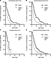
Autoimmune polyendocrinopathy-candidiasis-ectodermal dystrophy (APECED/APS-1) is a monogenic autoimmune disorder of impaired central tolerance classically diagnosed by the presence of 2 out of 3 classic triad manifestations: chronic mucocutaneous candidiasis, hypoparathyroidism, and adrenal insufficiency. However, many patients develop non-triad manifestations years earlier, delaying recognition and care. In 2016, we proposed expanded diagnostic criteria incorporating 3 early clinical manifestations — APECED rash, autoimmune enteritis, and enamel hypoplasia — based on observations in 35 North American patients. Here, we provide further support for the clinical utility of these expanded diagnostic criteria in independent cohorts of 57 American and 12 European patients enrolled in a prospective natural history study at the NIH. Across all cohorts, the expanded diagnostic criteria decreased the time to diagnosis by half relative to the classic diagnostic criteria. Patients exhibited an enrichment of early non-endocrine autoimmune manifestations, underscoring disease heterogeneity and the potential for developing organ-specific autoimmunity before endocrine failure. These findings demonstrate the clinical utility of the expanded APECED diagnostic criteria and support the notion that their adoption might enable earlier disease recognition and timely immunomodulatory therapy to improve long-term outcomes.
Authors
Elise M.N. Ferré, Joseph Pechacek, Monica M. Schmitt, Taura Webb, Heather Moorman, Thomas DiMaggio, Princess Barber, Vasielios Oikonomou, Stacey R. Rose, Peter D. Burbelo, Lindsey B. Rosen, Amy P. Hsu, Jennifer Stoddard, Shakuntala Rampertaap, Sergio D. Rosenzweig, Anjali Rai, Maria Teresa Magone, Chantal Cousineau-Krieger, Niki M. Moutsopoulos, Pamela J. Gardner, Heidi H. Kong, Leslie Castelo-Soccio, Ariane Soldatos, Katherine R. Calvo, Meryl Waldman, Behdad Afzali, Stefania Pittaluga, David Kleiner, Steven M. Holland, Kevin Fennelly, Jill Rothschild, Bryce A. Seifert, Magdalena Walkiewicz-Yvon, Theo Heller, Karen Winer, Michail S. Lionakis
Abstract
Systemic sclerosis (SSc) is a rare autoimmune disease characterized by vasculopathy and fibrosis of the skin and internal organs. Individuals with SSc often suffer from chronic acid reflux and dysphagia due to loss of esophageal motility. However, the pathogenesis of esophageal dysmotility in SSc is poorly understood. To determine whether distinct changes in esophageal epithelial cells contribute to esophageal involvement in SSc, we investigated the stratified squamous esophageal epithelium from proximal and distal biopsies using single-cell RNA sequencing (n=306,372 cells) in individuals with SSc compared those with gastroesophageal reflux disease (GERD) and healthy controls. The proportion of epithelial cells in the apical, superficial compartment of the esophageal epithelium was reduced in SSc (9.4% vs 21.6% in HCs). Differential gene expression in SSc was primarily limited to the superficial compartment (3,572 genes vs. 232 in all other compartments, based on pseudobulk analysis), with significant upregulation of extracellular matrix and keratinization genes. These cellular and molecular changes in SSc were highly correlated with those seen in GERD, indicating they were secondary to reflux; however, their magnitudes were more pronounced in the proximal esophagus, suggesting that esophageal dysmotility leads to greater proximal acid exposure, which may contribute to aspiration. SSc-specific gene dysregulation implicated immunoregulatory pathways likely pertinent to pathogenic mechanisms. Ligand-receptor interaction analysis revealed enhanced pro-fibrotic signaling between fibroblasts and epithelial cells in SSc. Cell type localization and SSc-specific changes were confirmed by spatial molecular imaging. By offering a comprehensive view of transcriptional dysregulation at single-cell resolution in human esophageal epithelial cells in SSc compared to GERD and healthy tissue, this work clarifies the state of epithelial cells in SSc-induced esophageal dysfunction.
Authors
Matthew Dapas, Margarette H. Clevenger, Hadijat-Kubura M. Makinde, Tyler Therron, Dustin A. Carlson, Mary Carns, Kathleen Aren, Cenfu Wei, Kainat Mian, Lutfiyya N. Muhammad, Carrie L. Richardson, Parambir S Dulai, Monique Hinchcliff, John E. Pandolfino, Harris R. Perlman, Deborah R. Winter, Marie-Pier Tetreault
No posts were found with this tag.


