
Pulmonology
Abstract
Patients who survive sepsis are predisposed to new hospitalizations for respiratory failure, but the underlying mechanisms are unknown. Using a murine model in which prior sepsis predisposes to enhanced lung injury, we previously discovered that classical monocytes persist in the lungs after long-term recovery from sepsis and exhibit enhanced cytokine expression after secondary challenge with intra-nasal lipopolysaccharide. Here, we hypothesized that immune reprogramming of post-sepsis monocytes and altered ontogeny predispose to enhanced lung injury. Monocyte depletion and/or adoptive transfer was performed three weeks and three months after sepsis. Monocytes from post-sepsis mice were necessary and sufficient for enhanced LPS-induced lung injury and promoted neutrophil degranulation. Prior sepsis enhanced JAK-STAT signaling and AP-1 accessibility in monocytes and shifted monocytes toward the neutrophil-like monocyte lineage. Neutrophil-like monocytes demonstrated a pro-inflammatory phenotype with enhanced IL-1β expression and reduced phagocytic capacity. In human sepsis and/or pneumonia survivors, monocytes were predictive of 90-day mortality and exhibit transcriptional and proteomic neutrophil-like signatures. We conclude that sepsis reprograms monocytes into a pro-inflammatory phenotype and skews bone marrow progenitors and monocytes toward the neutrophil-like lineage, predisposing them to induce neutrophil degranulation and lung injury.
Authors
Scott J. Denstaedt, Breanna McBean, Alan P. Boyle, Brett C. Arenberg, Matthias Mack, Bethany B. Moore, Michael W. Newstead, Yamei Deng, Alexey I. Nesvizhskii, Benjamin H. Singer, Jennifer Cano, Hallie C. Prescott, Helen S. Goodridge, Rachel L. Zemans
Abstract
Sex-specific mechanisms in chronic obstructive pulmonary disease (COPD) remain poorly defined. Sphingolipids are bioactive mediators that regulate airway epithelial integrity, inflammation, and bronchial smooth muscle tone, and their dysregulation contributes to airway hyperresponsiveness. We tested whether sphingolipid metabolism is regulated by sex in COPD. Lung tissue and paired plasma from female and male never-smoking controls (NSC), ever-smoking controls (SC), and COPD subjects were analyzed for sphingolipid enzyme expression and metabolite levels. Bronchial rings from mice and human bronchial smooth muscle cells (HBSMCs) were exposed to estradiol or cigarette smoke extract (CSE) ± ceramidase or sphingosine kinase inhibitors. Expression of sphingolipid regulators ASAH1 and ORMDL3 was increased in COPD airways, significantly higher in women, and correlated with airway smooth muscle remodeling. Plasma from females with COPD showed increased sphingosine-1-phosphate (S1P), whereas males exhibited higher ceramide levels. Female mice displayed greater CSE-induced bronchial reactivity, attenuated by sphingosine kinase inhibition. In male-donor HBSMCs, estradiol increased ASAH1, ORMDL3, S1P, and smooth muscle protein expression and enhanced contractility. CSE further augmented molecular responses in estradiol-treated cells but reduced collagen-gel contraction, whereas ceramidase inhibition attenuated estradiol-dependent effects. These findings suggest that estrogen-dependent alterations in sphingolipid metabolism contribute to airway hyperreactivity and remodeling in females with COPD.
Authors
Elisabetta Granato, Xiaoyun Wang, Yun Zhang, Joselyn Rojas-Quintero, Ida Cerqua, Scott A. Ochsner, Jeff Thomas Kue, Luca Cecchetto, Maor Sauler, Farrah Kheradmand, Joshua Malo, Fiorentina Roviezzo, Irina Petrache, Francesca Polverino
Abstract
Type 2 innate lymphoid cells (ILC2) are a population of lineage-negative cells in the lung, gastrointestinal tract, and skin which have emerged as a significant component of type 2 allergic inflammation. The regulation of cyclooxygenase-2 (COX-2) metabolites is critical to the pathophysiology of many inflammatory disorders, including allergic asthma. While COX-2 regulates Th9 and Th17 cell differentiation and function during allergic lung inflammation, it remains unknown whether COX-2 also regulates ILC2 cell differentiation and function under similar conditions. To address this question, we examined lung ILC2 cells from COX-2+/+ and COX-2-/- mice after ovalbumin (OVA)- or Alternaria-induced allergic lung inflammation. ILC2 cells were significantly increased in COX-2-/- lungs compared with COX-2+/+ lungs after OVA exposure in vivo. The increase in ILC2 cells was accompanied by an increase in expression of the cytokines IL-5 and IL-13, and the transcription factor GATA3. Both genetic disruption and selective inhibition of COX-2 significantly increased ILC2 cell differentiation from isolated common lymphoid progenitor cells (CLP) in vitro. Furthermore, COX-2-derived PGE2 acting via EP2 receptors significantly reduced IL-33 and TSLP expression, and attenuated ILC2 cell differentiation in vitro and in vivo. Thus, during allergic lung inflammation, COX-2-derived PGE2 signals through the EP2 receptor to negatively regulate lung ILC2 cell differentiation and function.
Authors
Hong Li, Matthew L. Edin, Daniel Menendez, J. Alyce Bradbury, Joan P. Graves, Artiom Gruzdev, Gregory S. Whitehead, Maria I Sifre, Laura M. DeGraff, Darryl C. Zeldin
Abstract
Both elevated plasma cell-free hemoglobin (CFH) and heparanase-driven endothelial glycocalyx shedding are contributors to microvascular dysfunction and organ injury in sepsis. However, the mechanisms governing heparanase activation, and the potential role of CFH in this process, are not understood. Utilizing patient samples, mice with cecal slurry-induced (CS) peritonitis and elevated CFH, and human lung microvascular endothelial cells (HLMVECs), we tested the hypothesis that CFH upregulates heparanase production to drive endothelial glycocalyx degradation. In human sepsis, elevated circulating CFH was associated with higher heparanase and heparan sulfate levels, which in turn correlated with adverse clinical outcomes. CS+CFH-treated mice had increased plasma heparanase, glycocalyx degradation, and pulmonary and systemic inflammation; endothelial heparanase deletion abrogated these effects. Additionally, in both pulmonary endothelial cells isolated from CS+CFH-treated mice and HLMVECs exposed to CFH and TNF, heparanase transcription and active enzyme production were increased. The deleterious effects of CFH were attenuated by acetaminophen, a hemoprotein reductant. In summary, we demonstrate that CFH oxidation stimulates endothelial heparanase expression and activation during sepsis, leading to endothelial glycocalyx degradation, which may disrupt the endothelial barrier and result in organ injury. Our findings highlight the CFH-heparanase axis as a potential therapeutic target for endothelial glycocalyx preservation in sepsis.
Authors
Avery M. Bogart, Anisa S. Haffizulla, Jason Lin, Nathan D. Putz, Han Noo Ri Lee, David M. Aslaner, Samantha K. Gonski, Nancy Wickersham, Kyle Riedmann, Jamie E. Meegan, Kaori Oshima, Ciara M. Shaver, Julie A. Bastarache, Eric P. Schmidt, Lorraine B. Ware
Abstract
Authors
Jianfei Ji, Wenjing You, Xiaoli Sun, Peng Zhao
Abstract
Despite advances in treatment approaches for lung cancer, the morbidity and survival of lung cancer patients with malignant pleural effusions (MPE) remain poor. This is in part due to gaps in understanding the role of immune cells in the pleural fluid microenvironment. We performed single cell analysis with flow cytometry validation of CD45+ cells in eight malignant and five benign pleural fluid (BPE) specimens to identify changes in the transcriptomic landscape of immune cells across disease states. We found upregulation of pro-inflammatory signaling pathways, including interferon and TNF signaling, in T cells, B cells, and macrophages in benign compared to malignant pleural effusions. Pro-inflammatory HLA-DR+ macrophages were associated with good survival outcomes while pro-tumorigenic HLA-DR- macrophages with upregulation of angiogenesis, TGFβ, and fibronectin signaling were associated with poor survival outcomes in patients with MPE. We also validated these findings with macrophage cell surface expression markers using flow cytometry in 14 MPE and 7 BPE specimens. Finally, we performed multiplex cytokine analysis which showed enrichment of the type 3 inflammatory cytokine, IL17A, in MPE as a putative mechanism for macrophage reprogramming. These data provide a rich resource for interrogating the immune cell types and states present across the spectrum of pleural disease. They offer not only prognostic value for patient outcomes at the time of pleural fluid collection, but also insights into novel immunotherapy targets.
Authors
Aaditya Khatri, Huimin Wang, Zhicheng Ji, Prekshaben Patel, Smita K. Nair, Javid P. Mohammed, Beth H. Shaz, Andrew B. Nixon, Scott M. Palmer, Kamran Mahmood
Abstract
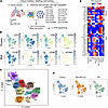
Idiopathic pulmonary fibrosis (IPF) is a fatal interstitial lung disease characterized by progressive scarring and respiratory failure. While T cells are elevated in IPF lungs, their contributions to fibrosis beyond inflammation remain poorly understood. Here, we performed multiplex imaging and single-cell RNA and protein profiling on about 90,000 CD3+ T cells from control and fibrotic lungs, revealing 11 distinct subsets of CD4+ and CD8+ T cells, including a rare CD56+ regulatory T cell. In addition to increased T cell numbers in severely fibrotic lungs compared with non-diseased controls, we observed CD4+ and CD8+ T cells localized near epithelial cells and in niches of abnormal epithelium. CXCR4/MIF signaling emerged as a central axis mediating T cell–epithelial interactions, while epidermal growth factor receptor (EGFR) and TGF-β pathways dominated in multiple T cell subsets. Our findings support the concept that T cells in IPF adopt nonclassical activation patterns that are driven by epithelial interactions within the fibrotic microenvironment. These studies provide a foundation for exploring alternative therapeutic strategies in IPF lungs by modulating T cell behavior and communication networks.
Authors
Ana P.M. Serezani, Julia M.R. Bazzano, Bruno D. Pascoalino, Ludmilla da Silva, Abigail J. Dietrich, Chase J. Taylor, Taylor Sherrill, Annika Vannan, Carla L. Calvi, Paula I. Gonzalez-Ericsson, Erin M. Wilfong, Matthew Bacchetta, Ciara M. Shaver, Lorraine B. Ware, Margaret L. Salisbury, Luc Van Kaer, Nicholas E. Banovich, Jonathan A. Kropski, Timothy S. Blackwell
Abstract
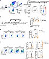
The cholinergic antiinflammatory pathway attenuates lung inflammation via the α7 nicotinic acetylcholine receptor (α7 nAChR) on immune cells. However, the role of α7 nAChR on lung megakaryocytes (Mks) in allergic airway inflammation remains unknown. In this study, allergen-challenged mouse models were used with conditional Mk-specific Chrna7 knockout, pharmacological activation (GTS-21), and Mk reconstitution. IL-33 expression and p38 MAPK signaling were assessed. We found that allergen challenge upregulated α7 nAChR specifically in lung Mks. Mk-specific Chrna7 deletion significantly alleviated allergic airway inflammation, whereas GTS-21 exacerbated inflammation via an Mk-dependent mechanism. Reconstitution with α7 nAChR+ Mks restored airway inflammatory responses. Mechanistically, α7 nAChR activation promoted Mk IL-33 synthesis and secretion through p38 MAPK signaling. Taken together, our results show that α7 nAChR on lung Mks plays a proinflammatory role in allergic airway inflammation, challenging its classical antiinflammatory paradigm and revealing pathogenic mechanisms.
Authors
Hang Wu, Rujia Tao, Shitao Xie, Yao Zhou, Jin-Fu Xu, Zhenwei Xia, Xiao Su

Abstract
Authors
Lori Asarian, Jianlong Jia, Dmytro Sirokha, Lara Paulini, Daniela Dietel, Mircea Gabriel Stoleriu, Marion Frankenberger, Ali Önder Yildirim, Katharina S. Götze, Juergen Behr, Gary M. Hunninghake, Isis E. Fernandez
Abstract
Authors
Allen Duong, Sajad Moshkelgosha, Tereza Martinu, Stephen Juvet
No posts were found with this tag.


