

Abstract
Autoimmune Addison’s disease (AD) is a rare but life-threatening disorder caused by immune-mediated destruction of the adrenal cortex, and progress in therapy has been limited by insufficient mechanistic insight. Here, we establish a model of experimental autoimmune adrenalitis that recapitulates key features of AD and reveals sex-dependent differences in disease manifestation within the model. Immunization with peptides derived from the adrenal self-antigen CYP11A1 induces corticosterone insufficiency. We show that autoimmune adrenalitis is driven by IFN-γ produced by self-reactive CD4+ T cells, promoting granulomatous inflammation in the adrenal cortex. Together, these findings identify IFN-γ as a central effector of autoimmune adrenalitis and suggest that targeting the IFN-γ pathway may represent a potential therapeutic strategy for AD.
Authors
Arina Andreyeva, Juraj Michalik, Veronika Niederlova, Veronika Cimermanova, Ales Drobek, Radislav Sedlacek, Jan Prochazka, Juraj Labaj, Olha Fedosieieva, Waldemar Kanczkowski, Peter Draber, André Sulen, Ondrej Stepanek, Aleš Neuwirth

Abstract
Right ventricular pressure overload (RVPO) is a critical pathophysiological feature of numerous pediatric cardiovascular diseases. Transverse tubules (T-tubules) form the foundation for efficient excitation-contraction coupling in mature cardiomyocytes. We hypothesized that RVPO impairs T-tubule maturation through the regulatory protein bridging integrator 1 (BIN1). In right ventricular samples from children with tetralogy of Fallot, characterized by RVPO, and in a neonatal rat RVPO model induced by pulmonary artery banding (PAB), T-tubule maturation was disrupted. RNA-seq revealed significant downregulation of T-tubule–associated genes, with Bin1 among the most suppressed. Bin1 overexpression restored T-tubule maturation in PAB rats. ATAC-seq showed reduced chromatin accessibility at Bin1 loci; motif analysis identified Mef2d (myocyte enhancer factor 2D) as the top enriched transcription factor. Mef2d knockdown rescued Bin1 expression and T-tubule maturation, and mutation of the Mef2d binding sites within the Bin1 promoter abolished the inhibitory effect of Mef2d on Bin1 promoter activity. This study delineates a phenomenon and a mechanism of cardiomyocyte maturation under pathological stress. The findings not only advance our understanding of this most pivotal event in postnatal cardiac development but also unveil a potential therapeutic direction for pediatric cardiovascular diseases associated with RVPO.
Authors
Yuqing Hu, Yiting Xue, Xudong Chen, Linghui Kong, Debao Li, Zheng Wang, Sixie Zheng, Siqi She, Hao Li, Sijuan Sun, Hao Chen, Lijun Chen, Peisen Ruan, Kai Wang, Lincai Ye
Abstract
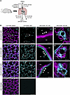
CFTR in the lung epithelium contributes to the secretion of a surface liquid layer that is essential to lung homeostasis and defense. The understanding of how liquid is secreted in the lung is derived largely from studies of the airway epithelium. Comparatively little is known about liquid secretion mechanisms in the alveolar epithelium, including its cellular source. To define which cell type drives alveolar liquid secretion, we generated transgenic mice that expressed a Cftr-null allele in alveolar type 1 (AT1) cells, type 2 (AT2) cells, or both, then viewed liquid secretion in live alveoli using confocal microscopy of isolated, perfused lungs. Our findings show that liquid secretion was blocked in alveoli of all 3 transgenic mice, indicating that both AT1 and AT2 cells contribute to alveolar liquid secretion. Cftr-null expression in AT1 cells also blocked the secretion-mediated clearance of small particle and bacterial clusters from alveolar walls, indicating that AT1 cell CFTR contributes to alveolar defense. Together, these findings show alveolar liquid secretion depends on both AT1 and AT2 cell CFTR, and that CFTR in AT1 cells — a cell type not traditionally considered in liquid secretion mechanisms or CFTR-related lung diseases — contributes to lung liquid dynamics and host defense.
Authors
Sayahi Suthakaran, Sonya Homami, Deebly Chavez, Stephanie Tang, Sarah K.L. Moore, Chaya Sussman, Jimmy Zhang, Clemente J. Britto, Alice Prince, Alison J. May, Jaymin J. Kathiriya, Jaime L. Hook
Abstract
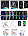
The glymphatic-meningeal pathway, important for brain homeostasis, depends on the drainage function of the cervical lymphatic system. Although new therapies aim to modulate this pathway, a lack of methods for quantifying lymphatic drainage function hinders our ability to understand how targeting the cervical lymph nodes may benefit brain health. To address this, we developed and applied a fluid transport model to dynamic contrast-enhanced MRI (DCE-MRI) data to visualize and quantify tracer-tagged lymph through the deep cervical lymph nodes (dcLNs). The model incorporated physical principles of solute transport to provide a biologically interpretable framework for analyzing microflows in real time. We applied this model to investigate the effects of chronic hypertension on dcLN drainage by comparing normotensive Wistar-Kyoto rats with spontaneously hypertensive stroke-prone (SHRSP) rats. In normal rats, the model revealed complex and tortuous lymph streams of a 200 kDa tracer transported through the sinus system of the dcLNs. In contrast, SHRSP rats exhibited significantly altered fluid dynamics, characterized by simpler stream patterns and reduced flow through the dcLNs. These findings demonstrated that untreated chronic hypertension adversely affects lymph node drainage function. This study provides potentially new insight into impaired lymphatic drainage as a mechanism linking systemic disease to brain health.
Authors
Kaiming Xu, Ankita Bhardwaj, Sunil Koundal, Qin Ren, Chenyu You, Xenophon Papademetris, Helene Benveniste, Tryphon T. Georgiou
Abstract
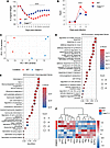
Severe SARS-CoV-2 infection is characterized by lung hyperinflammation, impaired IFN responses, and defective T cell activation, yet the molecular drivers of these immune dysregulations remain incompletely understood. Caspase-11 (CASP11), a key effector of the noncanonical inflammasome, has been shown to mediate an innate hyperinflammatory response and cytokine release in a mild SARS-CoV-2 infection model. However, the role played by CASP11 in severe SARS-CoV-2 disease and how it affects adaptive immunity has not been identified. Here, we found that CASP11 exacerbates severe SARS-CoV-2 pathogenesis by amplifying early innate immune responses while concurrently impairing antiviral CD8+ T cell immunity. Using global KO mice, hematopoietic BM chimeras, and Cx3cr1-expressing mononuclear phagocyte system cell–specific CASP11 deletion models, we show that targeting CASP11 reduces lung inflammation, promotes early NK cell–mediated IFN-γ production, and enhances robust virus-specific effector CD8+ T cell responses. This was associated with enhanced viral clearance and improved survival, even under lethal infection conditions. Importantly, CASP11-KO mice also exhibited faster resolution of postviral inflammation. These findings position CASP11 as a promising immunomodulatory target for acute and delayed manifestations of severe SARS-CoV-2 infection.
Authors
Mostafa M. Eltobgy, Mohamed M. Shamseldin, Owen D. Whitham, Heba M. Amer, Jeffrey R. Atkinson, Asmaa Badr, Jesse M. Hall, Gauruv Gupta, Yara Y. Hassan, Rabab El-Mergawy, Richard Perez, Sarah E. Faber, Maciej Pietrzak, Amy Webb, Xiaoli Zhang, Adam D. Kenney, Destiny Bissell, Jihad I. Omran, Shady Estfanous, Kylene P. Daily, Amir Yousif, Marisa R. Joldrichsen, Andrew McNamara, Mahesh KC, Mark E. Peeples, Emily A. Hemann, Hazem E. Ghoneim, Shahid M. Nimjee, Estelle Cormet-Boyaka, Jianrong Li, Prosper N. Boyaka, Jacob S. Yount, Benjamin M. Segal, Purnima Dubey, Amal O. Amer
Abstract
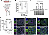
ACE2 is a membrane-bound monocarboxypeptidase strongly expressed in the renal proximal tubule (PT) with high affinity to degrade the vasopressor angiotensin II (AngII). We employed a mouse model of PT-specific ACE2 deletion (PT ACE2–KO) to demonstrate that the renal PT is a critical site for ACE2 regulation of blood pressure (BP) via modulation of the intrarenal renin-angiotensin system (RAS). While deletion of ACE2 from the PT had a minimal effect on baseline physiology, PT ACE2–KO mice were more susceptible to AngII hypertension than control mice. At day 5 of AngII infusion, the enhanced BP response was associated with cardiac hypertrophy, increased renal AngII levels, failure to suppress epithelial sodium channel (ENaC) γ cleavage, and increased sodium pump activity in PT ACE2–KO mice. Control mice instead increased renal ACE2 expression to reduce renal AngII accumulation and suppress intrarenal RAS activation, which offered protection from hypertension and complications. Transcriptional analysis corroborated changes in intrarenal RAS components and revealed alterations in distinct physiological pathways during AngII hypertension in PT ACE2–KO mice. Our studies provide evidence for alterations in ENaC regulation to contribute to the development of AngII hypertension and support PT-derived ACE2 as an integral member of the intrarenal RAS.
Authors
Jacqueline M. Emathinger, Zhidan Xiang, Jonathan W. Nelson, Matthew W. Hagen, Nakyung Kim, David I. Ortiz-Melo, Natalie Mattocks, Jorge F. Giani, Dexter L. Lee, Hannah Hartman-Houstman, Donna L. Ralph, Alicia A. McDonough, Brianna Chen, Stan G. Louie, Thomas M. Coffman, Susan B. Gurley
Abstract
Mycoplasma pneumoniae pneumonia (MPP) can cause serious extrapulmonary complications, including life-threatening thrombosis. This study aimed to elucidate the roles of neutrophils and neutrophil extracellular traps (NETs) in vascular endothelial cell (EC) activation in pediatric MPP-associated thrombosis. We analyzed the relationship between neutrophils and thrombosis in children with MPP and used mouse models of neutrophilia (Csf3 plasmid injection), neutropenia (Csf3 deficient, Csf3–/–), and defective NETs formation (Pad4 deficient, Pad4–/–). The effects of neutrophils and NETs on EC activation were further examined in vivo, in vitro, and in human samples. Elevated neutrophil count was observed in patients with thrombosis and functioned as a potential diagnostic marker as well as a risk factor for MPP-associated thrombosis. EC activation was enhanced in MPP mice with neutrophilia but attenuated in neutropenic or Pad4–/– mice. NETs activated ECs through TLR2 and JAK/STAT3 signaling, and inhibition of NETs formation (Cl-amidine), TLR2 (C29), and JAK (upadacitinib) each attenuated this response. Strong correlations among neutrophils, NETs, EC activation, and thrombosis were observed in pediatric patients. These findings suggest that neutrophils promoted thrombosis in MPP via NETs-mediated EC activation involving TLR2 and JAK/STAT3 signaling. This study provides mechanistic insights into the inflammatory-thrombotic processes in MPP-associated thrombosis and offers a rationale for further investigation of neutrophils, NETs, TLR2, and JAK/STAT3 signaling in this context.
Authors
Xia Huang, Yifan Zhu, Yun Guo, Tian Lv, Haiyan Gu, Yingying Luo, Dan Li, Hang Sun, Deyu Zhao, Feng Liu

Mitochonic acid 5 alleviates amyotrophic lateral sclerosis phenotypes via mitochondrial augmentation
Abstract
Amyotrophic lateral sclerosis (ALS) is a fatal neurodegenerative disease that urgently requires effective treatment. Mitochondrial dysfunction underlies ALS pathology and represents a potential therapeutic target. Here, we demonstrated the therapeutic potential of mitochonic acid 5 (MA-5), a novel mitochondria-targeted compound that ameliorated ALS phenotypes by enhancing mitochondrial function. In a Drosophila ALS model expressing a mutant human SOD1 (G85R), MA-5 significantly improved locomotor activity, with a trend toward restoration of mitochondrial integrity. In skin fibroblasts derived from ALS patients and motor neurons derived from induced pluripotent stem cells, MA-5 restored ATP production and increased mitochondrial motility. Multiomics analyses suggested that MA-5 modulated mitochondria-linked gene expression and downregulated the glycerophosphate shuttle, contributing to mitochondrial reactive oxygen species production. Transcriptomic analysis identified C7orf31 as a potential marker for monitoring the therapeutic effects of MA-5 and diagnosing ALS subtypes. These findings support MA-5 as a promising therapeutic candidate for ALS and propose C7orf31 as a potential biomarker for treatment monitoring and for disease subtyping.
Authors
Yoshitsugu Oikawa, Yuhan Luo, Naoki Suzuki, Tomoko Kasahara, Yoshiyasu Tongu, Yuki Yoshida, Tsukasa Tominari, Shogo Tanabe, Yoshiko Suto, Hitomi Kashiwagi, Saki Saito, Kensuke Ikeda, Chitose Suzuki, Arata Kuranaga, Tetsuya Akiyama, Satoru Morimoto, Yoshitsugu Aoki, Rieko Muramatsu, Tomoyoshi Soga, Masashi Aoki, Hideyuki Okano, Tetsuhiro Tanaka, Takaaki Abe, Erina Kuranaga, Takafumi Toyohara

Abstract
Small molecules that modulate myofibril ATPase activity via the myosin regulatory light chain (RLC) display a broad spectrum of activity in their ability to enhance relaxation and slow contraction. EDG-7500 exhibits features consistent with a ‘diastolic-selective’ cardiac sarcomere modulator (d-CSM), and its ability to treat HCM was explored in engineered human tissue (EHT), human HCM cardiac strips, and an R403Q mutation swine model. In fibers, EDG-7500 preferentially inhibited myofibril ATPase activity and force at diastolic calcium levels, retained length-dependent force activation, accelerated relaxation, and exhibited a shallow, self-limiting inhibitory-exposure response to LV fractional shortening. Compared to CMIs, EDG-7500 moved myosin heads towards the thin filament and accelerated relaxation without decreasing force in mutated EHTs (R403Q). In human HCM cardiac strips, EDG-7500 did not alter myosin SRX state, but decreased Ca2+-sensitivity of force independent of mutation. In R403Q swine, chronic EDG-7500 normalized LV filling pressure and prevented pathological cardiac remodeling while preserving normal systolic function and cardiac reserve. EDG-7500 differentiates itself from CMIs by uniquely targeting both phases of the cardiac cycle, improving ventricular relaxation while preserving systolic function. This suggests optimal diastolic efficacy can be reached without balancing systolic impairment.
Authors
Craig A. Emter, Marcus Henze, Mike DuVall, Sarah Lehman, Lindsey Lee, Ben Barthel, Natalie A. Hawryluk, Molly Madden, Yangsong Wu, Amy Perry, Martin Beyer, Eric Wei, Cassady Rupert, Steve Roof, Angela Peter, Emily DiNatale, Sara Cantrell, Jessica Tolley, Stephen Schlachter, Jolanda van der Velden, Michelle Michels, Christine Seidman, Weikang Ma, Leslie Leinwand, Stuart Campbell, Julien Ochala, David Bluemke, Darla Tharp, Jonathan Seidman, Carlos L. del Rio, Marc Semigran, Marc Evanchik, Kevin Koch, Alan Russell
Abstract
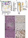
Metastases in renal cell carcinoma (RCC) typically arise from large primary tumors. However, a subset of patients with small renal masses (SRMs; ≤4 cm) can develop metastatic disease. Identifying these tumors is clinically important, as many SRMs are managed with active surveillance, and their study may provide insight into the early acquisition of metastatic competence. It remains unclear whether these tumors acquire distinct metastatic programs or instead show premature activation of the same aggressive programs typically associated with larger tumors. Here, we performed integrated morphological and molecular profiling of a multiinstitutional cohort of metastatic SRMs, including whole-exome sequencing and RNA-Seq, using nonmetastatic primary tumors as controls. Among metastatic, non–clear cell SRMs, we identified NF2-altered tumors, ELOC-mutated RCC, and an mTOR-driven eosinophilic vacuolated tumor. Metastatic clear cell SRMs were enriched by multi-hit aggressive genotypes, including recurrent losses of chromosomes 8p, 9, and 14q, as well as co-occurring driver alterations (≥2 events), including BAP1 and mTOR pathway genes. Transcriptomic analyses revealed enrichment of the non-negative matrix factorization 3 (NMF3) subtype from the IMmotion151 trial-based taxonomy, along with metabolic rewiring and reduced cytotoxic immune effector function. Collectively, these findings identify molecular programs associated with metastatic competence in SRMs, highlight the importance of genomic studies of equivocal non-clear cell SRMs, and provide a biological framework for risk stratification in patients often considered for active surveillance.
Authors
Payal Kapur, Daria Beshnova, Hua Zhong, Ruby Sharma, Pooja Ghatalia, Angela Yoo, Daniel D. Le, Ratna Mukhopadhyay, Alana Christie, Jeffrey Miyata, Shuanzeng Wei, Rana R. McKay, Dinesh Rakheja, Satwik Rajaram, Robert G. Uzzo, A. Ari Hakimi, Zora Modrusan, James Brugarolas
No posts were found with this tag.


