
Genetics
Abstract
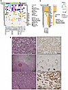
Metastases in renal cell carcinoma (RCC) typically arise from large primary tumors. However, a subset of patients with small renal masses (SRMs; ≤4 cm) can develop metastatic disease. Identifying these tumors is clinically important, as many SRMs are managed with active surveillance, and their study may provide insight into the early acquisition of metastatic competence. It remains unclear whether these tumors acquire distinct metastatic programs or instead show premature activation of the same aggressive programs typically associated with larger tumors. Here, we performed integrated morphological and molecular profiling of a multiinstitutional cohort of metastatic SRMs, including whole-exome sequencing and RNA-Seq, using nonmetastatic primary tumors as controls. Among metastatic, non–clear cell SRMs, we identified NF2-altered tumors, ELOC-mutated RCC, and an mTOR-driven eosinophilic vacuolated tumor. Metastatic clear cell SRMs were enriched by multi-hit aggressive genotypes, including recurrent losses of chromosomes 8p, 9, and 14q, as well as co-occurring driver alterations (≥2 events), including BAP1 and mTOR pathway genes. Transcriptomic analyses revealed enrichment of the non-negative matrix factorization 3 (NMF3) subtype from the IMmotion151 trial-based taxonomy, along with metabolic rewiring and reduced cytotoxic immune effector function. Collectively, these findings identify molecular programs associated with metastatic competence in SRMs, highlight the importance of genomic studies of equivocal non-clear cell SRMs, and provide a biological framework for risk stratification in patients often considered for active surveillance.
Authors
Payal Kapur, Daria Beshnova, Hua Zhong, Ruby Sharma, Pooja Ghatalia, Angela Yoo, Daniel D. Le, Ratna Mukhopadhyay, Alana Christie, Jeffrey Miyata, Shuanzeng Wei, Rana R. McKay, Dinesh Rakheja, Satwik Rajaram, Robert G. Uzzo, A. Ari Hakimi, Zora Modrusan, James Brugarolas
Abstract
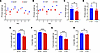
Prader-Willi syndrome (PWS) is a complex genetic disorder resulting from the deficiency of several maternally imprinted genes, including SNORD116, in the 15q11-q13 region. Loss of Snord116 in mice recapitulates some of the most salient clinical features of PWS, including growth hormone (GH) deficiency and hypogonadism. This study explored the impact of Snord116 deficiency on early postnatal pituitary development and growth in Snord116-KO mice. Snord116 was found to be expressed in both the anterior and posterior pituitary. Pituitary transcriptomes of Snord116-KO and WT mice at 2 developmental stages, P0 and 4 weeks of age, were interrogated and related to ex vivo analyses of GH secretion in the pituitaries of 5-week-old mice. Significant differences in pituitary transcriptomes were detected between Snord116-KO and WT mice at 4 weeks of age but not at P0. The differentially expressed genes and affected molecular pathways play important roles in regulating embryonic and postnatal pituitary development. Our results suggested that PWS GH deficiency was mainly due to pituitary hypoplasia and decreased GH production but not to reduced GH secretory function per se, implicating Snord116 in the specific molecular/cellular pathways that account for impaired postnatal pituitary development and GH deficiency in PWS.
Authors
Gabriel F. Batzli, Kaiying Guo, Fahrünisa Meryem Betül Erol, Charles A. LeDuc, Lisa C. Burnett, Rudolph L. Leibel, Yiying Zhang

Abstract
BACKGROUND. Loss of the Y chromosome (LOY) is a frequent event in male tumors and has been linked to cancer progression. However, the degree of mosaic LOY (mLOY) within normal tissues from men with or without cancer remains uncharacterized. METHODS. Here we used a FISH-based assay targeting X- and Y-chromosome centromeres to perform a pan-organ analysis of mLOY in 1,000 male tissue samples from 405 individuals representing 11 organs. Automated image processing generated a quantitative FISH-based mLOY score (YchrFISH) that we validated against a transcriptomic surrogate of Y-chromosome dosage from RNA-seq data. RESULTS. mLOY burden varied by tumor type, with highest degree in colorectal carcinoma. Across tissue groups, YchrFISH scores declined progressively from normal tissues of cancer-free men to histologically normal tissues adjacent to cancer and carcinoma (P < 0.0001). Paired analyses confirmed consistently greater mLOY in malignant compared with tumor-adjacent histologically normal tissue in different organs. Spatially resolved RNA-seq maps of bladders removed for cancer demonstrated a transcriptional gradient of Y-chromosome loss from normal urothelium through intraepithelial neoplasia to invasive carcinoma. CONCLUSION. mLOY gradients exist across histologically normal and malignant tissues, consistent with the concept of field cancerization. Our findings support epithelial mLOY as a biomarker of early malignant transformation and, to our knowledge, a previously unrecognized hallmark of male oncogenesis. FUNDING. NIH grants R35CA294022, P01CA163227, and P50CA97186 (the Pacific Northwest Prostate Cancer SPORE) and the Institute for Prostate Cancer Research.
Authors
Arkadiusz Gertych, Huihui Ye, Xingyu Chen, Eric Vail, V. Krishnan Ramanujan, Lauren Brady, Lawrence D. True, Peter S. Nelson, Peter R. Carroll, Dan Theodorescu
Abstract
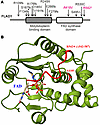
The FLAD1 gene codes for flavin adenine dinucleotide (FAD) synthase. FAD is a cofactor for many redox enzymes involved in vital processes from respiration to signal transduction. In this work, we described a clinical case of 2 siblings carrying compound heterozygous mutations in the FLAD1 gene resulting in the substitutions A418V and R542* at the protein level. The patients demonstrate adrenal insufficiency, which has not previously been associated with FLAD1 protein defects. To verify that adrenal insufficiency is caused by FLAD1 mutations, we created a personalized mouse model carrying the mutations found in the patients. The mutation in the FLAD1 gene, leading to the A418V substitution, appeared viable in the homozygous state, with minimal difference from the WT. The FLAD1 gene mutation leading to the R542* truncation is lethal when homozygous. The mouse model of the compound heterozygous FLAD1A418V/R542* mutations recapitulated the physiological, biochemical, and endocrine manifestations of FLAD1 mutations in patients. The mouse model created demonstrates the causal effect of FLAD1 mutations on the described pathology and potentially paves the way for understanding the disease’s molecular mechanism and developing better therapies.
Authors
Olga A. Averina, Natalia Yu. Kalinchenko, Vitaly A. Ioutsi, Andrey V. Pirogov, Alexander V. Bogachev, Oleg A. Permyakov, Vitaly S. Buev, Ekaterina A. Guseva, Anastasia V. Priymak, Olga A. Bazhanova, Mariia A. Emelianova, Olga O. Grigoryeva, Galina V. Baydakova, Maxim A. Abakumov, Vasily N. Manskikh, Olga A. Dontsova, Petr V. Sergiev, Anatoly N. Tiulpakov
Abstract
Mutations in mitochondrial DNA (mtDNA) cause various mitochondrial diseases that are currently incurable. Allotopic expression of nuclear-recoded mitochondrial genes represents a promising therapeutic strategy, given its demonstrated capacity to restore mitochondrial function in human cell models harboring mtDNA mutations. However, the in vivo evaluation of allotopic gene therapy has been hindered by optimization challenges and the lack of appropriate animal models. Here, we overcome these limitations by utilizing an optimized AAV2-ND6 construct with codon optimization and mitochondrial targeting sequence in a mouse model bearing the homoplasmic ND6P25L mutation, which recapitulates Leber hereditary optic neuropathy (LHON). High-dose administration of the AAV2-ND6 construct resulted in robust, sustained expression within the retina and optic nerve without apparent systemic toxicity. Strikingly, We compared the therapeutic efficacy in mutant mice at different ages and pre-symptomatic intervention with AAV2-ND6 effectively attenuated disease progression, mitigated retinal cellular deficiencies and optic nerve damage, and restored visual function in ND6P25L mice. Mechanistically, allotopic ND6 expression markedly rescued the mitochondrial dysfunction, corrected dysregulated retinol metabolism and phototransduction pathways, and suppressed apoptotic processes in the mutant retina. Our study validates the safety and therapeutic potential of allotopic expression in vivo and provide critical mechanistic insights into its role in treating LHON and other mitochondrial diseases.
Authors
Cheng Ai, Huiying Li, Jing Wu, Tianwei Zhou, Jing Wang, Shao-Hui Pan, Jun Yu, Douglas C. Wallace, Min-Xin Guan

Abstract
Haploinsufficiency of TBX1, which occurs in 22q11.2 deletion syndrome (22q11.2DS), leads to a heterogeneous spectrum of clinical manifestations, including craniofacial anomalies, immunodeficiency, and congenital heart defects. The variability in syndromic presentation between patients may be partially explained by variants in chromatin regulatory genes that act to further modify TBX1 function. To investigate this relationship, we selected KMT2D as a candidate gene because of its role in the etiology of Kabuki syndrome, which shares overlapping features with 22q11.2DS. We demonstrate that conditional inactivation of Kmt2d in the Tbx1 lineage in Tbx1-heterozygous mice leads to fully penetrant perinatal lethality and increased incidence of craniofacial dysmorphism, thymus and parathyroid gland hypoplasia, and aortic arch anomalies. At early stages, mutant embryos were found to have defects of the caudal pharyngeal apparatus, including abnormal patterning of the third pouch endoderm, hypoplastic fourth arches, and defective fourth arch arteries. Finally, analysis of single-cell RNA sequencing revealed dysregulation, and largely downregulation, of genes involved in basic cellular functions, suggesting that Tbx1 and Kmt2d developmentally converge upon essential biological processes. Overall, these results indicate that reduced dosage of Kmt2d perturbs the developmental landscape of the Tbx1 heterozygote, eliciting phenotypes that are shared between 22q11.2DS and Kabuki syndrome.
Authors
Daniella Miller, Kevyn Jackson, Timothy C. Cox, Bernice E. Morrow
Abstract
Autosomal dominant leukodystrophy (ADLD) is a fatal-adult-onset CNS demyelinating disorder for which no treatment exists. The majority of ADLD cases are caused by duplications of the lamin B1 (LMNB1) gene, resulting in increased LMNB1 expression. While reducing LMNB1 levels represents a logical therapeutic strategy, its efficacy has not been previously demonstrated in any in vivo model. Mouse models with oligodendrocyte-specific human LMNB1 (hLMNB1) overexpression recapitulate salient features of ADLD. Using a modified version of this model, where hLMNB1 can be inducibly downregulated, we demonstrated that hLMNB1 reduction can prevent or substantially ameliorate disease progression. Therapeutic effects were maximized when hLMNB1 reduction was induced before expected symptom onset, resulting in improvements in behavioral, biochemical, histopathological, and survival measures relative to untreated animals. Reducing hLMNB1 levels after symptom onset led to improved survival, but mixed results for other disease phenotypes. In addition, we identified potential biomarkers that track disease progression. Furthermore, we demonstrated that near-complete knockdown of murine LMNB1 expression in adulthood did not result in any overt CNS phenotype. Together, these results provide a proof of concept supporting LMNB1 reduction as a therapeutic strategy and offer a rationale for treatments aimed at lowering levels of this protein in ADLD.
Authors
Nathan Herdman, Kaveh Moradi, Bruce Nmezi, Anushe Munir, Krizchelle A. Magtoto, Fang Liu, Mara Sullivan, Xuemei Zeng, Thomas K. Karikari, Quasar S. Padiath

Abstract
In α-1 antitrypsin (AAT) deficiency (AATD), emphysema is classically linked to protease-antiprotease imbalance caused by decreased antiprotease AAT due levels and function. This decrease is secondary to the impaired release of Z-AAT polymers from hepatocytes carrying Pi*Z, E342K mutation in SERPENA1 gene. Whether the accumulation of Z-AAT polymers in distal lungs contributes directly to emphysema pathogenesis has remained unexplored due to the lack of suitable model systems. We characterized lung injury and airspace enlargement in a Z-AAT–overexpressing murine model. We generated Z-AAT Serpina1Null mice overexpressing human (E342K) SERPENA1 in Serpina1Null mice and analyzed pulmonary phenotypes in young and aged animals, complemented by translational studies using primary cells, bronchoalveolar lavage fluid (BALf), and lung tissue from individuals who have never smoked and individuals with AATD. Young Z-AAT Serpina1Null mice accumulated Z-AAT polymers in hepatocytes, plasma, and BALf, exhibited spontaneous neutrophilic lung inflammation, increased alveolo-capillary permeability, and premature airspace enlargement, which was worse in older Z-AAT Serpina1Null mice. Moreover, Z-AAT polymers accumulated in alveolar type-2 epithelial (AT2) cells and lung macrophages, associated with endoplasmic reticulum (ER) stress, mitochondria dysfunction, and incomplete autophago-lysosomal fusion, which we recapitulated in lung samples from individuals with AATD. These findings support the pathogenic role of Z-AAT polymer accumulation in distal lung epithelium as a driver of epithelial, endothelial, and macrophage dysfunction linked to AATD emphysema.
Authors
Maria Magallón Serrano, Nazli Khodayari, William Bowers, Edward P. Manning, Xinran Liu, Jungnam Lee, Tammy O. Flagg, Regina Oshins, Aidan Griffin, Sahil Patel, Jorge E. Lascano, Divay Chandra, Susan M. Majka, Irina Petrache, Mark L. Brantly, Karina A. Serban
Abstract
Authors
Ewa A. Ziółkowska, Letitia L. Williams, Elizabeth M. Eultgen, Grace R. Kick, Xukai Ding, Alexander Sorensen, Steven Q. Le, Balraj Doray, Patricia I. Dickson, Robert O. Heuckeroth, Jonathan D. Cooper
Abstract
BACKGROUND. Primary laminopathies are a heterogeneous group of rare diseases caused by nuclear lamina dysfunction due to pathogenic LMNA variants. However, despite their ubiquitous expression, LMNA variants have rarely been linked to chronic kidney disease (CKD). Here, we systematically investigate clinical implications and functional underpinnings of a distinct LMNA missense variant (lamin A/C p.(Arg349Trp)) that has sporadically been found in patients with a complex phenotype including lipodystrophy, proteinuria, and focal segmental glomerulosclerosis (FSGS). METHODS. In clinical and functional terms, we compare lamin A/C Arg349Trp with missense changes at Arg482, the most common hotspot residue for type 2 familial partial lipodystrophy (FPLD2). In particular, we assess renal endpoints in corresponding patient cohorts and investigate disease-associated alterations in vitro. RESULTS. In contrast to FPLD2 patients, individuals with lamin A/C Arg349Trp experience high-grade proteinuria and a rapid decline of glomerular filtration rate with kidney failure at a median age of 43 years. Mechanistically, we demonstrate that Arg349Trp associates with an abrogation of the structural interaction between lamin A/C and nucleoporin 155, nuclear pore complex aggregation, and an alteration of TGF-β1-dependent signaling. CONCLUSIONS. While patients with Lamin A/C Arg482 missense changes are at very low risk for progressive CKD, patients harboring Arg349Trp show nephrotic range proteinuria and kidney failure in midlife. Hence, high-grade proteinuric kidney disease is genotype-specific and patients with the Arg349Trp substitution require early renoprotective intervention to potentially halt progression and prevent kidney failure. FUNDING. German Research Foundation, project IDs 502928386, 445703531, and grants HA 9779/2-1, HA 6908/4-1, HA 6908/7-1, HA 6908/8-1, HA 6908/12-1.
Authors
Sebastian Sewerin, Charlotte Aurnhammer, Mohamed Hamed, Gwladys Revêchon, Ria Schönauer, Christin Findeisen, Konstanze Miehle, Šárka Tesařová, Theodoros Georgomanolis, Carsten Bergmann, Constantin A. Wolff, Marek Kollár, Baris Akinci, David Araujo-Vilar, Giovanni Ceccarini, Éva Csajbók, Alessandra Gambineri, Martin Heni, Thomas Scherer, Iztok Štotl, Ekaterina Sorkina, Marie-Christine Vantyghem, Elena Vorona, Martin Wabitsch, Julia von Schnurbein, Camille Vatier, Joëlle Roume, Yves Reznik, Maria Eriksson, Wolfram Antonin, Corinne Vigouroux, Jan Halbritter
No posts were found with this tag.


