
Immunology
Abstract
Ubiquitination is an important post-translational modification associated with essential cellular processes and implicated in regulation of immunity. Here we show that deletion of the E3 ubiquitin ligase Hectd3, germline or in hematopoietic compartment, including in CD11c+ cells, causes a more severe DSS-induced colitis and increased production of proinflammatory cytokines. Hectd3 mRNA levels were found reduced in colonic tissues of patients with ulcerative colitis (UC), which highlights a potential role for Hectd3 in regulating inflammatory responses in UC. We identified Myd88, a central adaptor in the TLR/IL1R signaling and inflammatory response, as a target for Hectd3 ubiquitination. We demonstrate that Hectd3 directly ubiquitinates Myd88 through K27-linked Poly-Ub chains in a nondegradative manner. Hectd3 KO GM-CSF-bone marrow derived cells treated with the TLR4 ligand lipopolysaccharide (LPS) produced more proinflammatory cytokines, show elevated phosphorylation of NF-κB and IRAK4, as well as elevated association of Myd88 with IRAK4, demonstrating that Hectd3 controls Myd88-IRAK4-NF-κB axis. Inhibition of Myd88 activity rescued colitis severity in the Hectd3 KO mice, including the elevated proinflammatory cytokine production. Thus, our results establish Myd88 as a new target for Hectd3 non-degradative polyubiquitination and restriction of immune colonic inflammation.
Authors
Shamima Islam, Shahnewaj Mannan, Ashley Zuniga, Upasana Parthasarathy, Valeriu B. Cismasiu, Leonardo Silvane, Sayan Chakraborty, Divya Priyanka Talada, Raghwendra Pratap Singh, Tomas Zelenka, Amreen Naveen, Sephra Chyanne Vickers, Michael G.M. Grant, Jonathan J. Cho, Theodore Drashansky, Alexander J. Kwiatkowski, Xintong Liu, Ross Tomaino, Olga A. Guryanova, Mariola J. Ferraro, Sang Yong Kim, Christian Jobin, Benjamin G. Keselowsky, Hongmin Li, Paulo C. Rodriguez, Martina Molgora, Amer A. Beg, Timothy I. Shaw, Zheng Ruan, Lixin Wan, Dorina Avram
Abstract
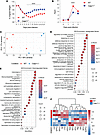
Severe SARS-CoV-2 infection is characterized by lung hyperinflammation, impaired IFN responses, and defective T cell activation, yet the molecular drivers of these immune dysregulations remain incompletely understood. Caspase-11 (CASP11), a key effector of the noncanonical inflammasome, has been shown to mediate an innate hyperinflammatory response and cytokine release in a mild SARS-CoV-2 infection model. However, the role played by CASP11 in severe SARS-CoV-2 disease and how it affects adaptive immunity has not been identified. Here, we found that CASP11 exacerbates severe SARS-CoV-2 pathogenesis by amplifying early innate immune responses while concurrently impairing antiviral CD8+ T cell immunity. Using global KO mice, hematopoietic BM chimeras, and Cx3cr1-expressing mononuclear phagocyte system cell–specific CASP11 deletion models, we show that targeting CASP11 reduces lung inflammation, promotes early NK cell–mediated IFN-γ production, and enhances robust virus-specific effector CD8+ T cell responses. This was associated with enhanced viral clearance and improved survival, even under lethal infection conditions. Importantly, CASP11-KO mice also exhibited faster resolution of postviral inflammation. These findings position CASP11 as a promising immunomodulatory target for acute and delayed manifestations of severe SARS-CoV-2 infection.
Authors
Mostafa M. Eltobgy, Mohamed M. Shamseldin, Owen D. Whitham, Heba M. Amer, Jeffrey R. Atkinson, Asmaa Badr, Jesse M. Hall, Gauruv Gupta, Yara Y. Hassan, Rabab El-Mergawy, Richard Perez, Sarah E. Faber, Maciej Pietrzak, Amy Webb, Xiaoli Zhang, Adam D. Kenney, Destiny Bissell, Jihad I. Omran, Shady Estfanous, Kylene P. Daily, Amir Yousif, Marisa R. Joldrichsen, Andrew McNamara, Mahesh KC, Mark E. Peeples, Emily A. Hemann, Hazem E. Ghoneim, Shahid M. Nimjee, Estelle Cormet-Boyaka, Jianrong Li, Prosper N. Boyaka, Jacob S. Yount, Benjamin M. Segal, Purnima Dubey, Amal O. Amer
Abstract
Mycoplasma pneumoniae pneumonia (MPP) can cause serious extrapulmonary complications, including life-threatening thrombosis. This study aimed to elucidate the roles of neutrophils and neutrophil extracellular traps (NETs) in vascular endothelial cell (EC) activation in pediatric MPP-associated thrombosis. We analyzed the relationship between neutrophils and thrombosis in children with MPP and used mouse models of neutrophilia (Csf3 plasmid injection), neutropenia (Csf3 deficient, Csf3–/–), and defective NETs formation (Pad4 deficient, Pad4–/–). The effects of neutrophils and NETs on EC activation were further examined in vivo, in vitro, and in human samples. Elevated neutrophil count was observed in patients with thrombosis and functioned as a potential diagnostic marker as well as a risk factor for MPP-associated thrombosis. EC activation was enhanced in MPP mice with neutrophilia but attenuated in neutropenic or Pad4–/– mice. NETs activated ECs through TLR2 and JAK/STAT3 signaling, and inhibition of NETs formation (Cl-amidine), TLR2 (C29), and JAK (upadacitinib) each attenuated this response. Strong correlations among neutrophils, NETs, EC activation, and thrombosis were observed in pediatric patients. These findings suggest that neutrophils promoted thrombosis in MPP via NETs-mediated EC activation involving TLR2 and JAK/STAT3 signaling. This study provides mechanistic insights into the inflammatory-thrombotic processes in MPP-associated thrombosis and offers a rationale for further investigation of neutrophils, NETs, TLR2, and JAK/STAT3 signaling in this context.
Authors
Xia Huang, Yifan Zhu, Yun Guo, Tian Lv, Haiyan Gu, Yingying Luo, Dan Li, Hang Sun, Deyu Zhao, Feng Liu
Abstract
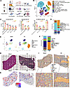
Surgical stress, such as liver ischemia/reperfusion (I/R) injury characterized by robust neutrophil infiltration and immune activation, induces sterile inflammation that reshapes tissue immunity and contributes to organ dysfunction, yet the intercellular circuits that spatially orchestrate these responses within the hepatic immune microenvironment remain incompletely defined. Here, we integrate single-cell RNA sequencing, spatial transcriptomics, high-dimensional spectral flow cytometry, and metabolomics to resolve the hepatic immune landscape following I/R at cellular and spatial resolution. While confirming extensive immune remodeling, we identify a dominant neutrophil–Kupffer cell communication axis mediated by neutrophil-derived thrombospondin-1 (TSP-1), which selectively engages CD36 on Kupffer cells. This interaction drives coordinated immune-metabolic reprogramming in Kupffer cells, characterized by suppression of oxidative phosphorylation, enhanced glycolysis, and remodeling of sphingolipid metabolism, including accumulation of hexosylceramides. Spatial analyses reveal preferential neutrophil–Kupffer cell colocalization within necrotic niches, and cross-species integration with human liver transplant datasets demonstrates conserved upregulation of the TSP-1/CD36 axis following reperfusion. Pharmacologic inhibition of TSP-1 attenuates liver injury and inflammatory responses in vivo. Together, these findings define a spatially organized, neutrophil-driven immune-metabolic circuit that governs functional reprogramming of Kupffer cells during surgical stress and identify the TSP-1/CD36 pathway as a conserved and targetable mediator of sterile liver injury.
Authors
Hongji Zhang, Xingping Huang, Preethi Jayakumar, Yunwei Zhang, Ti Yang, Xiaorong Guo, Grace Wu, Amrendra Kumar, Vijaya Bharti, Amblessed Onuma, Chengli Shen, Dequan Lou, Patricia S. Latham, James M. Crawford, Kong Chen, Anna E. Vilgelm, Lopa Mishra, Allan Tsung, Meihong Deng, Hai Huang
Abstract
Type 2 innate lymphoid cells (ILC2) are a population of lineage-negative cells in the lung, gastrointestinal tract, and skin which have emerged as a significant component of type 2 allergic inflammation. The regulation of cyclooxygenase-2 (COX-2) metabolites is critical to the pathophysiology of many inflammatory disorders, including allergic asthma. While COX-2 regulates Th9 and Th17 cell differentiation and function during allergic lung inflammation, it remains unknown whether COX-2 also regulates ILC2 cell differentiation and function under similar conditions. To address this question, we examined lung ILC2 cells from COX-2+/+ and COX-2-/- mice after ovalbumin (OVA)- or Alternaria-induced allergic lung inflammation. ILC2 cells were significantly increased in COX-2-/- lungs compared with COX-2+/+ lungs after OVA exposure in vivo. The increase in ILC2 cells was accompanied by an increase in expression of the cytokines IL-5 and IL-13, and the transcription factor GATA3. Both genetic disruption and selective inhibition of COX-2 significantly increased ILC2 cell differentiation from isolated common lymphoid progenitor cells (CLP) in vitro. Furthermore, COX-2-derived PGE2 acting via EP2 receptors significantly reduced IL-33 and TSLP expression, and attenuated ILC2 cell differentiation in vitro and in vivo. Thus, during allergic lung inflammation, COX-2-derived PGE2 signals through the EP2 receptor to negatively regulate lung ILC2 cell differentiation and function.
Authors
Hong Li, Matthew L. Edin, Daniel Menendez, J. Alyce Bradbury, Joan P. Graves, Artiom Gruzdev, Gregory S. Whitehead, Maria I Sifre, Laura M. DeGraff, Darryl C. Zeldin
Abstract
Sterile tissue injury triggers a rapid neutrophil response that can be either pathogenic or protective, reflecting substantial functional heterogeneity of neutrophils; however, the neutrophil subsets underlying these divergent functions remain poorly defined. Here, using a well-established sterile corneal injury model, we delineate the time-dependent functional and transcriptional heterogeneity of neutrophils following sterile injury. Temporal neutrophil depletion revealed that neutrophils recruited at day 1, but not day 7, are essential for suppressing inflammation, promoting epithelial healing, and preserving nerve density. Single-cell RNA sequencing uncovered substantial transcriptional heterogeneity among circulating neutrophils and their rapid reprogramming within 24 hours after injury. Specifically, Il1r2 was highly enriched in these early injury-responsive neutrophils. Functional validation demonstrated that adoptive transfer of IL-1R2+ neutrophils markedly attenuated inflammation and accelerated epithelial and nerve repair, restoring tissue integrity, whereas IL-1R2- neutrophils exacerbated inflammatory responses. Together, these findings identify IL-1R2+ neutrophils as an early protective neutrophil subset expanded by sterile injury that restrains excessive inflammation and preserves tissue homeostasis, providing mechanistic insight into injury-induced neutrophil reprogramming and highlighting a potential therapeutic target for enhancing tissue repair.
Authors
Hyun Ju Lee, Jung Hwa Ko, Joo Youn Oh
Abstract
Tregs play an essential role in immune tolerance, and Treg-promoting therapies are in development for the treatment of many inflammatory disorders. Interleukin-2 (IL-2)-based therapies increase Treg frequency, but little is known about impacts on Treg heterogeneity and function. We extended analyses of an IL-2 mutein (MK-6194) single–ascending-dose trial in healthy human participants by comprehensively defining Treg subsets and gene expression changes in vitro and in vivo. We found highly specific and dose-dependent activation and expansion of Tregs in clinical and pre-clinical studies. Following a single subcutaneous dose in humans, thymic-derived Tregs were selectively activated and expanded, while peripherally induced Tregs were unaffected. Expanded Tregs had increased expression of genes and proteins consistent with activation, suppressor function, and homing to non-lymphoid tissue, as well as increased transendocytosis activity, as measured by CTLA-4–dependent capture of CD80 and CD86 from non-Tregs. These results shed light onto underlying mechanisms by which Treg-targeted therapy may promote immune tolerance.
Authors
Laura A. Cooney, Mitch Fahning, Liliane Khoryati, Anna Kus, Sheila Scheiding, Lori Blanchfield, Matthew Lawrance, Basilin Benson, Kristina M. Harris, Gretchen A. Baltus, Shiuli Agarwal, Richard Wnek, Johannes F. Scheid, Kiki Cunningham-Bussel, Nancy D. Kim, S. Aubrey Stoch, Jyothsna Visweswaraiah, Nathan Higginson-Scott, Katalin Kis-Toth, Joanne L. Viney, Kevin L. Otipoby, Erik Sampson, Bridget Larkin, Daniel J. Campbell, S. Alice Long
Abstract
Authors
Jianfei Ji, Wenjing You, Xiaoli Sun, Peng Zhao
Abstract
Autophagy is a critical host defense mechanism that restricts intracellular pathogens such as Mycobacterium tuberculosis (Mtb). A key step in this process is the ubiquitination of Mtb or Mtb-associated structures. The E3 ligase SMURF1 catalyzes K48-linked ubiquitination, promoting bacterial clearance. However, the function of its homolog, SMURF2, in host defense remains undefined. Here, we demonstrate that Smurf2 deletion in murine macrophages increases SMURF1 levels, enhances LC3B lipidation, augments K48 ubiquitination of Mtb-associated structures, and reduces intracellular Mtb replication. These effects are reversed by Smurf1 deletion, supporting a role for SMURF1 in SMURF2-dependent control of Mtb. Mice with myeloid-specific Smurf2 deletion exhibit modestly prolonged survival following aerosol Mtb infection. In human macrophages, SMURF2 knockdown or its pharmacological inhibition with the HECT E3-ligase inhibitor Heclin reduces Mtb replication. Together, our findings identify SMURF2 as a negative regulator of macrophage control of Mtb and support further investigation of SMURF2 as a potential target for host-directed therapy in tuberculosis.
Authors
Priscila C. Campos, Kathryn C. Rahlwes, Victoria A. Ektnitphong, Beatriz R.S. Dias, Kubra F. Naqvi, Samuel Alvarez-Arguedas, Michael U. Shiloh

Abstract
In rheumatoid arthritis (RA), CD4+ T cells specific for citrullinated antigens (cit-antigens) are key drivers of disease, but knowledge about epitopes and phenotypes remains limited. We characterized the frequency and phenotype of cit-specific CD4+ T cells in peripheral blood using HLA class II tetramers combined with computational analysis of phenotypic clusters to simultaneously detect peptides derived from 5 cit-antigens (aggrecan, vimentin, fibrinogen, cartilage intermediate layer protein, and α-enolase) previously implicated in RA pathogenesis. In a cross-sectional cohort, cit-aggrecan–, cit-vimentin–, and cit-fibrinogen–specific T cells were more frequent in participants with RA than healthy volunteers, associated with active disease, and had Th1-like and stem-like lineages in RA. In a longitudinal cohort investigating response to therapy, the frequency of cit-aggrecan–, cit-vimentin–, and cit-fibrinogen–specific CD4+ T cells was significantly higher at baseline and further elevated in responders. Furthermore, the frequency of cit-specific Th1-like cells in responders decreased over time. In contrast, the frequency of Th1-like cells in non-responders increased over time. Collectively, these findings demonstrate that cit-specific CD4+ T cells are expanded in RA and target a broad number of antigens across a breadth of phenotypes. Furthermore, the predominant antigen specificities associate with disease activity and exhibit dynamic changes in phenotype that reflect response to therapy.
Authors
Cliff Rims, Hannah A. DeBerg, Sylvia E. Posso, Virginia S. Muir, Hannes Uchtenhagen, Anne M. Hocking, Heather Bukiri, Jeffrey Carlin, Bernard Ng, Peter S. Linsley, Eddie A. James, Jane H. Buckner
No posts were found with this tag.


