
Go to The Journal of Clinical Investigation
  • About
  • Editors
  • Consulting Editors
  • For authors
  • Journal stats
  • Publication ethics
  • Publication alerts by email
  • Transfers
  • Advertising
  • Job board
  • Contact
  • Physician-Scientist Development
  • Current issue
  • Past issues
  • By specialty
    • COVID-19
    • Cardiology
    • Immunology
    • Metabolism
    • Nephrology
    • Oncology
    • Pulmonology
    • All ...
  • Videos
  • Collections
    • In-Press Preview
    • Resource and Technical Advances
    • Clinical Research and Public Health
    • Research Letters
    • Editorials
    • Perspectives
    • Physician-Scientist Development
    • Reviews
    • Top read articles

  • Current issue
  • Past issues
  • Specialties
  • In-Press Preview
  • Resource and Technical Advances
  • Clinical Research and Public Health
  • Research Letters
  • Editorials
  • Perspectives
  • Physician-Scientist Development
  • Reviews
  • Top read articles
  • About
  • Editors
  • Consulting Editors
  • For authors
  • Journal stats
  • Publication ethics
  • Publication alerts by email
  • Transfers
  • Advertising
  • Job board
  • Contact
Top
  • View PDF
  • Download citation information
  • Send a comment
  • Terms of use
  • Standard abbreviations
  • Need help? Email the journal
  • Top
  • Abstract
  • Introduction
  • Results
  • Discussion
  • Methods
  • Author contributions
  • Conflict of interest
  • Funding support
  • Supplemental material
  • Acknowledgments
  • Footnotes
  • References
  • Version history
  • Article usage
  • Citations to this article
Advertisement

Research ArticleDevelopmentInflammationPulmonology Open Access | 10.1172/jci.insight.193625

Mesenchyme-derived inflammation during the saccular stage recruits macrophages and alters lung development

Benjamin C. Crawford,1 Jessica Chauviere Lee,1 Bertha C. Elias,1 Shivangi Dave,1 Riet van der Meer,1 Wei Han,2,3 Alexandria L. Sharkey,1 David S. Nichols,2 Charles Shissias,1 Lauren Pate,1 Hayden Tan,1 Dawn C. Newcomb,2 Wei Shi,4 Lawrence S. Prince,5 Erin J. Plosa,1,6 Bradley W. Richmond,2,7 Timothy S. Blackwell,2,3 Susan H. Guttentag,1 and John T. Benjamin1,5

1Department of Pediatrics, Division of Neonatology, and

2Department of Medicine, Division of Allergy, Pulmonary, and Critical Care Medicine, Vanderbilt University Medical Center, Nashville, Tennessee, USA.

3Department of Internal Medicine, Division of Critical Care and Pulmonary Medicine, University of Michigan Medical School, Ann Arbor, Michigan, USA.

4Department of Internal Medicine, Division of Pulmonary, Critical Care and Sleep Medicine, University of Cincinnati College of Medicine, Cincinnati, Ohio, USA.

5Department of Pediatrics, Division of Neonatal and Developmental Medicine, Stanford University School of Medicine, Palo Alto, California, USA.

6Department of Pediatrics, Division of Neonatal-Perinatal Medicine, University of North Carolina at Chapel Hill, Chapel Hill, North Carolina, USA.

7Department of Veterans Affairs Medical Center, Nashville, Tennessee, USA.

Address correspondence to: Benjamin C. Crawford, Monroe Carell Jr. Children’s Hospital, Vanderbilt University Medical Center, 2215B Garland Avenue, 1125 MRB4/Light Hall, Nashville, Tennessee, 37232, USA. Phone: 615.343.5666; Email: Benjamin.crawford@vumc.org.

Find articles by Crawford, B. in: PubMed | Google Scholar

1Department of Pediatrics, Division of Neonatology, and

2Department of Medicine, Division of Allergy, Pulmonary, and Critical Care Medicine, Vanderbilt University Medical Center, Nashville, Tennessee, USA.

3Department of Internal Medicine, Division of Critical Care and Pulmonary Medicine, University of Michigan Medical School, Ann Arbor, Michigan, USA.

4Department of Internal Medicine, Division of Pulmonary, Critical Care and Sleep Medicine, University of Cincinnati College of Medicine, Cincinnati, Ohio, USA.

5Department of Pediatrics, Division of Neonatal and Developmental Medicine, Stanford University School of Medicine, Palo Alto, California, USA.

6Department of Pediatrics, Division of Neonatal-Perinatal Medicine, University of North Carolina at Chapel Hill, Chapel Hill, North Carolina, USA.

7Department of Veterans Affairs Medical Center, Nashville, Tennessee, USA.

Address correspondence to: Benjamin C. Crawford, Monroe Carell Jr. Children’s Hospital, Vanderbilt University Medical Center, 2215B Garland Avenue, 1125 MRB4/Light Hall, Nashville, Tennessee, 37232, USA. Phone: 615.343.5666; Email: Benjamin.crawford@vumc.org.

Find articles by Lee, J. in: PubMed | Google Scholar

1Department of Pediatrics, Division of Neonatology, and

2Department of Medicine, Division of Allergy, Pulmonary, and Critical Care Medicine, Vanderbilt University Medical Center, Nashville, Tennessee, USA.

3Department of Internal Medicine, Division of Critical Care and Pulmonary Medicine, University of Michigan Medical School, Ann Arbor, Michigan, USA.

4Department of Internal Medicine, Division of Pulmonary, Critical Care and Sleep Medicine, University of Cincinnati College of Medicine, Cincinnati, Ohio, USA.

5Department of Pediatrics, Division of Neonatal and Developmental Medicine, Stanford University School of Medicine, Palo Alto, California, USA.

6Department of Pediatrics, Division of Neonatal-Perinatal Medicine, University of North Carolina at Chapel Hill, Chapel Hill, North Carolina, USA.

7Department of Veterans Affairs Medical Center, Nashville, Tennessee, USA.

Address correspondence to: Benjamin C. Crawford, Monroe Carell Jr. Children’s Hospital, Vanderbilt University Medical Center, 2215B Garland Avenue, 1125 MRB4/Light Hall, Nashville, Tennessee, 37232, USA. Phone: 615.343.5666; Email: Benjamin.crawford@vumc.org.

Find articles by Elias, B. in: PubMed | Google Scholar

1Department of Pediatrics, Division of Neonatology, and

2Department of Medicine, Division of Allergy, Pulmonary, and Critical Care Medicine, Vanderbilt University Medical Center, Nashville, Tennessee, USA.

3Department of Internal Medicine, Division of Critical Care and Pulmonary Medicine, University of Michigan Medical School, Ann Arbor, Michigan, USA.

4Department of Internal Medicine, Division of Pulmonary, Critical Care and Sleep Medicine, University of Cincinnati College of Medicine, Cincinnati, Ohio, USA.

5Department of Pediatrics, Division of Neonatal and Developmental Medicine, Stanford University School of Medicine, Palo Alto, California, USA.

6Department of Pediatrics, Division of Neonatal-Perinatal Medicine, University of North Carolina at Chapel Hill, Chapel Hill, North Carolina, USA.

7Department of Veterans Affairs Medical Center, Nashville, Tennessee, USA.

Address correspondence to: Benjamin C. Crawford, Monroe Carell Jr. Children’s Hospital, Vanderbilt University Medical Center, 2215B Garland Avenue, 1125 MRB4/Light Hall, Nashville, Tennessee, 37232, USA. Phone: 615.343.5666; Email: Benjamin.crawford@vumc.org.

Find articles by Dave, S. in: PubMed | Google Scholar

1Department of Pediatrics, Division of Neonatology, and

2Department of Medicine, Division of Allergy, Pulmonary, and Critical Care Medicine, Vanderbilt University Medical Center, Nashville, Tennessee, USA.

3Department of Internal Medicine, Division of Critical Care and Pulmonary Medicine, University of Michigan Medical School, Ann Arbor, Michigan, USA.

4Department of Internal Medicine, Division of Pulmonary, Critical Care and Sleep Medicine, University of Cincinnati College of Medicine, Cincinnati, Ohio, USA.

5Department of Pediatrics, Division of Neonatal and Developmental Medicine, Stanford University School of Medicine, Palo Alto, California, USA.

6Department of Pediatrics, Division of Neonatal-Perinatal Medicine, University of North Carolina at Chapel Hill, Chapel Hill, North Carolina, USA.

7Department of Veterans Affairs Medical Center, Nashville, Tennessee, USA.

Address correspondence to: Benjamin C. Crawford, Monroe Carell Jr. Children’s Hospital, Vanderbilt University Medical Center, 2215B Garland Avenue, 1125 MRB4/Light Hall, Nashville, Tennessee, 37232, USA. Phone: 615.343.5666; Email: Benjamin.crawford@vumc.org.

Find articles by van der Meer, R. in: PubMed | Google Scholar

1Department of Pediatrics, Division of Neonatology, and

2Department of Medicine, Division of Allergy, Pulmonary, and Critical Care Medicine, Vanderbilt University Medical Center, Nashville, Tennessee, USA.

3Department of Internal Medicine, Division of Critical Care and Pulmonary Medicine, University of Michigan Medical School, Ann Arbor, Michigan, USA.

4Department of Internal Medicine, Division of Pulmonary, Critical Care and Sleep Medicine, University of Cincinnati College of Medicine, Cincinnati, Ohio, USA.

5Department of Pediatrics, Division of Neonatal and Developmental Medicine, Stanford University School of Medicine, Palo Alto, California, USA.

6Department of Pediatrics, Division of Neonatal-Perinatal Medicine, University of North Carolina at Chapel Hill, Chapel Hill, North Carolina, USA.

7Department of Veterans Affairs Medical Center, Nashville, Tennessee, USA.

Address correspondence to: Benjamin C. Crawford, Monroe Carell Jr. Children’s Hospital, Vanderbilt University Medical Center, 2215B Garland Avenue, 1125 MRB4/Light Hall, Nashville, Tennessee, 37232, USA. Phone: 615.343.5666; Email: Benjamin.crawford@vumc.org.

Find articles by Han, W. in: PubMed | Google Scholar

1Department of Pediatrics, Division of Neonatology, and

2Department of Medicine, Division of Allergy, Pulmonary, and Critical Care Medicine, Vanderbilt University Medical Center, Nashville, Tennessee, USA.

3Department of Internal Medicine, Division of Critical Care and Pulmonary Medicine, University of Michigan Medical School, Ann Arbor, Michigan, USA.

4Department of Internal Medicine, Division of Pulmonary, Critical Care and Sleep Medicine, University of Cincinnati College of Medicine, Cincinnati, Ohio, USA.

5Department of Pediatrics, Division of Neonatal and Developmental Medicine, Stanford University School of Medicine, Palo Alto, California, USA.

6Department of Pediatrics, Division of Neonatal-Perinatal Medicine, University of North Carolina at Chapel Hill, Chapel Hill, North Carolina, USA.

7Department of Veterans Affairs Medical Center, Nashville, Tennessee, USA.

Address correspondence to: Benjamin C. Crawford, Monroe Carell Jr. Children’s Hospital, Vanderbilt University Medical Center, 2215B Garland Avenue, 1125 MRB4/Light Hall, Nashville, Tennessee, 37232, USA. Phone: 615.343.5666; Email: Benjamin.crawford@vumc.org.

Find articles by Sharkey, A. in: PubMed | Google Scholar

1Department of Pediatrics, Division of Neonatology, and

2Department of Medicine, Division of Allergy, Pulmonary, and Critical Care Medicine, Vanderbilt University Medical Center, Nashville, Tennessee, USA.

3Department of Internal Medicine, Division of Critical Care and Pulmonary Medicine, University of Michigan Medical School, Ann Arbor, Michigan, USA.

4Department of Internal Medicine, Division of Pulmonary, Critical Care and Sleep Medicine, University of Cincinnati College of Medicine, Cincinnati, Ohio, USA.

5Department of Pediatrics, Division of Neonatal and Developmental Medicine, Stanford University School of Medicine, Palo Alto, California, USA.

6Department of Pediatrics, Division of Neonatal-Perinatal Medicine, University of North Carolina at Chapel Hill, Chapel Hill, North Carolina, USA.

7Department of Veterans Affairs Medical Center, Nashville, Tennessee, USA.

Address correspondence to: Benjamin C. Crawford, Monroe Carell Jr. Children’s Hospital, Vanderbilt University Medical Center, 2215B Garland Avenue, 1125 MRB4/Light Hall, Nashville, Tennessee, 37232, USA. Phone: 615.343.5666; Email: Benjamin.crawford@vumc.org.

Find articles by Nichols, D. in: PubMed | Google Scholar

1Department of Pediatrics, Division of Neonatology, and

2Department of Medicine, Division of Allergy, Pulmonary, and Critical Care Medicine, Vanderbilt University Medical Center, Nashville, Tennessee, USA.

3Department of Internal Medicine, Division of Critical Care and Pulmonary Medicine, University of Michigan Medical School, Ann Arbor, Michigan, USA.

4Department of Internal Medicine, Division of Pulmonary, Critical Care and Sleep Medicine, University of Cincinnati College of Medicine, Cincinnati, Ohio, USA.

5Department of Pediatrics, Division of Neonatal and Developmental Medicine, Stanford University School of Medicine, Palo Alto, California, USA.

6Department of Pediatrics, Division of Neonatal-Perinatal Medicine, University of North Carolina at Chapel Hill, Chapel Hill, North Carolina, USA.

7Department of Veterans Affairs Medical Center, Nashville, Tennessee, USA.

Address correspondence to: Benjamin C. Crawford, Monroe Carell Jr. Children’s Hospital, Vanderbilt University Medical Center, 2215B Garland Avenue, 1125 MRB4/Light Hall, Nashville, Tennessee, 37232, USA. Phone: 615.343.5666; Email: Benjamin.crawford@vumc.org.

Find articles by Shissias, C. in: PubMed | Google Scholar

1Department of Pediatrics, Division of Neonatology, and

2Department of Medicine, Division of Allergy, Pulmonary, and Critical Care Medicine, Vanderbilt University Medical Center, Nashville, Tennessee, USA.

3Department of Internal Medicine, Division of Critical Care and Pulmonary Medicine, University of Michigan Medical School, Ann Arbor, Michigan, USA.

4Department of Internal Medicine, Division of Pulmonary, Critical Care and Sleep Medicine, University of Cincinnati College of Medicine, Cincinnati, Ohio, USA.

5Department of Pediatrics, Division of Neonatal and Developmental Medicine, Stanford University School of Medicine, Palo Alto, California, USA.

6Department of Pediatrics, Division of Neonatal-Perinatal Medicine, University of North Carolina at Chapel Hill, Chapel Hill, North Carolina, USA.

7Department of Veterans Affairs Medical Center, Nashville, Tennessee, USA.

Address correspondence to: Benjamin C. Crawford, Monroe Carell Jr. Children’s Hospital, Vanderbilt University Medical Center, 2215B Garland Avenue, 1125 MRB4/Light Hall, Nashville, Tennessee, 37232, USA. Phone: 615.343.5666; Email: Benjamin.crawford@vumc.org.

Find articles by Pate, L. in: PubMed | Google Scholar

1Department of Pediatrics, Division of Neonatology, and

2Department of Medicine, Division of Allergy, Pulmonary, and Critical Care Medicine, Vanderbilt University Medical Center, Nashville, Tennessee, USA.

3Department of Internal Medicine, Division of Critical Care and Pulmonary Medicine, University of Michigan Medical School, Ann Arbor, Michigan, USA.

4Department of Internal Medicine, Division of Pulmonary, Critical Care and Sleep Medicine, University of Cincinnati College of Medicine, Cincinnati, Ohio, USA.

5Department of Pediatrics, Division of Neonatal and Developmental Medicine, Stanford University School of Medicine, Palo Alto, California, USA.

6Department of Pediatrics, Division of Neonatal-Perinatal Medicine, University of North Carolina at Chapel Hill, Chapel Hill, North Carolina, USA.

7Department of Veterans Affairs Medical Center, Nashville, Tennessee, USA.

Address correspondence to: Benjamin C. Crawford, Monroe Carell Jr. Children’s Hospital, Vanderbilt University Medical Center, 2215B Garland Avenue, 1125 MRB4/Light Hall, Nashville, Tennessee, 37232, USA. Phone: 615.343.5666; Email: Benjamin.crawford@vumc.org.

Find articles by Tan, H. in: PubMed | Google Scholar

1Department of Pediatrics, Division of Neonatology, and

2Department of Medicine, Division of Allergy, Pulmonary, and Critical Care Medicine, Vanderbilt University Medical Center, Nashville, Tennessee, USA.

3Department of Internal Medicine, Division of Critical Care and Pulmonary Medicine, University of Michigan Medical School, Ann Arbor, Michigan, USA.

4Department of Internal Medicine, Division of Pulmonary, Critical Care and Sleep Medicine, University of Cincinnati College of Medicine, Cincinnati, Ohio, USA.

5Department of Pediatrics, Division of Neonatal and Developmental Medicine, Stanford University School of Medicine, Palo Alto, California, USA.

6Department of Pediatrics, Division of Neonatal-Perinatal Medicine, University of North Carolina at Chapel Hill, Chapel Hill, North Carolina, USA.

7Department of Veterans Affairs Medical Center, Nashville, Tennessee, USA.

Address correspondence to: Benjamin C. Crawford, Monroe Carell Jr. Children’s Hospital, Vanderbilt University Medical Center, 2215B Garland Avenue, 1125 MRB4/Light Hall, Nashville, Tennessee, 37232, USA. Phone: 615.343.5666; Email: Benjamin.crawford@vumc.org.

Find articles by Newcomb, D. in: PubMed | Google Scholar

1Department of Pediatrics, Division of Neonatology, and

2Department of Medicine, Division of Allergy, Pulmonary, and Critical Care Medicine, Vanderbilt University Medical Center, Nashville, Tennessee, USA.

3Department of Internal Medicine, Division of Critical Care and Pulmonary Medicine, University of Michigan Medical School, Ann Arbor, Michigan, USA.

4Department of Internal Medicine, Division of Pulmonary, Critical Care and Sleep Medicine, University of Cincinnati College of Medicine, Cincinnati, Ohio, USA.

5Department of Pediatrics, Division of Neonatal and Developmental Medicine, Stanford University School of Medicine, Palo Alto, California, USA.

6Department of Pediatrics, Division of Neonatal-Perinatal Medicine, University of North Carolina at Chapel Hill, Chapel Hill, North Carolina, USA.

7Department of Veterans Affairs Medical Center, Nashville, Tennessee, USA.

Address correspondence to: Benjamin C. Crawford, Monroe Carell Jr. Children’s Hospital, Vanderbilt University Medical Center, 2215B Garland Avenue, 1125 MRB4/Light Hall, Nashville, Tennessee, 37232, USA. Phone: 615.343.5666; Email: Benjamin.crawford@vumc.org.

Find articles by Shi, W. in: PubMed | Google Scholar

1Department of Pediatrics, Division of Neonatology, and

2Department of Medicine, Division of Allergy, Pulmonary, and Critical Care Medicine, Vanderbilt University Medical Center, Nashville, Tennessee, USA.

3Department of Internal Medicine, Division of Critical Care and Pulmonary Medicine, University of Michigan Medical School, Ann Arbor, Michigan, USA.

4Department of Internal Medicine, Division of Pulmonary, Critical Care and Sleep Medicine, University of Cincinnati College of Medicine, Cincinnati, Ohio, USA.

5Department of Pediatrics, Division of Neonatal and Developmental Medicine, Stanford University School of Medicine, Palo Alto, California, USA.

6Department of Pediatrics, Division of Neonatal-Perinatal Medicine, University of North Carolina at Chapel Hill, Chapel Hill, North Carolina, USA.

7Department of Veterans Affairs Medical Center, Nashville, Tennessee, USA.

Address correspondence to: Benjamin C. Crawford, Monroe Carell Jr. Children’s Hospital, Vanderbilt University Medical Center, 2215B Garland Avenue, 1125 MRB4/Light Hall, Nashville, Tennessee, 37232, USA. Phone: 615.343.5666; Email: Benjamin.crawford@vumc.org.

Find articles by Prince, L. in: PubMed | Google Scholar

1Department of Pediatrics, Division of Neonatology, and

2Department of Medicine, Division of Allergy, Pulmonary, and Critical Care Medicine, Vanderbilt University Medical Center, Nashville, Tennessee, USA.

3Department of Internal Medicine, Division of Critical Care and Pulmonary Medicine, University of Michigan Medical School, Ann Arbor, Michigan, USA.

4Department of Internal Medicine, Division of Pulmonary, Critical Care and Sleep Medicine, University of Cincinnati College of Medicine, Cincinnati, Ohio, USA.

5Department of Pediatrics, Division of Neonatal and Developmental Medicine, Stanford University School of Medicine, Palo Alto, California, USA.

6Department of Pediatrics, Division of Neonatal-Perinatal Medicine, University of North Carolina at Chapel Hill, Chapel Hill, North Carolina, USA.

7Department of Veterans Affairs Medical Center, Nashville, Tennessee, USA.

Address correspondence to: Benjamin C. Crawford, Monroe Carell Jr. Children’s Hospital, Vanderbilt University Medical Center, 2215B Garland Avenue, 1125 MRB4/Light Hall, Nashville, Tennessee, 37232, USA. Phone: 615.343.5666; Email: Benjamin.crawford@vumc.org.

Find articles by Plosa, E. in: PubMed | Google Scholar |

1Department of Pediatrics, Division of Neonatology, and

2Department of Medicine, Division of Allergy, Pulmonary, and Critical Care Medicine, Vanderbilt University Medical Center, Nashville, Tennessee, USA.

3Department of Internal Medicine, Division of Critical Care and Pulmonary Medicine, University of Michigan Medical School, Ann Arbor, Michigan, USA.

4Department of Internal Medicine, Division of Pulmonary, Critical Care and Sleep Medicine, University of Cincinnati College of Medicine, Cincinnati, Ohio, USA.

5Department of Pediatrics, Division of Neonatal and Developmental Medicine, Stanford University School of Medicine, Palo Alto, California, USA.

6Department of Pediatrics, Division of Neonatal-Perinatal Medicine, University of North Carolina at Chapel Hill, Chapel Hill, North Carolina, USA.

7Department of Veterans Affairs Medical Center, Nashville, Tennessee, USA.

Address correspondence to: Benjamin C. Crawford, Monroe Carell Jr. Children’s Hospital, Vanderbilt University Medical Center, 2215B Garland Avenue, 1125 MRB4/Light Hall, Nashville, Tennessee, 37232, USA. Phone: 615.343.5666; Email: Benjamin.crawford@vumc.org.

Find articles by Richmond, B. in: PubMed | Google Scholar

1Department of Pediatrics, Division of Neonatology, and

2Department of Medicine, Division of Allergy, Pulmonary, and Critical Care Medicine, Vanderbilt University Medical Center, Nashville, Tennessee, USA.

3Department of Internal Medicine, Division of Critical Care and Pulmonary Medicine, University of Michigan Medical School, Ann Arbor, Michigan, USA.

4Department of Internal Medicine, Division of Pulmonary, Critical Care and Sleep Medicine, University of Cincinnati College of Medicine, Cincinnati, Ohio, USA.

5Department of Pediatrics, Division of Neonatal and Developmental Medicine, Stanford University School of Medicine, Palo Alto, California, USA.

6Department of Pediatrics, Division of Neonatal-Perinatal Medicine, University of North Carolina at Chapel Hill, Chapel Hill, North Carolina, USA.

7Department of Veterans Affairs Medical Center, Nashville, Tennessee, USA.

Address correspondence to: Benjamin C. Crawford, Monroe Carell Jr. Children’s Hospital, Vanderbilt University Medical Center, 2215B Garland Avenue, 1125 MRB4/Light Hall, Nashville, Tennessee, 37232, USA. Phone: 615.343.5666; Email: Benjamin.crawford@vumc.org.

Find articles by Blackwell, T. in: PubMed | Google Scholar

1Department of Pediatrics, Division of Neonatology, and

2Department of Medicine, Division of Allergy, Pulmonary, and Critical Care Medicine, Vanderbilt University Medical Center, Nashville, Tennessee, USA.

3Department of Internal Medicine, Division of Critical Care and Pulmonary Medicine, University of Michigan Medical School, Ann Arbor, Michigan, USA.

4Department of Internal Medicine, Division of Pulmonary, Critical Care and Sleep Medicine, University of Cincinnati College of Medicine, Cincinnati, Ohio, USA.

5Department of Pediatrics, Division of Neonatal and Developmental Medicine, Stanford University School of Medicine, Palo Alto, California, USA.

6Department of Pediatrics, Division of Neonatal-Perinatal Medicine, University of North Carolina at Chapel Hill, Chapel Hill, North Carolina, USA.

7Department of Veterans Affairs Medical Center, Nashville, Tennessee, USA.

Address correspondence to: Benjamin C. Crawford, Monroe Carell Jr. Children’s Hospital, Vanderbilt University Medical Center, 2215B Garland Avenue, 1125 MRB4/Light Hall, Nashville, Tennessee, 37232, USA. Phone: 615.343.5666; Email: Benjamin.crawford@vumc.org.

Find articles by Guttentag, S. in: PubMed | Google Scholar |

1Department of Pediatrics, Division of Neonatology, and

2Department of Medicine, Division of Allergy, Pulmonary, and Critical Care Medicine, Vanderbilt University Medical Center, Nashville, Tennessee, USA.

3Department of Internal Medicine, Division of Critical Care and Pulmonary Medicine, University of Michigan Medical School, Ann Arbor, Michigan, USA.

4Department of Internal Medicine, Division of Pulmonary, Critical Care and Sleep Medicine, University of Cincinnati College of Medicine, Cincinnati, Ohio, USA.

5Department of Pediatrics, Division of Neonatal and Developmental Medicine, Stanford University School of Medicine, Palo Alto, California, USA.

6Department of Pediatrics, Division of Neonatal-Perinatal Medicine, University of North Carolina at Chapel Hill, Chapel Hill, North Carolina, USA.

7Department of Veterans Affairs Medical Center, Nashville, Tennessee, USA.

Address correspondence to: Benjamin C. Crawford, Monroe Carell Jr. Children’s Hospital, Vanderbilt University Medical Center, 2215B Garland Avenue, 1125 MRB4/Light Hall, Nashville, Tennessee, 37232, USA. Phone: 615.343.5666; Email: Benjamin.crawford@vumc.org.

Find articles by Benjamin, J. in: PubMed | Google Scholar

Published June 2, 2026 - More info

Published in Volume 11, Issue 14 on July 22, 2026
JCI Insight. 2026;11(14):e193625. https://doi.org/10.1172/jci.insight.193625.
© 2026 Crawford et al. This work is licensed under the Creative Commons Attribution 4.0 International License. To view a copy of this license, visit http://creativecommons.org/licenses/by/4.0/.
Published June 2, 2026 - Version history
Received: April 28, 2025; Accepted: May 26, 2026
View PDF
Abstract

Fibroblasts in the lung mesenchyme produce growth factors and extracellular matrix components that guide formation of distal airspaces during the saccular stage of lung development. Inflammation in preterm infants disrupts this process, leading to bronchopulmonary dysplasia (BPD). To examine how mesenchymal inflammation contributes to BPD pathogenesis, we developed a transgenic mouse model (IKKβTbx4) in which expression of activated human IκB kinase β (IKKβ), an upstream activator of NF-κB, was induced in Tbx4 lung enhancer–positive mesenchymal cells during the saccular stage of lung development (P0–P5). Saccular-stage IKKβTbx4 mice exhibited a BPD-like phenotype with interstitial thickening and reduced distal airspaces at P5, progressing to emphysematous enlargement of the distal lung at 2 months of age. Mesenchymal NF-κB activity upregulated the chemokines CCL2 and CCL7, recruiting CCR2pos monocyte-derived macrophages to the lung. Recruited macrophages disrupted the elastin scaffold and impaired microvascular organization with reductions in CAP2 endothelial cells and pericytes. Blocking CCR2-dependent monocyte recruitment with a small-molecule CCR2 antagonist rescued the abnormal lung phenotype. These findings identify mesenchyme-macrophage crosstalk as a mechanism by which inflammation disrupts saccular-stage lung development, suggesting a role for this signaling axis in BPD pathogenesis.

Graphical Abstract
graphical abstract
Introduction

The saccular stage of lung development marks a critical period during which lung mesenchymal cells guide the formation of primitive air sacs in the distal lung. During sacculation, fibroblasts produce growth factors and extracellular matrix components that direct epithelial and endothelial organization and provide the structural support for alveolar development (1–3). Disrupting these critical events can permanently alter the form and function of the developing lung and lead to bronchopulmonary dysplasia (BPD) (4–8), a chronic respiratory condition that primarily affects preterm infants born before 30 weeks of gestation (9). BPD is characterized by alveolar simplification and dysmorphic lung structure (10–15). Clinically, BPD is defined by an extended need for respiratory support, and affected individuals are at increased risk of pulmonary impairment throughout life (16–18). Despite improvements in neonatal clinical care over the past several decades, effective therapeutic options for affected infants are still lacking, and the incidence of BPD has continued to increase (19, 20).

Both clinical and laboratory data demonstrate that antenatal and early postnatal inflammation can alter saccular-stage lung development and contribute to BPD. In utero exposure to chorioamnionitis is an independent risk factor for developing BPD (21–23). Further, early life exposures that cause pulmonary inflammation in preterm infants, such as mechanical ventilation and oxygen supplementation, can injure the lung and disrupt its development (24–29). Animal studies corroborate these findings, demonstrating that inflammation in the immature lung leads to a BPD-like phenotype (30–34). However, the mechanisms linking inflammatory signaling to the molecular events mediating abnormal lung morphogenesis remain unclear.

In addition to their well-described role in lung organogenesis, cells of the distal lung mesenchyme also participate in immune signaling. Multiple mesenchymal subpopulations express TLRs and other pathogen-recognition receptors that activate inflammation via the NF-κB pathway (35–38). Upon activation by inflammatory stimuli, the lung mesenchyme produces chemokines and cytokines that amplify inflammatory signaling and recruit immune cells to the lung (39–44). Recent transcriptomic profiling of BPD models has identified proinflammatory populations of fibroblasts in the lung mesenchyme (34, 45, 46). Predicted to function as central hubs of paracrine signaling, inflammatory fibroblasts exhibit robust chemokine expression that correlates with immune cell recruitment. Yet, whether inflammation originating from the lung mesenchyme is sufficient to disrupt saccular-stage development has not been tested.

To examine the cell-specific effects of mesenchymal inflammation on the developing postnatal lung, we generated a transgenic mouse model (IKKβTbx4) in which we induced expression of IκB kinase β (IKKβ) in Tbx4 lung enhancer–expressing mesenchymal cells during the saccular stage of lung development (P0–P5). We demonstrated that mesenchymal-derived inflammation during this critical developmental window was sufficient to recruit CCR2pos monocyte-derived macrophages and alter distal lung structure. Our findings establish that mesenchymal-macrophage crosstalk disrupts saccular-stage development, leading to characteristic features observed in the BPD lung.

Results

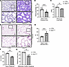
Mesenchymal inflammation alters saccular-stage lung development. We developed a transgenic mouse model (IKKβTbx4) to investigate the effects of mesenchymal inflammation on the developing saccular-stage lung. In this model, an activated form of human IKKβ, the upstream activator of NF-κB, is expressed in cells with Tbx4 lung enhancer activity in a doxycycline-inducible (Dox-inducible) manner. The breeding strategy used to generate IKKβTbx4 and littermate control mice is shown in Supplemental Figure 1A (supplemental material available online with this article; https://doi.org/10.1172/jci.insight.193625DS1). Lactating dams were administered Dox from P0 to P5 (Supplemental Figure 1B) to selectively induce expression of the IKKβ transgene and activate NF-κB in the lung mesenchyme of transgenic pups (Supplemental Figure 2, A–C). In littermate control mice, the distal lung parenchyma demonstrated arborized and well-distributed distal airspaces separated by a thin interstitium. In comparison, lungs from Dox-exposed IKKβTbx4 mice were markedly hypercellular with interstitial thickening and a significant reduction in distal airspace area (Figure 1, A and B). These morphological changes were not attributable to pulmonary edema, as the wet-to-dry lung weight ratio was not increased in transgenic mice compared with controls (Figure 1C).
NF-κB activation in the lung mesenchyme alters saccular-stage lung developmFigure 1

NF-κB activation in the lung mesenchyme alters saccular-stage lung development. Doxycycline was added to drinking water of lactating dams from P0 to P5, and neonatal lungs were harvested on P5 (A and B) or at 2 months of age (D–G). (A and D) Representative H&E-stained lung sections of littermate control (CTRL) and IKKβTbx4 mice at P5 (A) and 2 months of age (D). (B and E) Morphometric quantification of airspace volume density (ASVD) at P5 (B) and mean linear intercept (MLI) at 2 months of age (E). (C) Wet-to-dry lung weight ratios in CTRL and IKKβTbx4 lungs at P5. (F and G) Lung tissue (F) and respiratory system (G) elastance were measured at 2 months of age using flexiVent in CTRL and IKKβTbx4 mice. Data are expressed as mean ± SEM. n = 10 to 12 nonoverlapping lung fields per lung (biological replicate) (B and E); n = 9 to 10 mice per group (B); n = 3 to 5 mice per group (C); n = 5 to 7 mice per group (E); and n = 4 mice per group (F and G); scale bar: 100 μm (A and D); **P < 0.01, ***P < 0.001, and ****P < 0.0001 by unpaired 2-tailed t test (B, C, and E–G).

Despite the severe structural abnormalities at P5, IKKβTbx4 mice survived into adulthood but exhibited long-term consequences from neonatal inflammation. Adult IKKβTbx4 lungs appeared emphysematous, with markedly enlarged distal airspaces compared with littermate controls (Figure 1, D and E). Consistent with this structural phenotype, IKKβTbx4 mice demonstrated impaired lung mechanics, with reduced tissue and total lung elastance (Figure 1, F and G). Transgene induction was required for these abnormalities, as Dox-naive IKKβTbx4 mice and Dox-exposed littermate controls exhibited normal lung morphology at both P5 and 2 months of age (Supplemental Figure 3, A–D).

To identify the mesenchymal cell populations targeted by the Tbx4 lung enhancer in our model, we crossed a Tbx4 lung enhancer–driven rtTA TetO-Cre Dox-inducible line with a Cre-dependent tdTomato reporter line. After Dox administration from P0 to P5, the distal lung of P5 progeny showed tdTomato fluorescence that colocalized with PDGFR-αpos/α-SMApos in myofibroblasts and in pericytes identified by NG2pos membrane staining (Supplemental Figure 4, B and C). tdTomato fluorescence did not colocalize with α-SMApos smooth muscle cells or with PDGFR-αpos adventitial fibroblasts found in discrete concentric layers in pulmonary artery walls (Supplemental Figure 4A). We corroborated these findings by querying a previously published single-cell RNA-Seq (scRNA-Seq) dataset of WT mouse lung parenchymal cells (47). In the P5 lung, Tbx4 expression was nearly exclusive to the mesenchymal compartment (99.5% Tbx4pos cells, Supplemental Table 1), with myofibroblasts accounting for 61.0% and pericytes for 10.8% of total Tbx4pos cells. Few smooth muscle cells and adventitial fibroblasts were Tbx4pos at P5 in this lung atlas. Thus, Tbx4 lung enhancer activity is restricted to specific mesenchymal subpopulations during the saccular stage, as previously reported (48–50).

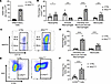
Mesenchymal inflammation recruits macrophages to the saccular-stage lung. We hypothesized that the hypercellular interstitium of P5 IKKβTbx4 lungs reflected inflammatory cell recruitment. To evaluate this hypothesis, we used flow cytometry to quantify myeloid cell populations in IKKβTbx4 and littermate control lungs (Supplemental Figure 5A). IKKβTbx4 lungs had a nearly 10-fold increase in CD45pos inflammatory cells compared with controls at P5 (Figure 2A). Notably, recruitment of circulating monocytes increased the CD11bpos/MERTKpos macrophage population in IKKβTbx4 lungs, consistent with interstitial and monocyte-derived macrophages (Figure 2, B–D). Many recruited cells expressed CCR2, a chemokine receptor expressed on BM-derived monocytes and macrophages (Figure 2, E and F) (51–53). Recruited macrophages primarily localized to the lung interstitium of IKKβTbx4 lungs (Supplemental Figure 5B).
Mesenchymal-derived inflammation recruits macrophages to the saccular-stageFigure 2

Mesenchymal-derived inflammation recruits macrophages to the saccular-stage lung. Doxycycline was added to drinking water of lactating dams from P0 to P5, and neonatal lungs were harvested on P5. Flow cytometry was used to quantify cells. (A) Quantification of viable CD45pos cells in lungs of control (CTRL) and IKKβTbx4 mice at P5. (B) Quantification of myeloid populations in lungs of CTRL and IKKβTbx4 mice at P5. (C and E) Flow cytometry plots demonstrating interstitial macrophages and monocyte-derived macrophages (IM & Mo-ϕ [MERTKpos, CD11bpos]) and monocytes (Mono [MERTKneg, CD11bpos]) (C) and CCR2pos cells (E) in CTRL and IKKβTbx4 lungs at P5 (SSC, side scatter). (D and F) Quantification of interstitial and monocyte-derived macrophages (D) and CCR2pos cells (F) in lungs of CTRL and IKKβTbx4 mice at P5. Data are expressed as mean ± SEM. n = 5 to 9 mice per group (B, D, and F); *P < 0.05, ***P < 0.001, and ****P < 0.0001 by unpaired 2-tailed t test (A, B, D, and F).

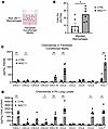
Saccular-stage fibroblasts produce chemokines that recruit macrophages. We postulated that mesenchymal chemokine production contributed to monocyte and macrophage recruitment to lungs of IKKβTbx4 mice. To test this, we generated primary lung fibroblast cultures from IKKβTbx4 and littermate control mice at P2. After in vitro transgene induction in these cells (Dox 3 μg/mL for 24 hours in serum-free conditions), fibroblast conditioned media was collected for use in macrophage chemotaxis assays using a modified Boyden chamber (Figure 3A). Conditioned media from IKKβTbx4 fibroblasts increased macrophage chemotaxis in comparison to media from control fibroblasts (Figure 3B), indicating that inflammatory signaling in lung mesenchymal cells released paracrine factors facilitating the recruitment of monocyte-derived macrophages to the saccular-stage lung. Of note, IKKβ activation in primary lung fibroblasts did not induce expression of markers of myofibroblast differentiation (Supplemental Figure 6A).
IKKβTbx4 mouse lung fibroblasts produce chemokines that recruit macrophagesFigure 3

IKKβTbx4 mouse lung fibroblasts produce chemokines that recruit macrophages. (A and B) Primary lung fibroblasts were isolated from IKKβTbx4 and littermate control (CTRL) lungs on P2 and were treated with doxycycline (3 μg/mL in media) at passage 4 or 5. Conditioned media was collected after 24 hours. Conditioned media from CTRL and IKKβTbx4 lung fibroblasts was added to the bottom well of a modified Boyden chamber. RAW 264.7 cells (50,000 per insert) were added to the top chamber (A) and migrated macrophages were fixed, stained, imaged, and counted (B). (C) Chemokine protein levels were quantified in fibroblast conditioned media using a 32-cytokine/chemokine multiplex assay and CCL7 ELISA. (D) Doxycycline was added to drinking water of lactating dams from P0 to P5. Lungs from CTRL and IKKβTbx4 mice were harvested on P5. Chemokine protein levels were quantified in lung lysate using a 32-cytokine/chemokine multiplex assay and CCL7 ELISA. Full multiplex and ELISA results are shown in Supplemental Tables 2 and 3. Inflammatory protein concentrations in lung lysate were normalized to total lung protein content. Data are expressed as mean ± SEM. n = 6 Transwells per group and n = 5 fields per Transwell membrane (B); n = 4 biological replicates per group (C); n = 5 mice per group (D); † = analyte measured using ELISA; *P < 0.05, **P < 0.01, ***P < 0.001, and ****P < 0.0001 by unpaired 2-tailed t test (B–D).

To determine whether specific chemokines mediated macrophage recruitment, we next quantified chemokine/cytokine expression in fibroblast-conditioned media. Among the cytokines and chemokines that were increased in conditioned media from IKKβTbx4 fibroblasts (Supplemental Table 2), we measured high levels of CCL2 and CCL7, both ligands of CCR2 (Figure 3C). Lung tissue lysates from transgenic IKKβTbx4 mice contained higher concentrations of CCL2 and CCL7 compared with controls (Figure 3D, Supplemental Figure 6B, and Supplemental Table 3), and expression of Ccl2 and Ccl7 transcripts was also upregulated in IKKβTbx4 lungs. By comparison, mRNA expression of other known murine CCR2 ligands, Ccl8 and Ccl12 (54), was not increased (Supplemental Figure 6C). Together, these data suggest that select CCR2 chemokines produced by the IKKβTbx4 lung mesenchyme mediate monocyte and macrophage recruitment into the saccular-stage lung.

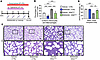
Inhibiting recruitment of CCR2pos monocytes prevents structural abnormalities in IKKβTbx4 lungs. Having observed that CCR2pos cells were increased in transgenic lungs, we next tested whether blocking recruitment of CCR2pos monocytes to IKKβTbx4 lungs would inhibit the observed structural abnormalities. For these experiments, from P1 to P4 we administered i.p. injections daily of vehicle (DMSO) or the small-molecule CCR2 antagonist RS504393 to block recruitment of circulating CCR2pos monocytes prior to lung harvest at P5 (Figure 4A). RS504393 administration reduced CCR2pos monocytes and macrophages in IKKβTbx4 lungs and rescued the morphometric abnormalities (Figure 4, B–D). Although vehicle-treated IKKβTbx4 mouse lungs demonstrated interstitial thickening and reduced distal airspace area, lungs of RS504393-treated IKKβTbx4 mice were indistinguishable from littermate controls. These findings demonstrate that CCR2-mediated monocyte recruitment drives the abnormal lung phenotype in IKKβTbx4 mice.
Inhibiting recruitment of CCR2pos monocyte-derived macrophages rescues struFigure 4

Inhibiting recruitment of CCR2pos monocyte-derived macrophages rescues structural abnormalities in IKKβTbx4 lungs. (A) Schematic of experimental time points. For these experiments, doxycycline was added to drinking water of lactating dams from P1 to P5, and i.p. injections of either vehicle (DMSO, 20 μL) or CCR2 antagonist (RS504393, 2 μg/g) were administered daily from P1 to P4. Lungs were harvested on P5. (B) Flow cytometry was used to quantify viable CCR2pos monocytes and macrophages in lungs of vehicle- or RS504393-treated littermate control (CTRL) and IKKβTbx4 mice at P5. (C) Representative H&E-stained lung sections from vehicle- and RS504393-treated mice at P5. (D) Morphometric quantification of airspace volume density (ASVD) of vehicle- and RS504393-treated mouse lungs at P5. Data are expressed as mean ± SEM. n = 10 to 12 nonoverlapping lung fields per lung (biological replicate) (D), n = 5 to 17 mice per group (B); n = 4 to 8 mice per group (D); scale bar: 100 μm (C); *P < 0.05, **P < 0.01, ***P < 0.001, and ****P < 0.0001 by 1-way ANOVA with Tukey’s multiple-comparison test (B and D).

Elastic fiber organization is altered in IKKβTbx4 lungs. Precise arrangement of elastic fibers around developing saccules is critical for normal saccular-stage lung development, and loss of this process leads to an emphysema phenotype later in life (55, 56). Because adult IKKβTbx4 lungs had an emphysematous phenotype with reduced tissue elastance (Figure 1, D–G), we examined elastic fiber deposition in the distal saccular-stage lung. At P5, elastic fibers were organized as cord-like structures around distal airspaces in control lungs, whereas fibers appeared sparse, thin, and discontinuous around airspaces in IKKβTbx4 lungs (Figure 5A). Abnormalities in elastin deposition persisted into adulthood, and IKKβTbx4 lungs continued to demonstrate a markedly disorganized elastin network at 2 months of age (Supplemental Figure 7). Importantly, treatment with the CCR2 antagonist RS504393 preserved the structure and organization of elastic fibers in P5 IKKβTbx4 lungs (Figure 5A).
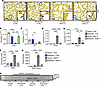
Elastic fiber organization is altered in IKKβTbx4 lungs and rescued with CCFigure 5

Elastic fiber organization is altered in IKKβTbx4 lungs and rescued with CCR2 antagonism. Doxycycline was added to drinking water of lactating dams from P1 to P5, and i.p. injections of either vehicle (DMSO, 20 μL) or CCR2 antagonist (RS504393, 2 μg/g) were given daily on P1–P4. Lungs were harvested on P5. (A) Representative photomicrographs of Hart’s-stained P5 lung sections from vehicle- or RS504393-treated littermate control (CTRL) and IKKβTbx4 mice. Arrows denote normal elastic fibers. Arrowheads denote fragmented elastic fibers in vehicle-treated IKKβTbx4 lungs. Experiments were repeated 4–5 times per group. (B–E) Gene expression of elastin assembly components (B and C) and macrophage elastases (D and E) in the lungs of vehicle- or RS504393-treated CTRL and IKKβTbx4 mice at P5 measured by qRT-PCR. (F–H) Lungs from vehicle- and RS504393-treated CTRL and IKKβTbx4 mice at P5 were homogenized and used to quantify MMP-9 protein levels and enzymatic activity using a mouse MMP-9 ELISA (F) and gelatin zymography (G and H), respectively. Data are expressed as mean ± SEM. n = 4 to 6 mice per group (B–E); n = 3 to 4 mice per group (F); n = 3 mice per group (H); scale bar: 100 μm (A); *P < 0.05, **P < 0.01, ***P < 0.001, and ****P < 0.0001 by 1-way ANOVA with Tukey’s multiple-comparison test (B–G).

We next investigated mechanisms by which recruited macrophages could disrupt elastic fiber organization in IKKβTbx4 lungs. We first investigated components of elastic fiber assembly. Expression of 2 critical components of elastin assembly, Fbln5 and Eln, was downregulated in lungs of vehicle-treated IKKβTbx4 mice and rescued with CCR2-antagonist treatment (Figure 5, B and C), indicating that recruited CCR2pos macrophages may alter elastic fiber assembly in the saccular-stage lung. Because macrophages produce MMPs that degrade elastic fibers, we also examined whether macrophage-derived MMP-9 or MMP-12 were increased in IKKβTbx4 lungs. Both Mmp9 and Mmp12 were upregulated in lungs of vehicle-treated transgenic mice compared with controls (Figure 5, D and E) and returned to control levels after RS504393 treatment. MMP9 protein levels and enzymatic activity quantified by zymography mirrored the gene expression data and were similarly normalized by RS504393 treatment (Figure 5, F–H). Collectively, these data indicated that monocyte-derived macrophages may disrupt elastin networks through 2 separate mechanisms: by altering elastic fiber assembly or by degrading preassembled elastic fibers through MMP activity.

Monocyte-derived macrophages disrupt saccular-stage microvascular development. During the transition to postnatal life, immature capillary endothelial cells differentiate alongside proliferating pericytes to expand a functional microvascular network for gas exchange (47, 57–59). Disrupting vascular patterning during this critical period leads to alveolar simplification (60–64). We therefore examined microvascular development in IKKβTbx4 mice. Lung endothelial cells were quantified at P5 by immunostaining for the pan-endothelial nuclear marker ERG. IKKβTbx4 lungs exhibited a marked reduction in endothelial cell numbers compared with littermate controls (Figure 6, A and B). Microvascular organization was also impaired, with the appearance of fewer Endomucinpos vessel structures in transgenic lungs (Supplemental Figure 8A). These vascular abnormalities persisted into adulthood, as IKKβTbx4 lungs continued to demonstrate reduced microvascular complexity at 2 months of age (Supplemental Figure 8B).
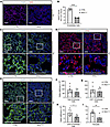
CAP2 endothelial cells and pericytes are reduced in IKKβTbx4 lungs.Figure 6

CAP2 endothelial cells and pericytes are reduced in IKKβTbx4 lungs. Doxycycline was added to drinking water of lactating dams from P0 to P5, and neonatal lungs were harvested on P5. (A–E) Immunostained lung sections from control (CTRL) and IKKβTbx4 mice at P5. Immunostaining (A) and quantification (B) of lung endothelial cell (EC) nuclear marker ERG. Immunostaining for CAP1 EC marker KIT (C), CAP2 EC marker CAR4 (D), and pericyte marker NG2 (E). Nuclei labeled with DAPI (A–E). Fluorescent imaging was performed using either widefield (A and D) or confocal microscopy (C and E). (F–I) Lung microvascular populations were quantified using flow cytometry. Number of total ECs (F), CAP1 ECs (G), CAP2 ECs (H), and pericytes (I) in the P5 lung. Gating strategy to identify capillary subpopulations and pericytes is outlined in Supplemental Figure 9B. Data are expressed as mean ± SEM; 10 to 12 nonoverlapping tissue fields were evaluated per lung (biological replicate), n = 4 or 5 mice per group (B); n = 7 mice per group (F–H); n = 4 to 7 mice per group (I); scale bar: 100 μm (A and C–E); *P < 0.05, **P < 0.01, and ****P < 0.0001 by unpaired 2-tailed t test (B and F–I).

To determine whether microvascular subpopulations contributed to this phenotype, P5 lung sections were immunostained for endothelial subpopulations and capillary-associated pericytes. Staining for both CAP1 (general capillary [gCap]) and CAP2 (alveolar capillary [aCap]) endothelial cell populations was reduced in transgenic lungs, as assessed by KIT and CAR4 staining, respectively (Figure 6, C and D). Similarly, staining for the pericyte markers NG2 and PDGFR-β was decreased in IKKβTbx4 lungs compared with controls (Figure 6E and Supplemental Figure 9A). Although flow cytometry (Supplemental Figure 9B) did not detect a significant decrease in total PECAMpos/Ve-Cadherinpos endothelial or KITpos CAP1 cells (Figure 6, F and G), both CAR4pos/KITneg CAP2s and PDGFR-βpos/CD146pos pericytes were decreased in lungs of IKKβTbx4 mice (Figure 6, H and I).

CCR2-blockade with RS504393 restored ERGpos endothelial cell numbers in P5 IKKβTbx4 lungs (Figure 7, A and B). Microvascular architecture was also improved, as demonstrated by Endomucin and CAR4 staining of capillary structures (Supplemental Figure 10, A and B). Given the reduction in CAP2 endothelial cells and pericytes, we examined expression of proangiogenic growth factors associated with these populations. Multiplex analysis of lung lysates revealed reduced levels of VEGF-A in IKKβTbx4 lungs compared with controls (Supplemental Table 3). Consistent with this, gene expression of Vegfa, Angpt1, and their cognate receptors Kdr and Tek, respectively, was decreased in vehicle-treated transgenic lungs and restored with RS504393 treatment (Figure 7, C–F). These data indicate that IKKβTbx4 mice exhibit abnormal microvascular development during the saccular stage, with disorganized capillary and pericyte architecture and reduced numbers of CAP2 endothelial cells and pericytes. CCR2 inhibition rescued microvascular architecture, endothelial cell numbers, and growth factor expression, indicating that recruitment of CCR2pos cells disrupts signaling required for saccular-stage microvascular maturation.
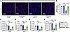
CCR2 antagonism rescues vascular development in IKKβTbx4 mice.Figure 7

CCR2 antagonism rescues vascular development in IKKβTbx4 mice. Doxycycline was added to drinking water of lactating dams from P1 to P5, and i.p. injections of either vehicle (20 μL of DMSO) or CCR2 antagonist (RS504393, 2 μg/g) were given daily on P1–P4. Lungs were harvested on P5. (A and B) Quantification of lung endothelial cells at P5. (A) Representative photomicrographs of lung sections from vehicle- and RS504393-treated control (CTRL) and IKKβTbx4 mice at P5 immunostained for endothelial cell nuclear marker ERG; nuclei are labeled with DAPI. Fluorescent imaging performed using Keyence BZX-800 widefield microscope. (B) Quantification of ERGpos nuclei at P5. (C–F) Gene expression of select proangiogenic growth factors and their cognate receptors in CTRL and IKKβTbx4 lungs at P5 measured by qRT-PCR. Data are expressed as mean ± SEM; 10 to 12 nonoverlapping tissue fields were evaluated per lung, n = 4 or 5 mice per group (B); n = 4 to 6 mice per group (C–F); scale bar: 100 μm (A); *P < 0.05, **P < 0.01, ***P < 0.001, and ****P < 0.0001 by 1-way ANOVA with Tukey’s multiple-comparison test (B–F).
Discussion

Despite the considerable morbidity associated with BPD (16, 18, 65), fundamental questions remain about how inflammation alters lung development. Although innate factors that increase the risk of developing BPD (such as gestational age) cannot be modified after birth, the mechanisms driving inflammatory lung injury may still be targetable. Identifying these critical mediators could inform strategies that improve short- and long-term outcomes in neonates at risk of developing BPD. With this goal, we developed the IKKβTbx4 mouse model, where inflammatory signaling was specifically and transiently activated in the saccular-stage lung mesenchyme. In this model, IKKβ transgene expression was sufficient to cause permanent abnormalities in lung structures, similar to those observed in the BPD lung. Further, mesenchymal inflammation recruited CCR2pos monocyte-derived macrophages, which disrupted the elastic fiber scaffold and impaired microvascular development. Blockade of CCR2-dependent immune cell recruitment rescued lung structure, establishing a mechanistic link between mesenchymal chemokine signaling and structural injury in the saccular-stage lung. The similarities between mouse IKKβTbx4 and human BPD lungs further support the lung mesenchyme as a potent amplifier of pathological inflammation and reinforce the saccular-stage lung’s vulnerability to inflammatory injury.

IKKβTbx4 mice recapitulated structural and inflammatory features of human BPD. Histologically, lungs of infants with BPD are characterized by simplified distal airspaces, irregular elastin organization, and dysmorphic capillary architecture (11–15, 66–68), all features demonstrated in the lungs of IKKβTbx4 mice. Lungs from transgenic mice also demonstrated robust macrophage infiltration, mirroring the increased macrophage burden observed in BPD lungs (10, 68–71). Macrophage-derived inflammation disrupts normal lung development (33, 72), and reducing macrophage recruitment improves alveolarization in animal models of neonatal lung injury (70, 73–77). Similarly, blocking CCR2-dependent macrophage infiltration using RS504393 rescued the lung phenotype of IKKβTbx4 mice, confirming that monocyte-derived macrophages, recruited by mesenchymal CCL2 and CCL7 production, are the primary drivers of structural injury. Proinflammatory macrophages are increased in human BPD lungs (69, 78, 79), and CCL2 and CCL7 levels are higher in plasma and tracheal aspirates of infants developing BPD (80–82). These parallels to human disease strengthen the evidence that mesenchymal NF-κB–driven inflammation recruits macrophages in BPD and further validate the translational relevance of the IKKβTbx4 model.

We previously explored the role of epithelial cell–associated inflammation using the IKTA (inhibitory κB kinase β transactivated) mouse model, in which the same activated IKKβ transgene was induced to increase NF-κB activity in the airway epithelium during the saccular stage of lung development (55, 56). Both the IKKβTbx4 mesenchymal and IKTA epithelial models exhibit a pronounced, but transient, inflammatory response after transgene induction during the saccular stage. However, whereas the IKKβTbx4 model was associated with robust macrophage recruitment to the lung and interstitial thickening, the IKTA model produced a neutrophil-predominant inflammatory response with dilated, emphysematous-appearing airspaces. Prior reports also indicate that in the developing lung endothelium, NF-κB activity is protective against exogenous inflammatory injury and required for normal vascular development (83–85). Thus, the effects of NF-κB activation are cell-type dependent. Developmental stage also influences the consequences of NF-κB–mediated inflammatory signaling. Constitutive expression of IKKβ in Twist2pos mesenchymal cells (Twist2-IKKβca mice) activated NF-κB signaling and recruited macrophages, yet no changes were observed in airway branching or elastin deposition in the canalicular-stage lung (86). Similarly, in IKTA mice, only saccular (but not alveolar) stage NF-κB activation altered lung development (56). The contrasting phenotypes across these models demonstrate how the context of NF-κB activation determines the downstream immune response and pattern of structural injury. Understanding how the cellular source and timing of NF-κB signaling shape its impact will be important when targeting this pathway using novel therapeutics.

IKKβTbx4 mice build upon prior evidence that the saccular stage is a period of developmental vulnerability, during which even brief inflammatory exposures can permanently alter lung structure (33, 55, 87). Despite distinct early phenotypes, both IKKβTbx4 and IKTA mice demonstrated lasting abnormalities in elastin deposition and emphysematous enlargement of the distal airspaces at 2 months of age, mirroring the durable structural changes observed in BPD lungs (56). Although often regarded as a disease of prematurity, BPD has consequences that extend well beyond infancy; affected individuals exhibit persistent reductions in lung function (18, 88–90), and those with severe disease are at risk for chronic obstructive pulmonary disease in adulthood (91–94). Defining how mesenchymal inflammation disrupts saccular-stage development programs may inform early interventions to prevent lifelong morbidity.

Elastin is assembled during a narrow window of lung development and, under normal conditions, undergoes minimal turnover throughout life (95). Thus, disruption during the saccular stage can permanently compromise the elastic fiber scaffold (56). IKKβTbx4 lungs demonstrated sparse, fragmented elastic fibers lining distal airspaces. Abnormalities in elastin assembly and degradation have been strongly implicated in the pathogenesis of BPD, with disordered elastin organization described in both animal models of neonatal lung inflammation and in preterm infants exposed to saccular-stage inflammation (31, 55, 96–100). Macrophage recruitment to IKKβTbx4 lungs reduced the expression of critical elastin assembly genes and increased production of macrophage-derived MMPs, disrupting the elastin network through both impaired assembly and enhanced degradation. Importantly, CCR2 inhibition with RS504393 prevented macrophage recruitment and rescued the elastin phenotype, confirming a direct role for recruited macrophages in elastin dysregulation.

Coordinated maturation of the lung microvasculature during the saccular stage is essential for normal alveolarization (60–63, 101, 102). Lungs from infants with BPD exhibit capillary rarefaction, dysmorphic capillary architecture, and aberrant endothelial patterning (11, 12, 68, 103–106). Recent scRNA-Seq studies have identified 2 major pulmonary capillary endothelial subpopulations: proliferative CAP1 cells that support vascular growth and repair, and CAP2 cells that differentiate from CAP1s during the saccular stage to facilitate gas exchange (34, 47, 63, 107, 108). IKKβTbx4 lungs demonstrated reduced and disorganized microvascular structure with decreased CAP2 and pericyte numbers. Expression of Vegfa and Angpt1, growth factors critical for CAP2 and pericyte development, was decreased in transgenic lungs. RS504393 rescued expression of both genes and restored ERGpos and CAR4pos endothelial cell staining at P5, demonstrating that CCR2-dependent macrophage recruitment impairs proangiogenic signaling directing saccular-stage microvascular development. In line with these findings, augmenting VEGF-A signaling improves distal lung development across multiple neonatal injury models (60, 101, 109) and is a future direction for our studies using the IKKβTbx4 model.

Capillary loss and pathological vascular remodeling during lung development can predispose to chronic pulmonary vascular disease (110–113); adult survivors of BPD remain at elevated risk across the lifespan (114). Pulmonary hypertension (PH) is a common comorbidity of BPD (BPD-PH), affecting as many as 40% of infants with severe disease (115). Infants with BPD-PH are at a higher risk of adverse clinical outcomes, demonstrating increased neurodevelopmental impairment, higher rates of tracheostomy placement, and a 5- to 6-fold increase in mortality compared with infants who have only BPD (115–118). Constitutive overexpression of IL-6 or TNF-α in the mouse lung during development is sufficient to cause PH (119, 120), and inhibition of cytokine signaling protects against PH in multiple neonatal injury models (110, 121). Macrophage depletion similarly prevents hyperoxia-induced PH in neonatal rodents (122). IKKβTbx4 mice exhibit persistent capillary rarefaction and a macrophage-dependent reduction in proangiogenic signaling, making PH a plausible long-term consequence. Experiments to address this are underway.

Recent studies demonstrate that inflammatory injury drives dynamic shifts in capillary endothelial subpopulations, though the specific patterns and timing of these changes remain an area of active investigation (45, 57, 102, 105, 123–125). IKKβTbx4 lungs exhibited decreased CAP2 and pericyte numbers at P5, but the fate of these populations beyond this early time point has not yet been characterized. Bidirectional signaling between endothelial cells and pericytes is required for normal capillary network formation. The interdependence of these cell populations has been illustrated by studies in which disrupting postnatal pericyte signaling was sufficient to cause alveolar simplification with reduced microvascular complexity (64), and in which primary endothelial dysfunction was shown to reduce pericyte numbers (63). The loss of CAP2 endothelial cells and pericytes in IKKβTbx4 lungs raises important questions about how mesenchymal-derived inflammation impairs coordinated microvascular development.

Although our data indicate that recruited macrophages mediate the observed vascular abnormalities in IKKβTbx4 lungs, direct effects of the mesenchymal secretome on vascular development cannot be fully excluded. Additionally, wet-to-dry lung weight ratios did not indicate pulmonary edema in IKKβTbx4 mice, but this measure reflects total lung water content relative to tissue mass and may have limited sensitivity for detecting changes in vascular permeability (126). Given the reduction in pericytes, which are critical regulators of capillary barrier function, a targeted assessment of vascular integrity will be a focus of future studies.

In summary, we demonstrate that mesenchymal-derived inflammation in the saccular-stage lung causes lasting disruptions of lung structure. Activation of NF-κB in the lung mesenchyme upregulated CCL2 and CCL7, recruiting CCR2pos macrophages. Despite a brief interval of inflammation, IKKβTbx4 mice exhibited durable impairment of lung development, specifically dysregulated elastic fiber deposition and impaired microvascular development. These findings establish mesenchyme-macrophage crosstalk as a central contributor to BPD pathogenesis. IKKβTbx4 mice model the core features of human BPD, enabling further investigation of how inflammation disrupts specific developmental programs during the saccular stage. Future work will address how recruited macrophages disrupt vascular development, how the microvasculature remodels after early endothelial and pericyte loss, and the impact of these features on adult cardiopulmonary disease.

Methods

Sex as a biological variable. Animals of both sexes were included in the analysis for all experiments completed at P5 and 2 months of age. No discernible sex differences were noted in the analysis.

Reagents. Immunostaining was completed using anti-Endomucin (ab106100), anti-α smooth muscle actin (ab5694), and anti-ERG (ab92513) from Abcam; anti-phospho-NF-κB p65 (Cell Signaling Technology, 3033); anti-CD117/c-Kit (KIT, AF1356), anti-carbonic anhydrase IV/CA4 (CAR4, AF2414), and anti-PDGFR-β (AF1042) from R&D Systems; and anti-NG2 (AB5320) from MilliporeSigma. All secondary antibodies used in immunofluorescent staining were highly cross-absorbed donkey IgG conjugated to Alexa Fluor Plus (Invitrogen) fluorophores. Autofluorescence was minimized in paraffin-embedded lung sections using TrueBlack Plus Autofluorescence Quencher (Biotium, 23014-T). Nuclei were stained with DAPI solution (Thermo Fisher Scientific, 62248). Coverslips were mounted using ProLong Gold Antifade reagent (Life Science Technologies, P36930).

The primary antibodies used for flow cytometry and FACS are listed in Supplemental Table 4. Otherwise, donkey anti-goat PE-conjugated cross-absorbed F(ab’)2 (Invitrogen, 31860) was used as a secondary antibody for anti-CAR4. DAPI (Thermo Fisher Scientific, 62248) labeling was used to assess cell viability in all flow experiments.

Mouse model design. All mice used in this study were maintained on a C57BL/6J background. WT C57BL/6J mice used for breeding were obtained from The Jackson Laboratory (strain 000664). To selectively induce expression of IKKβ and activate NF-κB in mesenchymal cells in the saccular-stage lung, we crossed hemizygous mice containing a FLAG-tagged form of activated human IKKβ under the control of the tetracycline operator-minimal CMV promoter (IKKβ mice) (127) with heterozygous mice expressing reverse tetracycline transactivator (rtTA) under the control of the 5.5 kb mouse Tbx4 lung enhancer (Tbx4-rtTA mice) (48). In the saccular-stage mouse, Tbx4 is primarily expressed in lung mesenchyme (48–50). Approximately 25% of mice from the above-described matings expressed both the IKKβ and Tbx4-rtTA transgenes; we designated these mice IKKβTbx4. The remaining littermate mice, which carried only IKKβ, only Tbx4-rtTA, or neither transgene, were used as controls. To induce transgene expression in neonatal lungs, lactating dams were administered Dox (Sigma-Aldrich, D9891) in drinking water (2 g/L of water) during the saccular stage from P0 to P5 (or from P1 to P5; see Monocyte depletion below). Lungs were then harvested at P5 or 2 months of age.

To assess cellular transgene expression patterns under the control of the Tbx4 lung enhancer, we crossed Tbx4-rtTA/TetO-Cre mice with a tdTomato reporter mouse (B6.Cg-Gt(ROSA)26Sortm14(CAG-tdTomato)Hze/J, [The Jackson Laboratory, JAX strain 007914]). Upon Dox administration, progeny from these crossings (referred to as Tbx4tdTomato mice), expressed cytosolic tdTomato fluorescence in cells expressing the Tbx4 lung enhancer. For fluorescent labeling of Tbx4pos cells in Tbx4tdTomato neonatal lungs (as with IKKβTbx4 mice), lactating dams were administered Dox (Sigma-Aldrich) in drinking water (2 g/L of water) from P0 to P5 and harvested at P5.

Monocyte depletion. To deplete recruited CCR2pos cells, neonatal mice received i.p. injections of a small-molecule CCR2 antagonist RS504393 (2 μg/g body weight) from Sigma-Aldrich (SML0711) or vehicle DMSO (20 μL, 28% DMSO in PBS) from Sigma-Aldrich daily from P1 to P4 (128, 129). For these experiments, Dox administration was delayed by 1 day (administered from P1 to P5) to align with injections for monocyte depletion. Lungs were then harvested at P5.

scRNA-Seq analysis. To determine the cellular expression of Tbx4 in the P5 mouse lung, we reexamined a previously published scRNA-Seq dataset of CD45- and Ter119-depleted WT mouse lung parenchymal cells using a web-based explorer (https://lungcells.app.vumc.org/) (47). Cells were previously sequenced using the 10x Genomics Chromium platform. For analysis, we queried Tbx4 expression among previously annotated cell populations.

Lung histology. For paraffin-embedded samples, lungs of P5 mice were perfused with PBS, isolated, and fixed for 24 hours in 10% neutral buffered formalin before automated tissue processing (Leica) and paraffin embedding using standard methods. Lungs from mice at 2 months of age (8–10 weeks) were inflation-fixed with 10% formalin (25 cm H2O inflation pressure) before harvest. Paraffin-embedded lung tissue was sequentially sectioned using a rotary microtome at 5 μm thickness and adhered to Superfrost Plus microscope slides (Thermo Fisher Scientific) for further processing. H&E staining was completed using paraffin-embedded tissue sections for assessment of lung morphology using standard methods before imaging using brightfield microscopy with a Keyence BZX-810 inverted widefield microscope (55, 56).

For frozen sections, lungs of P5 mice were perfused with PBS, isolated, and fixed in 4% paraformaldehyde at 4°C overnight before embedding and freezing in Tissue-Tek OCT media (Sakura). In samples expressing endogenous fluorescent protein, fixation was limited to 30 minutes at room temperature. Frozen tissue blocks were sequentially sectioned at –20°C using a manual cryostat at 8 μm thickness, plated on Superfrost Plus microscope slides, and allowed to adhere for 30 minutes at room temperature before storage at –80°C for future immunostaining.

Lung morphometry. To conduct morphometric analysis of H&E-stained lung sections from neonatal and adult mice, 10 to 12 nonoverlapping lung fields were imaged across multiple lobes within a single lung section from each mouse. Five to 9 mice were imaged per group. Tiff images of H&E-stained sections were exported and analyzed using Fiji (130) software (version 2.16.0/1.54p) by a trained investigator blinded to animal genotype and treatment status, as previously described (55, 56). Analysis was performed with the semiautomated tool AlveolEye as previously described (131). To determine the proportion of distal airspace area in P5 neonatal lung sections, airspace volume density of distal lung saccules was measured using images obtained at 40× original magnification. Mean linear intercept was measured using 3 test lines overlapped on images taken at 20× original magnification from mice at 2 months of age. With the assistance of structural screening by AlveolEye, large-caliber airspaces and large pulmonary vessels were manually identified and excluded from the distal airspace before measurements and analysis.

Lung wet-to-dry weight ratios. To determine the lung wet-to-dry weight ratio as a surrogate measure for pulmonary edema (132), P5 mouse lungs were perfused with PBS and isolated en bloc. Whole lung was weighed immediately after harvest and again after 16 hours of oven dehydration at 65°C (133).

Immunofluorescent staining and ERG quantification. Immunofluorescent staining was performed using both paraffin-embedded and frozen lung tissue sections. To prepare paraffin-embedded lung tissue for immunostaining, sections were rehydrated using standard techniques, subjected to antigen retrieval with pH 6.5 citrate buffer (Sigma-Aldrich, C9999), and blocked for 10 minutes at room temperature using Power Block per the manufacturer’s recommendations (Biogenex Labs, HK0855K). For frozen lung tissue immunostaining, sections were brought to room temperature, washed in PBS to remove excess OCT, permeabilized for 10 minutes using 0.1% Triton X-100 (Sigma-Aldrich), and blocked for 30 minutes with 1% BSA and 5% horse serum in PBS. After initial steps, all lung sections were incubated with primary antibody in PBS-Tween (0.05%) overnight at 4°C. Sections were subsequently stained with secondary antibody in PBS-Tween (0.05%) for 1 hour at room temperature and briefly incubated with DAPI nuclear stain. Before cover-slipping, paraffin-embedded lung sections were treated with TrueBlack Plus Autofluorescence Quencher per the manufacturer’s recommendations. All slides were mounted with ProLong Gold Antifade Mountant and allowed to cure for at least 24 hours before imaging. Imaging of ERG, Endomucin, and CAR4 staining was performed using a Keyence BZX-810 inverted fluorescent widefield microscope. Other fluorescent images were taken on a Nikon TiE inverted spinning disk confocal microscope equipped with a Yokogawa X1 spinning disk head and Photometrics Prime 95B camera.

To quantify endothelial cells in P5 lungs, lung sections were immunostained for endothelial nuclear marker ERG and imaged with a Keyence BZX-810 inverted fluorescent widefield microscope at 10× and 40× original magnification. Ten to 12 nonoverlapping high-power lung fields were imaged across multiple lobes within a single lung section from each mouse. Four to 5 mice were included per group. Tiff images of ERG-stained sections were exported, and ERGpos nuclei were quantified using Fiji software. Specifically, a threshold was uniformly applied to all ERG-fluorescent images to identify regions of positive staining; ERGpos cells were counted using the Analyze Particles function. The number of ERGpos cells was normalized to the DAPI-fluorescent tissue area (mm2) of each image. ERG fluorescence localized outside regions of DAPI staining was considered nonspecific and excluded from analysis. ERGpos nuclei per tissue area was averaged for each animal.

Hart’s elastic fiber staining. Elastic fibers were visualized using Hart’s elastin staining as previously described (55). Briefly, paraffin-embedded lung sections were rehydrated and incubated with resorcin-fuchsin (Electron Microscopy Sciences) overnight to stain elastic fibers. Tartrazine in saturated picric acid (Electron Microscopy Sciences) was used to counterstain elastin-stained sections. Slides were imaged using brightfield microscopy with a Keyence BZX-810 inverted widefield microscope.

Mouse lung fibroblast cultures and generation of conditioned media. Neonatal mouse lung fibroblasts were isolated using previously described methods (55). Briefly, neonatal mouse lungs at P2 were perfused with sterile PBS and harvested. Lungs were minced with scissors and enzymatically digested. The resulting cell suspension was centrifuged, resuspended in fresh medium, and sequentially filtered. Filtrate from the third filtration was plated on 60 mm cell culture dishes in DMEM containing 10% FBS overnight and passaged or cryopreserved for use in later experiments. All experiments utilizing fibroblast cultures were completed using cells from the fourth or fifth passage. For in vitro experiments, 3 × 10⁵ IKKβTbx4 or control lung fibroblasts were cultured on 6-well plastic dishes and treated with Dox (3 μg/mL) for 24 hours prior to harvest. The supernatant containing conditioned media was collected and centrifuged (500g) to pellet cellular debris. The remaining fibroblasts were harvested in TRIzol for later isolation of total RNA. Both components were frozen at –80°C for future use.

Mouse lung macrophage cultures. RAW 264.7 macrophages (ATCC, TIB-71) were cultured in ATCC-formulated DMEM (ATCC, 30-2002) with 10% FBS. Macrophage cultures were passaged (1:6) every 2–3 days, when cells reached 70%–90% confluency.

Macrophage migration assay. Experiments utilized a modified Boyden chamber with Transwell filters containing 5 μm pores (Corning, CLS3421) placed in a well of a 12-well tissue culture plate as previously described (134). RAW 264.7 macrophages (50,000 per insert) were placed on top of the Transwell filter in 0.1 mL serum-free DMEM. Conditioned media (0.4 mL) was collected (as above) from IKKβTbx4 and littermate control fibroblast cultures and added to the bottom well. After a 4-hour incubation at 37°C, macrophages still present on the top of the filter were removed by clearing with a sterile cotton swab. Migrated macrophages on the underside of the Transwell membrane were fixed in 4% paraformaldehyde and stained using the Hema 3 Manual Staining System (Thermo Fisher Scientific, 23-123869) per the manufacturer’s instructions. Five nonoverlapping 20× images of the membrane were taken using a brightfield microscope, and the number of migrated cells per field was quantified.

Lung function measurements. After transgene induction with Dox from P0 to P5, lung function was quantified by flexiVent apparatus (SCIREQ) in IKKβTbx4 and littermate control mice at 2 months of age using previously established methods (135, 136). Briefly, mice were anesthetized using pentobarbital sodium (85 mg/kg) before an 18-gauge tracheostomy tube was placed in the trachea. Mice were mechanically ventilated using the SCIREQ flexiVent apparatus at 150 breaths/min and a 10 mL/kg tidal volume before obtaining lung function measurements. Respiratory system elastance and compliance were assessed using the flexiVent Snapshot model. Tissue elastance was determined using a constant phase model, which distinguishes between airway and tissue mechanics. FEV0.1 (volume expired over initial 0.1 seconds of exhalation) was obtained using a forced expired volume maneuver, which mimics clinical spirometry measurements.

Flow cytometry and FACS. Flow cytometry to quantify myeloid cells was completed using previously described methods (56). Briefly, mouse lungs at P5 were perfused with PBS and harvested. Lungs were minced and enzymatically digested for 20 minutes at 37°C using collagenase XI (0.7 mg/mL; Sigma-Aldrich, C7657), type IV DNase (30 μg/mL; Sigma-Aldrich, D5025), and Dispase (0.25 mg/mL; Gibco, 17105-041). Digested cells were filtered through a 70 μm filter to obtain a single-cell suspension and RBCs were lysed using ACK lysing buffer (Gibco) for 3 minutes at room temperature before halting the reaction with cold PBS. Cells were labeled with conjugated primary antibodies and analyzed using a 5-laser LSRFortessa analytical flow cytometer and FlowJo software version 10.10.0 (both from BD Biosciences). DAPI was used to determine cell viability. The total number of cells per lung was determined by preparing a single-cell suspension (as above) and counting using a TC20 Automated Cell Counter per the manufacturer’s instructions (Bio-Rad).

To isolate individual populations of lung parenchyma, transgenic mouse lungs harvested at P4 were processed for flow cytometry as described above. Cells were sorted using FACS with a 5-Laser FACS Aria III (BD Biosciences) cell sorter into epithelial (CD45neg, EpCAMpos, CD31neg), mesenchymal (CD45neg, EpCAMneg, and CD31neg), and endothelial (CD45neg, EpCAMneg, CD31pos) cell populations. Sorted cells were pelleted and resuspended in TRIzol reagent (Invitrogen) before freezing at –80°C for future RNA extraction.

Flow cytometry was also used to quantify lung microvascular subpopulations. Samples were prepared as above with the following protocol modifications to enhance yield and viability (57, 137). Mouse lungs were harvested and minced as above, but were digested with Liberase TM (Roche, 540119001) and type IV DNase (100 μg/mL) for 30 minutes at 37°C before quenching with 10% FBS in PBS. To enhance antibody specificity, cells were blocked with CD-32 Fc block (BD Biosciences, 553141) according to the manufacturer’s recommendations. A subset of samples was stained with a fluorescent-conjugated secondary antibody when required. Finally, for precise quantification of cell populations, Precision Counting Beads (BioLegend, 424902) were added to stained single-cell suspensions immediately before analysis, which was completed with a 3-laser LSRFortessa analytical flow cytometer (BD Biosciences). Cells and counting beads were analyzed using FlowJo software, and lung cell populations were quantified per the manufacturer’s instructions.

RT-qPCR. Total RNA was isolated from whole neonatal lungs, cultured lung fibroblasts, or FACS-sorted lung parenchymal cells with TRIzol reagent (Invitrogen) using standard procedures. First-strand cDNA was synthesized using oligo-dT primers and the Superscript VILO cDNA Reverse Transcriptase kit, which uses a Moloney murine leukemia virus (MMLV) reverse transcriptase (Invitrogen, 11754050). PCR primers were designed using the PrimerQuest design tool (Integrated DNA Technologies). Target gene forward (sense) and reverse (antisense) primer sequences are listed in Supplemental Table 5. Two-step real-time PCR was performed using an IQ5 thermocycler and SYBR Green detection system (Bio-Rad). Target gene expression was normalized to housekeeping gene Actb in each sample. Fold-change in gene expression was calculated using the 2–ΔΔCt method (138). Gel electrophoresis was used to verify the size of each amplified gene product.

Protein quantification using ELISA and multiplex assay. Tissue lysates for protein analysis were generated by sonicating the right-upper lung lobe in 0.5× RIPA (Pierce, 89900) diluted in PBS containing protease and phosphatase inhibitor cocktails (Roche, 04-693-132-001 and 04-906-845-001). Samples were centrifuged, supernatant was collected, and total protein was quantified using a bicinchoninic acid protein assay kit (Pierce, 23225). Conditioned media was prepared as previously described (see Mouse lung fibroblast cultures and generation of conditioned media), and total protein was also quantified using a bicinchoninic acid protein assay kit.

Cytokines/chemokines in whole lung tissue lysates and fibroblast conditioned media were quantified using a magnetic bead 32-multiplex assay (MilliporeSigma, MCYTMAG-70K-PX32) in duplicate per the manufacturer’s instructions. The multiplex assay was performed on the Luminex MAGPIX platform in the Vanderbilt Hormone and Analytical Services Core. Mouse CCL7 (Abcam, ab205571) and MMP-9 ELISAs (Abcam, ab253227) were used to quantify CCL7 and MMP-9 expression per the manufacturer’s recommended methods. Cytokine concentrations were normalized to total protein content of the sample where indicated.

Gelatin zymography. Lung lysates were prepared as described above. First, 20 μg protein per sample was loaded onto an acrylamide gel containing 0.1% gelatin and subjected to electrophoresis. Gels were prepared and stained with Coomassie blue using standard methods (139). After destaining, zymograms were imaged, and MMP-9 bands (93 kDa) were quantified by densitometry analysis with Fiji software using standard methods (130).

Statistics. Pairwise comparisons were done using an unpaired 2-tailed Student’s t test or a 1-way ANOVA with Tukey’s correction for multiple comparisons. All analyses were performed using GraphPad Prism version 10.6.0. Data are shown as mean ± SEM unless otherwise noted. A P value less than 0.05 was used to determine statistical significance.

Study approval. The experiments in this study were conducted in accordance with the guidelines outlined by the US Public Health Service Policy on the Humane Care and Use of Laboratory Animals. All animal experimental protocols were approved by the IACUC at Vanderbilt University Medical Center (protocol M1800030).

Data availability. All reagents used in this study will be made available upon reasonable request to the corresponding author. Values for all data points in the graphs are provided in the Supporting Data Values file.

Author contributions

BCC, JCL, BCE, SD, RVDM, WH, ALS, CS, LP, EJP, DCN, BWR, SHG, and JTB designed and performed experiments and analyzed data. BCC, JCL, CS, LP, DSN, HT, and JTB performed histological analysis. WS, EJP, LSP, and BWR provided methodological support. WS provided the Tbx4-rtTA mouse line. BCC, BWR, TSB, SHG, and JTB conceived of and designed the experiments. BCC and JTB drafted the original manuscript. BCC, BWR, LSP, TSB, SHG, and JTB revised the manuscript. All authors reviewed and approved the final manuscript.

Conflict of interest

The authors have declared that no conflict of interest exists.

Funding support

This work is the result of NIH funding, in whole or in part, and is subject to the NIH Public Access Policy. Through acceptance of this federal funding, the NIH has been given a right to make the work publicly available in PubMed Central.

  • NIH grants HL094296, HL105334, and HD087023 (supporting BCC as a Research Scholar).
  • NIH grants HL122554 and HL136664 (to DCN).
  • NIH grant HL163195 (to EJP).
  • NIH grant HL150617 (to SHG).
  • NIH grant HL157373 (to JTB).
  • Department of Veterans Affairs grant IK2BX003841 (to BWR).
  • SHG is the Julia Carell Stadler Professor of Pediatrics at the Monroe Carell Jr. Children’s Hospital.
Supplemental material

View Supplemental data

View Unedited blot and gel images

View Supporting data values

Acknowledgments

Flow cytometry experiments were performed in the Vanderbilt University Medical Center Flow Cytometry Shared Resource (supported by the Vanderbilt Ingram Cancer Center P30 CA68485 and the Vanderbilt Digestive Disease Research Center DK058404). Additionally, the authors would like to thank Jennifer Sucre, Nicholas Negretti, Shawyon Shirazi, Peter Gulleman, and Chris Jeter for their technical support. The graphical abstract and components of Figure 3A, Figure 4A, and Supplemental Figure 1A were created with BioRender.

Address correspondence to: Benjamin C. Crawford, Monroe Carell Jr. Children’s Hospital, Vanderbilt University Medical Center, 2215B Garland Avenue, 1125 MRB4/Light Hall, Nashville, Tennessee, 37232, USA. Phone: 615.343.5666; Email: Benjamin.crawford@vumc.org.

Footnotes

Copyright: © 2026, Crawford et al. This is an open access article published under the terms of the Creative Commons Attribution 4.0 International License.

Authorship note: SHG and JTB are co–senior authors.

Reference information: JCI Insight. 2026;11(14):e193625.https://doi.org/10.1172/jci.insight.193625.

References
  1. Schittny JC. Development of the lung. Cell Tissue Res. 2017;367(3):427–444.
    View this article via: CrossRef PubMed Google Scholar
  2. McCulley D, et al. The pulmonary mesenchyme directs lung development. Curr Opin Genet Dev. 2015;32:98–105.
    View this article via: CrossRef PubMed Google Scholar
  3. Branchfield K, et al. A three-dimensional study of alveologenesis in mouse lung. Dev Biol. 2016;409(2):429–441.
    View this article via: CrossRef PubMed Google Scholar
  4. Chao CM, et al. A breath of fresh air on the mesenchyme: impact of impaired mesenchymal development on the pathogenesis of bronchopulmonary dysplasia. Front Med (Lausanne). 2015;2:27.
    View this article via: PubMed CrossRef Google Scholar
  5. Bourbon J, et al. Control mechanisms of lung alveolar development and their disorders in bronchopulmonary dysplasia. Pediatr Res. 2005;57(5 pt 2):38R–46R.
    View this article via: CrossRef PubMed Google Scholar
  6. Neptune ER, et al. Dysregulation of TGF-beta activation contributes to pathogenesis in Marfan syndrome. Nat Genet. 2003;33(3):407–411.
    View this article via: CrossRef PubMed Google Scholar
  7. Ruiz-Camp J, Morty RE. Divergent fibroblast growth factor signaling pathways in lung fibroblast subsets: where do we go from here? Am J Physiol Lung Cell Mol Physiol. 2015;309(8):L751–L755.
    View this article via: CrossRef PubMed Google Scholar
  8. Gouveia L, et al. PDGF-A signaling is required for secondary alveolar septation and controls epithelial proliferation in the developing lung. Development. 2018;145(7):dev161976.
    View this article via: CrossRef PubMed Google Scholar
  9. Thebaud B, et al. Bronchopulmonary dysplasia. Nat Rev Dis Primers. 2019;5(1):78.
    View this article via: CrossRef PubMed Google Scholar
  10. Ambalavanan N, et al. The evolving pathophysiology of bronchopulmonary dysplasia. Physiol Rev. 2026;106(1):197–237.
    View this article via: CrossRef PubMed Google Scholar
  11. Bhatt AJ, et al. Disrupted pulmonary vasculature and decreased vascular endothelial growth factor, Flt-1, and TIE-2 in human infants dying with bronchopulmonary dysplasia. Am J Respir Crit Care Med. 2001;164(10 pt 1):1971–1980.
    View this article via: CrossRef PubMed Google Scholar
  12. De Paepe ME, et al. Growth of pulmonary microvasculature in ventilated preterm infants. Am J Respir Crit Care Med. 2006;173(2):204–211.
    View this article via: CrossRef PubMed Google Scholar
  13. Coalson JJ. Pathology of bronchopulmonary dysplasia. Semin Perinatol. 2006;30(4):179–184.
    View this article via: CrossRef PubMed Google Scholar
  14. Callaway DA, et al. Lung biopsy in infants with severe bronchopulmonary dysplasia. Pediatr Pulmonol. 2023;58(7):2068–2075.
    View this article via: CrossRef PubMed Google Scholar
  15. Stone A, et al. Early histological changes of bronchopulmonary dysplasia and pulmonary hypertension may precede clinical diagnosis in preterm infants. Early Hum Dev. 2022;171:105612.
    View this article via: CrossRef PubMed Google Scholar
  16. Jensen EA, et al. Severity of bronchopulmonary dysplasia among very preterm infants in the United States. Pediatrics. 2021;148(1):e2020030007.
    View this article via: CrossRef PubMed Google Scholar
  17. Collaco JM, et al. Long-term pulmonary outcomes in BPD throughout the life-course. J Perinatol. 2024;46(2):127–135.
    View this article via: CrossRef PubMed Google Scholar
  18. Islam JY, et al. Understanding the short- and long-term respiratory outcomes of prematurity and bronchopulmonary dysplasia. Am J Respir Crit Care Med. 2015;192(2):134–156.
    View this article via: CrossRef PubMed Google Scholar
  19. Stoll BJ, et al. Neonatal outcomes of extremely preterm infants from the NICHD Neonatal Research Network. Pediatrics. 2010;126(3):443–456.
    View this article via: CrossRef PubMed Google Scholar
  20. Horbar JD, et al. Trends in mortality and morbidities for infants born 24 to 28 weeks in the US: 1997–2021. Pediatrics. 2024;153(1):e2023064153.
    View this article via: CrossRef PubMed Google Scholar
  21. Watterberg KL, et al. Chorioamnionitis and early lung inflammation in infants in whom bronchopulmonary dysplasia develops. Pediatrics. 1996;97(2):210–215.
    View this article via: CrossRef PubMed Google Scholar
  22. Villamor-Martinez E, et al. Association of chorioamnionitis with bronchopulmonary dysplasia among preterm infants: a systematic review, meta-analysis, and metaregression. JAMA Netw Open. 2019;2(11):e1914611.
    View this article via: CrossRef PubMed Google Scholar
  23. Liu WL, et al. Relationship between chorioamnionitis or funisitis and lung injury among preterm infants: meta-analysis involved 16 observational studies with 68,397 participants. BMC Pediatr. 2024;24(1):157.
    View this article via: CrossRef PubMed Google Scholar
  24. Kallapur SG, Jobe AH. Contribution of inflammation to lung injury and development. Arch Dis Child Fetal Neonatal Ed. 2006;91(2):F132–F135.
    View this article via: CrossRef PubMed Google Scholar
  25. Holzfurtner L, et al. When inflammation meets lung development-an update on the pathogenesis of bronchopulmonary dysplasia. Mol Cell Pediatr. 2022;9(1):7.
    View this article via: CrossRef PubMed Google Scholar
  26. Buczynski BW, et al. The role of hyperoxia in the pathogenesis of experimental BPD. Semin Perinatol. 2013;37(2):69–78.
    View this article via: CrossRef PubMed Google Scholar
  27. Keszler M, Sant’Anna G. Mechanical ventilation and bronchopulmonary dysplasia. Clin Perinatol. 2015;42(4):781–796.
    View this article via: CrossRef PubMed Google Scholar
  28. Moss TJ, et al. Early gestational intra-amniotic endotoxin: lung function, surfactant, and morphometry. Am J Respir Crit Care Med. 2002;165(6):805–811.
    View this article via: CrossRef PubMed Google Scholar
  29. Wai KC, et al. Early cumulative supplemental oxygen predicts bronchopulmonary dysplasia in high risk extremely low gestational age newborns. J Pediatr. 2016;177:97–102.
    View this article via: CrossRef PubMed Google Scholar
  30. Hilgendorff A, et al. Chronic lung disease in the preterm infant. Lessons learned from animal models. Am J Respir Cell Mol Biol. 2014;50(2):233–245.
    View this article via: CrossRef PubMed Google Scholar
  31. Albertine KH, et al. Chronic lung injury in preterm lambs. Disordered respiratory tract development. Am J Respir Crit Care Med. 1999;159(3):945–958.
    View this article via: CrossRef PubMed Google Scholar
  32. Liao J, et al. The NLRP3 inflammasome is critically involved in the development of bronchopulmonary dysplasia. Nat Commun. 2015;6:8977.
    View this article via: CrossRef PubMed Google Scholar
  33. Stouch AN, et al. IL-1β and inflammasome activity link inflammation to abnormal fetal airway development. J Immunol. 2016;196(8):3411–3420.
    View this article via: CrossRef PubMed Google Scholar
  34. Toth A, et al. Inflammatory blockade prevents injury to the developing pulmonary gas exchange surface in preterm primates. Sci Transl Med. 2022;14(638):eabl8574.
    View this article via: CrossRef PubMed Google Scholar
  35. Xia Y, et al. RelB regulation of chemokine expression modulates local inflammation. Am J Pathol. 1997;151(2):375–387.
    View this article via: PubMed Google Scholar
  36. Yang X, et al. Proinflammatory phenotype of vascular smooth muscle cells: role of efficient Toll-like receptor 4 signaling. Am J Physiol Heart Circ Physiol. 2005;289(3):H1069–H1076.
    View this article via: CrossRef PubMed Google Scholar
  37. Guijarro-Munoz I, et al. Lipopolysaccharide activates Toll-like receptor 4 (TLR4)-mediated NF-κB signaling pathway and proinflammatory response in human pericytes. J Biol Chem. 2014;289(4):2457–2468.
    View this article via: CrossRef PubMed Google Scholar
  38. Rayner SG, et al. Lung pericytes as mediators of inflammation. Am J Physiol Lung Cell Mol Physiol. 2023;325(1):L1–L8.
    View this article via: CrossRef PubMed Google Scholar
  39. Rolfe MW, et al. Pulmonary fibroblast expression of interleukin-8: a model for alveolar macrophage-derived cytokine networking. Am J Respir Cell Mol Biol. 1991;5(5):493–501.
    View this article via: CrossRef PubMed Google Scholar
  40. Smith RS, et al. Fibroblasts as sentinel cells. Synthesis of chemokines and regulation of inflammation. Am J Pathol. 1997;151(2):317–322.
    View this article via: PubMed Google Scholar
  41. Bautista-Hernandez LA, et al. Fibroblasts: the unknown sentinels eliciting immune responses against microorganisms. Eur J Microbiol Immunol (Bp). 2017;7(3):151–157.
    View this article via: CrossRef PubMed Google Scholar
  42. Medal RM, et al. The innate immune response in fetal lung mesenchymal cells targets VEGFR2 expression and activity. Am J Physiol Lung Cell Mol Physiol. 2017;312(6):L861–L872.
    View this article via: CrossRef PubMed Google Scholar
  43. Hawwa RL, et al. IL-10 inhibits inflammatory cytokines released by fetal mouse lung fibroblasts exposed to mechanical stretch. Pediatr Pulmonol. 2011;46(7):640–649.
    View this article via: CrossRef PubMed Google Scholar
  44. Soucy AM, et al. Transcriptomic responses of lung mesenchymal cells during pneumonia. JCI Insight. 2025;10(7):e177084.
    View this article via: JCI Insight CrossRef PubMed Google Scholar
  45. Hurskainen M, et al. Single cell transcriptomic analysis of murine lung development on hyperoxia-induced damage. Nat Commun. 2021;12(1):1565.
    View this article via: CrossRef PubMed Google Scholar
  46. Jackson CM, et al. A potent myeloid response is rapidly activated in the lungs of premature Rhesus macaques exposed to intra-uterine inflammation. Mucosal Immunol. 2022;15(4):730–744.
    View this article via: CrossRef PubMed Google Scholar
  47. Negretti NM, et al. A single-cell atlas of mouse lung development. Development. 2021;148(24):dev199512.
    View this article via: CrossRef PubMed Google Scholar
  48. Zhang W, et al. Spatial-temporal targeting of lung-specific mesenchyme by a Tbx4 enhancer. BMC Biol. 2013;11:111.
    View this article via: CrossRef PubMed Google Scholar
  49. Xie T, et al. Transcription factor TBX4 regulates myofibroblast accumulation and lung fibrosis. J Clin Invest. 2016;126(9):3626.
    View this article via: JCI CrossRef PubMed Google Scholar
  50. Cai Y, et al. RNA-Seq and ChIP-Seq identification of unique and overlapping target genes and pathways regulated by TBX4 in human pulmonary fibroblasts and pericytes. Pulm Circ. 2025;15(1):e70058.
    View this article via: CrossRef PubMed Google Scholar
  51. Opalek JM, et al. Alveolar macrophages lack CCR2 expression and do not migrate to CCL2. J Inflamm (lond). 2007;4:19.
    View this article via: CrossRef PubMed Google Scholar
  52. Oliveira VLS, et al. Absence of CCR2 promotes proliferation of alveolar macrophages that control lung inflammation in acute respiratory distress syndrome in mice. Int J Mol Sci. 2022;23(21):12920.
    View this article via: CrossRef PubMed Google Scholar
  53. Oz HH, et al. Recruited monocytes/macrophages drive pulmonary neutrophilic inflammation and irreversible lung tissue remodeling in cystic fibrosis. Cell Rep. 2022;41(11):111797.
    View this article via: CrossRef PubMed Google Scholar
  54. She S, et al. Functional roles of chemokine receptor ccr2 and its ligands in liver disease. Front Immunol. 2022;13:812431.
    View this article via: CrossRef PubMed Google Scholar
  55. Benjamin JT, et al. Epithelial-derived inflammation disrupts elastin assembly and alters saccular stage lung development. Am J Pathol. 2016;186(7):1786–1800.
    View this article via: CrossRef PubMed Google Scholar
  56. Benjamin JT, et al. Neutrophilic inflammation during lung development disrupts elastin assembly and predisposes adult mice to COPD. J Clin Invest. 2021;131(1):e139481.
    View this article via: JCI CrossRef PubMed Google Scholar
  57. Zanini F, et al. Developmental diversity and unique sensitivity to injury of lung endothelial subtypes during postnatal growth. iScience. 2023;26(3):106097.
    View this article via: CrossRef PubMed Google Scholar
  58. Zanini F, et al. Hyperoxia prevents the dynamic neonatal increases in lung mesenchymal cell diversity. Sci Rep. 2024;14(1):2033.
    View this article via: CrossRef PubMed Google Scholar
  59. Sharma A, Niethamer TK. Specialized pulmonary vascular cells in development and disease. Annu Rev Physiol. 2025;87(1):229–255.
    View this article via: CrossRef PubMed Google Scholar
  60. Thebaud B, et al. Vascular endothelial growth factor gene therapy increases survival, promotes lung angiogenesis, and prevents alveolar damage in hyperoxia-induced lung injury: evidence that angiogenesis participates in alveolarization. Circulation. 2005;112(16):2477–2486.
    View this article via: CrossRef PubMed Google Scholar
  61. Jakkula M, et al. Inhibition of angiogenesis decreases alveolarization in the developing rat lung. Am J Physiol Lung Cell Mol Physiol. 2000;279(3):L600–L607.
    View this article via: CrossRef PubMed Google Scholar
  62. Le Cras TD, et al. Treatment of newborn rats with a VEGF receptor inhibitor causes pulmonary hypertension and abnormal lung structure. Am J Physiol Lung Cell Mol Physiol. 2002;283(3):L555–L562.
    View this article via: CrossRef PubMed Google Scholar
  63. Vila Ellis L, et al. Epithelial Vegfa specifies a distinct endothelial population in the mouse lung. Dev Cell. 2020;52(5):617–630.
    View this article via: CrossRef PubMed Google Scholar
  64. Kato K, et al. Pulmonary pericytes regulate lung morphogenesis. Nat Commun. 2018;9(1):2448.
    View this article via: CrossRef PubMed Google Scholar
  65. Doyle LW, et al. Increasing airway obstruction from 8 to 18 years in extremely preterm/low-birthweight survivors born in the surfactant era. Thorax. 2017;72(8):712–719.
    View this article via: CrossRef PubMed Google Scholar
  66. Thibeault DW, et al. Lung elastic tissue maturation and perturbations during the evolution of chronic lung disease. Pediatrics. 2000;106(6):1452–1459.
    View this article via: CrossRef PubMed Google Scholar
  67. Thibeault DW, et al. Acinar arterial changes with chronic lung disease of prematurity in the surfactant era. Pediatr Pulmonol. 2003;36(6):482–489.
    View this article via: CrossRef PubMed Google Scholar
  68. Dylag AM, et al. New insights into the natural history of bronchopulmonary dysplasia from proteomics and multiplexed immunohistochemistry. Am J Physiol Lung Cell Mol Physiol. 2023;325(4):L419–L433.
    View this article via: CrossRef PubMed Google Scholar
  69. Hirani D, et al. Macrophage-derived IL-6 trans-signalling as a novel target in the pathogenesis of bronchopulmonary dysplasia. Eur Respir J. 2022;59(2):2002248.
    View this article via: CrossRef PubMed Google Scholar
  70. Hirani DV, et al. CXCL10 deficiency limits macrophage infiltration, preserves lung matrix, and enables lung growth in bronchopulmonary dysplasia. Inflamm Regen. 2023;43(1):52.
    View this article via: CrossRef PubMed Google Scholar
  71. Toti P, et al. Bronchopulmonary dysplasia of the premature baby: an immunohistochemical study. Pediatr Pulmonol. 1997;24(1):22–28.
    View this article via: CrossRef PubMed Google Scholar
  72. Blackwell TS, et al. NF-κB signaling in fetal lung macrophages disrupts airway morphogenesis. J Immunol. 2011;187(5):2740–2747.
    View this article via: CrossRef PubMed Google Scholar
  73. Eldredge LC, et al. Imbalance of Ly-6Chi and Ly-6Clo monocytes/macrophages worsens hyperoxia-induced lung injury and is rescued by IFN-γ. J Immunol. 2019;202(9):2772–2781.
    View this article via: CrossRef PubMed Google Scholar
  74. Kalymbetova TV, et al. Resident alveolar macrophages are master regulators of arrested alveolarization in experimental bronchopulmonary dysplasia. J Pathol. 2018;245(2):153–159.
    View this article via: CrossRef PubMed Google Scholar
  75. Shahzad T, et al. Anti-CCL2 therapy reduces oxygen toxicity to the immature lung. Cell Death Discov. 2024;10(1):311.
    View this article via: CrossRef PubMed Google Scholar
  76. Cui TX, et al. CCR2 mediates chronic LPS-induced pulmonary inflammation and hypoalveolarization in a murine model of bronchopulmonary dysplasia. Front Immunol. 2020;11:579628.
    View this article via: CrossRef PubMed Google Scholar
  77. Tu Z, et al. Macrophage pyroptosis mediates hyperoxia-induced inflammatory lung injury in neonates. Front Immunol. 2025;16:1546986.
    View this article via: CrossRef PubMed Google Scholar
  78. Mestan KK, et al. Bronchopulmonary dysplasia: signatures of monocyte-macrophage reactivity and tolerance define novel placenta-lung endotypes. Pediatr Res. 2025;98(6):2352–2362.
    View this article via: CrossRef PubMed Google Scholar
  79. Sahoo D, et al. Transcriptional profiling of lung macrophages identifies a predictive signature for inflammatory lung disease in preterm infants. Commun Biol. 2020;3(1):259.
    View this article via: CrossRef PubMed Google Scholar
  80. Baier RJ, et al. Monocyte chemoattractant protein-1 and interleukin-8 are increased in bronchopulmonary dysplasia: relation to isolation of Ureaplasma urealyticum. J Investig Med. 2001;49(4):362–369.
    View this article via: CrossRef PubMed Google Scholar
  81. Baier RJ, et al. Increased interleukin-8 and monocyte chemoattractant protein-1 concentrations in mechanically ventilated preterm infants with pulmonary hemorrhage. Pediatr Pulmonol. 2002;34(2):131–137.
    View this article via: CrossRef PubMed Google Scholar
  82. Baier RJ, et al. CC chemokine concentrations increase in respiratory distress syndrome and correlate with development of bronchopulmonary dysplasia. Pediatr Pulmonol. 2004;37(2):137–148.
    View this article via: CrossRef PubMed Google Scholar
  83. Iosef C, et al. Inhibiting NF-κB in the developing lung disrupts angiogenesis and alveolarization. Am J Physiol Lung Cell Mol Physiol. 2012;302(10):L1023–L1036.
    View this article via: CrossRef PubMed Google Scholar
  84. Hou Y, et al. Activation of the nuclear factor-κB pathway during postnatal lung inflammation preserves alveolarization by suppressing macrophage inflammatory protein-2. Am J Physiol Lung Cell Mol Physiol. 2015;309(6):L593–L604.
    View this article via: CrossRef PubMed Google Scholar
  85. Rao S, et al. Endothelial-specific loss of IKKβ disrupts pulmonary endothelial angiogenesis and impairs postnatal lung growth. Am J Physiol Lung Cell Mol Physiol. 2023;325(3):L299–L313.
    View this article via: CrossRef PubMed Google Scholar
  86. McCoy AM, et al. IKKβ activation in the fetal lung mesenchyme alters lung vascular development but not airway morphogenesis. Am J Pathol. 2017;187(12):2635–2644.
    View this article via: CrossRef PubMed Google Scholar
  87. Backstrom E, et al. Developmental stage is a major determinant of lung injury in a murine model of bronchopulmonary dysplasia. Pediatr Res. 2011;69(4):312–318.
    View this article via: CrossRef PubMed Google Scholar
  88. Kotecha SJ, et al. Effect of preterm birth on later FEV1: a systematic review and meta-analysis. Thorax. 2013;68(8):760–766.
    View this article via: CrossRef PubMed Google Scholar
  89. Simpson SJ, et al. Lung function trajectories throughout childhood in survivors of very preterm birth: a longitudinal cohort study. Lancet Child Adolesc Health. 2018;2(5):350–359.
    View this article via: CrossRef PubMed Google Scholar
  90. Doyle LW, et al. Expiratory airflow in late adolescence and early adulthood in individuals born very preterm or with very low birthweight compared with controls born at term or with normal birthweight: a meta-analysis of individual participant data. Lancet Respir Med. 2019;7(8):677–686.
    View this article via: CrossRef PubMed Google Scholar
  91. Bardsen T, et al. Tracking of lung function from 10 to 35 years after being born extremely preterm or with extremely low birth weight. Thorax. 2022;77(8):790–798.
    View this article via: CrossRef PubMed Google Scholar
  92. Bui DS, et al. Childhood lung function predicts adult chronic obstructive pulmonary disease and asthma-chronic obstructive pulmonary disease overlap syndrome. Am J Respir Crit Care Med. 2017;196(1):39–46.
    View this article via: CrossRef PubMed Google Scholar
  93. Bui DS, et al. Association between very to moderate preterm births, lung function deficits, and COPD at age 53 years: analysis of a prospective cohort study. Lancet Respir Med. 2022;10(5):478–484.
    View this article via: CrossRef PubMed Google Scholar
  94. Lange P, et al. Lung-function trajectories leading to chronic obstructive pulmonary disease. N Engl J Med. 2015;373(2):111–122.
    View this article via: CrossRef PubMed Google Scholar
  95. Mecham RP. Elastin in lung development and disease pathogenesis. Matrix Biol. 2018;73:6–20.
    View this article via: CrossRef PubMed Google Scholar
  96. Margraf LR, et al. Morphometric analysis of the lung in bronchopulmonary dysplasia. Am Rev Respir Dis. 1991;143(2):391–400.
    View this article via: CrossRef PubMed Google Scholar
  97. Bland RD. Neonatal chronic lung disease in the post-surfactant era. Biol Neonate. 2005;88(3):181–191.
    View this article via: CrossRef PubMed Google Scholar
  98. McCurnin DC, et al. Inhaled NO improves early pulmonary function and modifies lung growth and elastin deposition in a baboon model of neonatal chronic lung disease. Am J Physiol Lung Cell Mol Physiol. 2005;288(3):L450–L459.
    View this article via: CrossRef PubMed Google Scholar
  99. Bland RD, et al. Impaired alveolar development and abnormal lung elastin in preterm lambs with chronic lung injury: potential benefits of retinol treatment. Biol Neonate. 2003;84(1):101–102.
    View this article via: CrossRef PubMed Google Scholar
  100. Han W, et al. Aberrant elastin remodeling in the lungs of O2-exposed newborn mice; primarily results from perturbed interaction between integrins and elastin. Cell Tissue Res. 2015;359(2):589–603.
    View this article via: CrossRef PubMed Google Scholar
  101. Wallace B, et al. Anti-sFlt-1 therapy preserves lung alveolar and vascular growth in antenatal models of bronchopulmonary dysplasia. Am J Respir Crit Care Med. 2018;197(6):776–787.
    View this article via: CrossRef PubMed Google Scholar
  102. Vila Ellis L, et al. p53 maintains lineage fidelity during lung capillary injury-repair in neonatal hyperoxia. JCI Insight. 2025;10(17):e182880.
    View this article via: JCI Insight CrossRef PubMed Google Scholar
  103. Thibeault DW, et al. Lung microvascular adaptation in infants with chronic lung disease. Biol Neonate. 2004;85(4):273–282.
    View this article via: CrossRef PubMed Google Scholar
  104. Shirazi SP, et al. Bronchopulmonary dysplasia with pulmonary hypertension associates with semaphorin signaling loss and functionally decreased FOXF1 expression. Nat Commun. 2025;16(1):5004.
    View this article via: CrossRef PubMed Google Scholar
  105. Zhang D, et al. Postnatal abrogation of VEGFR2 blocks terminal Cap2 differentiation by preventing the developmental progression from a capillary intermediate cell state [preprint]. https://doi.org/10.1101/2025.07.23.666389 Posted on bioRxiv November 10, 2025.
  106. Chirculescu R, et al. The impact of antenatal and postnatal factors on the development of the pulmonary microvasculature in preterm infants. Children (Basel). 2025;12(5):650.
    View this article via: PubMed CrossRef Google Scholar
  107. Gillich A, et al. Capillary cell-type specialization in the alveolus. Nature. 2020;586(7831):785–789.
    View this article via: CrossRef PubMed Google Scholar
  108. Niethamer TK, et al. Defining the role of pulmonary endothelial cell heterogeneity in the response to acute lung injury. Elife. 2020;9:e53072.
    View this article via: CrossRef PubMed Google Scholar
  109. Kunig AM, et al. Recombinant human VEGF treatment transiently increases lung edema but enhances lung structure after neonatal hyperoxia. Am J Physiol Lung Cell Mol Physiol. 2006;291(5):L1068–L1078.
    View this article via: CrossRef PubMed Google Scholar
  110. Bui CB, et al. Interleukin-1 receptor antagonist protects newborn mice against pulmonary hypertension. Front Immunol. 2019;10:1480.
    View this article via: CrossRef PubMed Google Scholar
  111. Dias Maia P, et al. Development of late pulmonary hypertension after antenatal inflammation in experimental bronchopulmonary dysplasia. Pediatr Res. 2026;99(2):798–807.
    View this article via: CrossRef PubMed Google Scholar
  112. Gong J, et al. Endothelial to mesenchymal transition during neonatal hyperoxia-induced pulmonary hypertension. J Pathol. 2020;252(4):411–422.
    View this article via: CrossRef PubMed Google Scholar
  113. Yee M, et al. Neonatal hyperoxia causes pulmonary vascular disease and shortens life span in aging mice. Am J Pathol. 2011;178(6):2601–2610.
    View this article via: CrossRef PubMed Google Scholar
  114. Goss KN, et al. Early pulmonary vascular disease in young adults born preterm. Am J Respir Crit Care Med. 2018;198(12):1549–1558.
    View this article via: CrossRef PubMed Google Scholar
  115. Mascarenhas D, et al. Pulmonary hypertension in preterm neonates with bronchopulmonary dysplasia: a meta-analysis. Arch Dis Child Fetal Neonatal Ed. 2025;110(4):344–352.
    View this article via: CrossRef PubMed Google Scholar
  116. Choi EK, et al. Developmental outcomes of preterm infants with bronchopulmonary dysplasia-associated pulmonary hypertension at 18-24 months of corrected age. BMC Pediatr. 2019;19(1):26.
    View this article via: CrossRef PubMed Google Scholar
  117. Lagatta JM, et al. The impact of pulmonary hypertension in preterm infants with severe bronchopulmonary dysplasia through 1 year. J Pediatr. 2018;203:218–224.
    View this article via: CrossRef PubMed Google Scholar
  118. Arjaans S, et al. Identification of gaps in the current knowledge on pulmonary hypertension in extremely preterm infants: A systematic review and meta-analysis. Paediatr Perinat Epidemiol. 2018;32(3):258–267.
    View this article via: CrossRef PubMed Google Scholar
  119. Steiner MK, et al. Interleukin-6 overexpression induces pulmonary hypertension. Circ Res. 2009;104(2):236–244, 28p following 244.
    View this article via: CrossRef PubMed Google Scholar
  120. Fujita M, et al. Overexpression of tumor necrosis factor-alpha produces an increase in lung volumes and pulmonary hypertension. Am J Physiol Lung Cell Mol Physiol. 2001;280(1):L39–L49.
    View this article via: CrossRef PubMed Google Scholar
  121. Selle J, et al. Maternal and perinatal obesity induce bronchial obstruction and pulmonary hypertension via IL-6-FoxO1-axis in later life. Nat Commun. 2022;13(1):4352.
    View this article via: CrossRef PubMed Google Scholar
  122. Jankov RP, et al. Gadolinium chloride inhibits pulmonary macrophage influx and prevents O(2)-induced pulmonary hypertension in the neonatal rat. Pediatr Res. 2001;50(2):172–183.
    View this article via: CrossRef PubMed Google Scholar
  123. Xia S, et al. Neonatal hyperoxia induces activated pulmonary cellular states and sex-dependent transcriptomic changes in a model of experimental bronchopulmonary dysplasia. Am J Physiol Lung Cell Mol Physiol. 2023;324(2):L123–L140.
    View this article via: CrossRef PubMed Google Scholar
  124. Cantu A, et al. Remarkable sex-specific differences at single-cell resolution in neonatal hyperoxic lung injury. Am J Physiol Lung Cell Mol Physiol. 2023;324(1):L5–L31.
    View this article via: CrossRef PubMed Google Scholar
  125. Scaffa A, et al. Single-cell transcriptomics reveals lasting changes in the lung cellular landscape into adulthood after neonatal hyperoxic exposure. Redox Biol. 2021;48:102091.
    View this article via: CrossRef PubMed Google Scholar
  126. Parker JC. Acute lung injury and pulmonary vascular permeability: use of transgenic models. Compr Physiol. 2011;1(2):835–882.
    View this article via: CrossRef PubMed Google Scholar
  127. Cheng DS, et al. Airway epithelium controls lung inflammation and injury through the NF-kappa B pathway. J Immunol. 2007;178(10):6504–6513.
    View this article via: CrossRef PubMed Google Scholar
  128. Richmond BW, et al. Monocyte-derived dendritic cells link localized secretory IgA deficiency to adaptive immune activation in COPD. Mucosal Immunol. 2021;14(2):431–442.
    View this article via: CrossRef PubMed Google Scholar
  129. Xu N, Yutzey KE. Therapeutic CCR2 blockade prevents inflammation and alleviates myxomatous valve disease in Marfan syndrome. JACC Basic Transl Sci. 2022;7(11):1143–1157.
    View this article via: CrossRef PubMed Google Scholar
  130. Schindelin J, et al. Fiji: an open-source platform for biological-image analysis. Nat Methods. 2012;9(7):676–682.
    View this article via: CrossRef PubMed Google Scholar
  131. Hirsh J, et al. AlveolEye: Rapid and precise lung morphometry guided by computer vision[preprint]. https://doi.org/10.1101/2025.09.30.679300 Posted on bioRxiv October 2, 2025.
  132. Kulkarni HS, et al. Update on the features and measurements of experimental acute lung injury in animals: an official American Thoracic Society Workshop Report. Am J Respir Cell Mol Biol. 2022;66(2):e1–e14.
    View this article via: CrossRef PubMed Google Scholar
  133. Yeginsu A, Ergin M. Determination of optimal drying period in wet to dry weight ratio measurement. Tuberk Toraks. 2010;58(2):229–230.
    View this article via: PubMed Google Scholar
  134. Plosa EJ, et al. β1 Integrin regulates adult lung alveolar epithelial cell inflammation. JCI Insight. 2020;5(2):e129259.
    View this article via: JCI Insight CrossRef PubMed Google Scholar
  135. Polosukhin VV, et al. Intratracheal bleomycin causes airway remodeling and airflow obstruction in mice. Exp Lung Res. 2012;38(3):135–146.
    View this article via: CrossRef PubMed Google Scholar
  136. Lawson WE, et al. Endoplasmic reticulum stress enhances fibrotic remodeling in the lungs. Proc Natl Acad Sci U S A. 2011;108(26):10562–10567.
    View this article via: CrossRef PubMed Google Scholar
  137. Palumbo F, et al. Impact of different tissue dissociation protocols on endothelial cell recovery from developing mouse lungs. Cytometry A. 2024;105(7):521–535.
    View this article via: CrossRef PubMed Google Scholar
  138. Livak KJ, Schmittgen TD. Analysis of relative gene expression data using real-time quantitative PCR and the 2(-Delta Delta C(T)) Method. Methods. 2001;25(4):402–408.
    View this article via: CrossRef PubMed Google Scholar
  139. Toth M, et al. Assessment of gelatinases (MMP-2 and MMP-9) by gelatin zymography. Methods Mol Biol. 2012;878:121–135.
    View this article via: PubMed CrossRef Google Scholar
Version history
  • Version 1 (June 2, 2026): In-Press Preview
  • Version 2 (July 22, 2026): Electronic publication

Article tools

  • View PDF
  • Download citation information
  • Send a comment
  • Terms of use
  • Standard abbreviations
  • Need help? Email the journal

Metrics

  • Article usage
  • Citations to this article

Go to

  • Top
  • Abstract
  • Introduction
  • Results
  • Discussion
  • Methods
  • Author contributions
  • Conflict of interest
  • Funding support
  • Supplemental material
  • Acknowledgments
  • Footnotes
  • References
  • Version history
Advertisement
Advertisement

Copyright © 2026 American Society for Clinical Investigation
ISSN 2379-3708

Sign up for email alerts