
Research ArticleAIDS/HIVImmunologyNeuroscience
Open Access |  10.1172/jci.insight.194766
Monocyte correlates of neurocognitive impairment in chronic HIV infection include early, persistent changes with ART
Hai Duc Nguyen,1 Andrew K. Ding-Su,2 Caroline Soulas,3 Tricia H. Burdo,4 Patrick Autissier,2 Pasiri Sithinamsuwan,5 Nitiya Chomchey,6 Jintanat Ananworanich,6,7 Victor Valcour,8 Silvia Ratto-Kim,9 Woong-Ki Kim,1,10 and Kenneth C. Williams2
1Division of Microbiology, Tulane National Biomedical Research Center, Tulane University, Covington, Louisiana, USA.
2Biology Department, Boston College, Chestnut Hill, Massachusetts, USA.
3Innate Pharma, Marseille, France.
4Department of Medicine, Rutgers Institute of Translational Medicine and Science, Rutgers Biomedical Health and Sciences, Rutgers University, New Brunswick, New Jersey, USA.
5Division of Neurology, Department of Medicine, Phramongkutklao Hospital, Bangkok, Thailand.
6SEARCH Foundation, Bangkok, Thailand.
7Department of Global Health, Amsterdam Medical Center, University of Amsterdam, Amsterdam, Netherlands.
8Department of Neurology, University of California, San Francisco, San Francisco, California, USA.
9International Vaccine Institute, Seoul, South Korea.
10Department of Microbiology & Immunology, Tulane University School of Medicine, New Orleans, Louisiana, USA.
Address correspondence to: Kenneth Williams, Department of Biology, Boston College, 445 Higgins Hall, Chestnut Hill, Massachusetts 02745, USA. Phone: 617.552.1186; Email: Kenneth.williams.3@bc.edu. Or to: Woong-Ki Kim, Department of Microbiology, Tulane National Biomedical Research Center, 18703 Three Rivers Road, Covington, Louisiana 70433, USA. Phone: 985.892.2040; Email: wkim6@tulane.edu.
Find articles by Nguyen, H. in: PubMed | Google Scholar
1Division of Microbiology, Tulane National Biomedical Research Center, Tulane University, Covington, Louisiana, USA.
2Biology Department, Boston College, Chestnut Hill, Massachusetts, USA.
3Innate Pharma, Marseille, France.
4Department of Medicine, Rutgers Institute of Translational Medicine and Science, Rutgers Biomedical Health and Sciences, Rutgers University, New Brunswick, New Jersey, USA.
5Division of Neurology, Department of Medicine, Phramongkutklao Hospital, Bangkok, Thailand.
6SEARCH Foundation, Bangkok, Thailand.
7Department of Global Health, Amsterdam Medical Center, University of Amsterdam, Amsterdam, Netherlands.
8Department of Neurology, University of California, San Francisco, San Francisco, California, USA.
9International Vaccine Institute, Seoul, South Korea.
10Department of Microbiology & Immunology, Tulane University School of Medicine, New Orleans, Louisiana, USA.
Address correspondence to: Kenneth Williams, Department of Biology, Boston College, 445 Higgins Hall, Chestnut Hill, Massachusetts 02745, USA. Phone: 617.552.1186; Email: Kenneth.williams.3@bc.edu. Or to: Woong-Ki Kim, Department of Microbiology, Tulane National Biomedical Research Center, 18703 Three Rivers Road, Covington, Louisiana 70433, USA. Phone: 985.892.2040; Email: wkim6@tulane.edu.
Find articles by Ding-Su, A. in: PubMed | Google Scholar
1Division of Microbiology, Tulane National Biomedical Research Center, Tulane University, Covington, Louisiana, USA.
2Biology Department, Boston College, Chestnut Hill, Massachusetts, USA.
3Innate Pharma, Marseille, France.
4Department of Medicine, Rutgers Institute of Translational Medicine and Science, Rutgers Biomedical Health and Sciences, Rutgers University, New Brunswick, New Jersey, USA.
5Division of Neurology, Department of Medicine, Phramongkutklao Hospital, Bangkok, Thailand.
6SEARCH Foundation, Bangkok, Thailand.
7Department of Global Health, Amsterdam Medical Center, University of Amsterdam, Amsterdam, Netherlands.
8Department of Neurology, University of California, San Francisco, San Francisco, California, USA.
9International Vaccine Institute, Seoul, South Korea.
10Department of Microbiology & Immunology, Tulane University School of Medicine, New Orleans, Louisiana, USA.
Address correspondence to: Kenneth Williams, Department of Biology, Boston College, 445 Higgins Hall, Chestnut Hill, Massachusetts 02745, USA. Phone: 617.552.1186; Email: Kenneth.williams.3@bc.edu. Or to: Woong-Ki Kim, Department of Microbiology, Tulane National Biomedical Research Center, 18703 Three Rivers Road, Covington, Louisiana 70433, USA. Phone: 985.892.2040; Email: wkim6@tulane.edu.
Find articles by Soulas, C. in: PubMed | Google Scholar
1Division of Microbiology, Tulane National Biomedical Research Center, Tulane University, Covington, Louisiana, USA.
2Biology Department, Boston College, Chestnut Hill, Massachusetts, USA.
3Innate Pharma, Marseille, France.
4Department of Medicine, Rutgers Institute of Translational Medicine and Science, Rutgers Biomedical Health and Sciences, Rutgers University, New Brunswick, New Jersey, USA.
5Division of Neurology, Department of Medicine, Phramongkutklao Hospital, Bangkok, Thailand.
6SEARCH Foundation, Bangkok, Thailand.
7Department of Global Health, Amsterdam Medical Center, University of Amsterdam, Amsterdam, Netherlands.
8Department of Neurology, University of California, San Francisco, San Francisco, California, USA.
9International Vaccine Institute, Seoul, South Korea.
10Department of Microbiology & Immunology, Tulane University School of Medicine, New Orleans, Louisiana, USA.
Address correspondence to: Kenneth Williams, Department of Biology, Boston College, 445 Higgins Hall, Chestnut Hill, Massachusetts 02745, USA. Phone: 617.552.1186; Email: Kenneth.williams.3@bc.edu. Or to: Woong-Ki Kim, Department of Microbiology, Tulane National Biomedical Research Center, 18703 Three Rivers Road, Covington, Louisiana 70433, USA. Phone: 985.892.2040; Email: wkim6@tulane.edu.
Find articles by Burdo, T. in: PubMed | Google Scholar
1Division of Microbiology, Tulane National Biomedical Research Center, Tulane University, Covington, Louisiana, USA.
2Biology Department, Boston College, Chestnut Hill, Massachusetts, USA.
3Innate Pharma, Marseille, France.
4Department of Medicine, Rutgers Institute of Translational Medicine and Science, Rutgers Biomedical Health and Sciences, Rutgers University, New Brunswick, New Jersey, USA.
5Division of Neurology, Department of Medicine, Phramongkutklao Hospital, Bangkok, Thailand.
6SEARCH Foundation, Bangkok, Thailand.
7Department of Global Health, Amsterdam Medical Center, University of Amsterdam, Amsterdam, Netherlands.
8Department of Neurology, University of California, San Francisco, San Francisco, California, USA.
9International Vaccine Institute, Seoul, South Korea.
10Department of Microbiology & Immunology, Tulane University School of Medicine, New Orleans, Louisiana, USA.
Address correspondence to: Kenneth Williams, Department of Biology, Boston College, 445 Higgins Hall, Chestnut Hill, Massachusetts 02745, USA. Phone: 617.552.1186; Email: Kenneth.williams.3@bc.edu. Or to: Woong-Ki Kim, Department of Microbiology, Tulane National Biomedical Research Center, 18703 Three Rivers Road, Covington, Louisiana 70433, USA. Phone: 985.892.2040; Email: wkim6@tulane.edu.
Find articles by Autissier, P. in: PubMed | Google Scholar
1Division of Microbiology, Tulane National Biomedical Research Center, Tulane University, Covington, Louisiana, USA.
2Biology Department, Boston College, Chestnut Hill, Massachusetts, USA.
3Innate Pharma, Marseille, France.
4Department of Medicine, Rutgers Institute of Translational Medicine and Science, Rutgers Biomedical Health and Sciences, Rutgers University, New Brunswick, New Jersey, USA.
5Division of Neurology, Department of Medicine, Phramongkutklao Hospital, Bangkok, Thailand.
6SEARCH Foundation, Bangkok, Thailand.
7Department of Global Health, Amsterdam Medical Center, University of Amsterdam, Amsterdam, Netherlands.
8Department of Neurology, University of California, San Francisco, San Francisco, California, USA.
9International Vaccine Institute, Seoul, South Korea.
10Department of Microbiology & Immunology, Tulane University School of Medicine, New Orleans, Louisiana, USA.
Address correspondence to: Kenneth Williams, Department of Biology, Boston College, 445 Higgins Hall, Chestnut Hill, Massachusetts 02745, USA. Phone: 617.552.1186; Email: Kenneth.williams.3@bc.edu. Or to: Woong-Ki Kim, Department of Microbiology, Tulane National Biomedical Research Center, 18703 Three Rivers Road, Covington, Louisiana 70433, USA. Phone: 985.892.2040; Email: wkim6@tulane.edu.
Find articles by Sithinamsuwan, P. in: PubMed | Google Scholar
1Division of Microbiology, Tulane National Biomedical Research Center, Tulane University, Covington, Louisiana, USA.
2Biology Department, Boston College, Chestnut Hill, Massachusetts, USA.
3Innate Pharma, Marseille, France.
4Department of Medicine, Rutgers Institute of Translational Medicine and Science, Rutgers Biomedical Health and Sciences, Rutgers University, New Brunswick, New Jersey, USA.
5Division of Neurology, Department of Medicine, Phramongkutklao Hospital, Bangkok, Thailand.
6SEARCH Foundation, Bangkok, Thailand.
7Department of Global Health, Amsterdam Medical Center, University of Amsterdam, Amsterdam, Netherlands.
8Department of Neurology, University of California, San Francisco, San Francisco, California, USA.
9International Vaccine Institute, Seoul, South Korea.
10Department of Microbiology & Immunology, Tulane University School of Medicine, New Orleans, Louisiana, USA.
Address correspondence to: Kenneth Williams, Department of Biology, Boston College, 445 Higgins Hall, Chestnut Hill, Massachusetts 02745, USA. Phone: 617.552.1186; Email: Kenneth.williams.3@bc.edu. Or to: Woong-Ki Kim, Department of Microbiology, Tulane National Biomedical Research Center, 18703 Three Rivers Road, Covington, Louisiana 70433, USA. Phone: 985.892.2040; Email: wkim6@tulane.edu.
Find articles by Chomchey, N. in: PubMed | Google Scholar
1Division of Microbiology, Tulane National Biomedical Research Center, Tulane University, Covington, Louisiana, USA.
2Biology Department, Boston College, Chestnut Hill, Massachusetts, USA.
3Innate Pharma, Marseille, France.
4Department of Medicine, Rutgers Institute of Translational Medicine and Science, Rutgers Biomedical Health and Sciences, Rutgers University, New Brunswick, New Jersey, USA.
5Division of Neurology, Department of Medicine, Phramongkutklao Hospital, Bangkok, Thailand.
6SEARCH Foundation, Bangkok, Thailand.
7Department of Global Health, Amsterdam Medical Center, University of Amsterdam, Amsterdam, Netherlands.
8Department of Neurology, University of California, San Francisco, San Francisco, California, USA.
9International Vaccine Institute, Seoul, South Korea.
10Department of Microbiology & Immunology, Tulane University School of Medicine, New Orleans, Louisiana, USA.
Address correspondence to: Kenneth Williams, Department of Biology, Boston College, 445 Higgins Hall, Chestnut Hill, Massachusetts 02745, USA. Phone: 617.552.1186; Email: Kenneth.williams.3@bc.edu. Or to: Woong-Ki Kim, Department of Microbiology, Tulane National Biomedical Research Center, 18703 Three Rivers Road, Covington, Louisiana 70433, USA. Phone: 985.892.2040; Email: wkim6@tulane.edu.
Find articles by
Ananworanich, J.
in:
PubMed
|
Google Scholar
|

1Division of Microbiology, Tulane National Biomedical Research Center, Tulane University, Covington, Louisiana, USA.
2Biology Department, Boston College, Chestnut Hill, Massachusetts, USA.
3Innate Pharma, Marseille, France.
4Department of Medicine, Rutgers Institute of Translational Medicine and Science, Rutgers Biomedical Health and Sciences, Rutgers University, New Brunswick, New Jersey, USA.
5Division of Neurology, Department of Medicine, Phramongkutklao Hospital, Bangkok, Thailand.
6SEARCH Foundation, Bangkok, Thailand.
7Department of Global Health, Amsterdam Medical Center, University of Amsterdam, Amsterdam, Netherlands.
8Department of Neurology, University of California, San Francisco, San Francisco, California, USA.
9International Vaccine Institute, Seoul, South Korea.
10Department of Microbiology & Immunology, Tulane University School of Medicine, New Orleans, Louisiana, USA.
Address correspondence to: Kenneth Williams, Department of Biology, Boston College, 445 Higgins Hall, Chestnut Hill, Massachusetts 02745, USA. Phone: 617.552.1186; Email: Kenneth.williams.3@bc.edu. Or to: Woong-Ki Kim, Department of Microbiology, Tulane National Biomedical Research Center, 18703 Three Rivers Road, Covington, Louisiana 70433, USA. Phone: 985.892.2040; Email: wkim6@tulane.edu.
Find articles by Valcour, V. in: PubMed | Google Scholar
1Division of Microbiology, Tulane National Biomedical Research Center, Tulane University, Covington, Louisiana, USA.
2Biology Department, Boston College, Chestnut Hill, Massachusetts, USA.
3Innate Pharma, Marseille, France.
4Department of Medicine, Rutgers Institute of Translational Medicine and Science, Rutgers Biomedical Health and Sciences, Rutgers University, New Brunswick, New Jersey, USA.
5Division of Neurology, Department of Medicine, Phramongkutklao Hospital, Bangkok, Thailand.
6SEARCH Foundation, Bangkok, Thailand.
7Department of Global Health, Amsterdam Medical Center, University of Amsterdam, Amsterdam, Netherlands.
8Department of Neurology, University of California, San Francisco, San Francisco, California, USA.
9International Vaccine Institute, Seoul, South Korea.
10Department of Microbiology & Immunology, Tulane University School of Medicine, New Orleans, Louisiana, USA.
Address correspondence to: Kenneth Williams, Department of Biology, Boston College, 445 Higgins Hall, Chestnut Hill, Massachusetts 02745, USA. Phone: 617.552.1186; Email: Kenneth.williams.3@bc.edu. Or to: Woong-Ki Kim, Department of Microbiology, Tulane National Biomedical Research Center, 18703 Three Rivers Road, Covington, Louisiana 70433, USA. Phone: 985.892.2040; Email: wkim6@tulane.edu.
Find articles by Ratto-Kim, S. in: PubMed | Google Scholar
1Division of Microbiology, Tulane National Biomedical Research Center, Tulane University, Covington, Louisiana, USA.
2Biology Department, Boston College, Chestnut Hill, Massachusetts, USA.
3Innate Pharma, Marseille, France.
4Department of Medicine, Rutgers Institute of Translational Medicine and Science, Rutgers Biomedical Health and Sciences, Rutgers University, New Brunswick, New Jersey, USA.
5Division of Neurology, Department of Medicine, Phramongkutklao Hospital, Bangkok, Thailand.
6SEARCH Foundation, Bangkok, Thailand.
7Department of Global Health, Amsterdam Medical Center, University of Amsterdam, Amsterdam, Netherlands.
8Department of Neurology, University of California, San Francisco, San Francisco, California, USA.
9International Vaccine Institute, Seoul, South Korea.
10Department of Microbiology & Immunology, Tulane University School of Medicine, New Orleans, Louisiana, USA.
Address correspondence to: Kenneth Williams, Department of Biology, Boston College, 445 Higgins Hall, Chestnut Hill, Massachusetts 02745, USA. Phone: 617.552.1186; Email: Kenneth.williams.3@bc.edu. Or to: Woong-Ki Kim, Department of Microbiology, Tulane National Biomedical Research Center, 18703 Three Rivers Road, Covington, Louisiana 70433, USA. Phone: 985.892.2040; Email: wkim6@tulane.edu.
Find articles by Kim, W. in: PubMed | Google Scholar
1Division of Microbiology, Tulane National Biomedical Research Center, Tulane University, Covington, Louisiana, USA.
2Biology Department, Boston College, Chestnut Hill, Massachusetts, USA.
3Innate Pharma, Marseille, France.
4Department of Medicine, Rutgers Institute of Translational Medicine and Science, Rutgers Biomedical Health and Sciences, Rutgers University, New Brunswick, New Jersey, USA.
5Division of Neurology, Department of Medicine, Phramongkutklao Hospital, Bangkok, Thailand.
6SEARCH Foundation, Bangkok, Thailand.
7Department of Global Health, Amsterdam Medical Center, University of Amsterdam, Amsterdam, Netherlands.
8Department of Neurology, University of California, San Francisco, San Francisco, California, USA.
9International Vaccine Institute, Seoul, South Korea.
10Department of Microbiology & Immunology, Tulane University School of Medicine, New Orleans, Louisiana, USA.
Address correspondence to: Kenneth Williams, Department of Biology, Boston College, 445 Higgins Hall, Chestnut Hill, Massachusetts 02745, USA. Phone: 617.552.1186; Email: Kenneth.williams.3@bc.edu. Or to: Woong-Ki Kim, Department of Microbiology, Tulane National Biomedical Research Center, 18703 Three Rivers Road, Covington, Louisiana 70433, USA. Phone: 985.892.2040; Email: wkim6@tulane.edu.
Find articles by Williams, K. in: PubMed | Google Scholar
Published August 27, 2026 - More info
JCI Insight. 2026;11(19):e194766. https://doi.org/10.1172/jci.insight.194766.
© 2026 Nguyen et al. This work is licensed under the Creative Commons Attribution 4.0 International License. To view a copy of this license, visit http://creativecommons.org/licenses/by/4.0/.
Received: April 25, 2025; Accepted: August 17, 2026

-
Results
We studied 30 ART-naive individuals and 7 controls before treatment and at 6 and 12 months after ART initiation. All PLWH participants had CD4 counts of fewer than 350 cells/mm2, with detectable plasma RNA ranging from 1,985 to 750,000 copies/mL before ART. The average age was 35 years (range 22–54), with 11 males and 19 females. Participants were grouped by neurocognitive diagnosis: neurologically normal (NL) (n = 9), asymptomatic neurocognitive impairment (ANI) (n = 6), mild neurocognitive disorder (MND) (n = 6), and HIV-associated dementia (HAD) (n = 9) (Supplemental Table 1; supplemental material available online with this article; https://doi.org/10.1172/jci.insight.194766DS1). We also studied individuals grouped as asymptomatic (NL and ANI) and symptomatic (MND and HAD). Additionally, we used NPZglobal scores, a composite of individual psychological test scores converted to z scores that measure standard deviations of a person’s performance from a normative mean (based on age, education, and demographically matched healthy controls). Controls consisted of HIV-seronegative individuals (n = 7; average age 35, range 27–41; 4 male and 3 female). Blood samples and flow cytometric analyses of T lymphocytes and monocytes were conducted before ART and 6 and 12 months after ART initiation.
The neurocognitive status of the participants at the end of the study was as follows: all asymptomatic participants (NL and ANI) remained asymptomatic at follow-up. Six symptomatic patients (MND and HAD) improved to asymptomatic status, while 9 of the 15 impaired participants remained impaired. Thus, the final classification included 14 NL, 7 ANI, 7 MND, and 2 HAD. Correlations of neuropsychological performance (NPZ) composite scores for all HIV+ individuals prior to ART indicate an indirect correlation with plasma viral load (Spearman’s correlation r = –0.42, P = 0.02) (Figure 1A). Analysis at 6 and 12 months after ART demonstrated a significant improvement in NPZ composite (NPZcomp) scores (Figure 1B). The majority of individuals on ART had undetectable plasma virus after 6 and 12 months of ART (Supplemental Table 1). Three ART participants did not have decreased plasma virus (P001, P002, and P020) and were likely non-ART-compliant, while at least 3 individuals had detectable plasma virus at one of the time points after 6 months of ART.
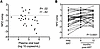 Figure 1
NPZ composite score correlation and response to ART. (A) Spearman’s correlation between NPZ composite scores and plasma viral load in 30 ART naive individuals before and 6 and 12 months after ART (r = –0.42, P = 0.02). (B) Changes in NPZ composite scores from baseline (pre-ART) to 6 and 12 months after ART, indicating significant improvement in neurocognitive performance over time. Statistical significance between time points was assessed using 1-way ANOVA with Tukey’s post hoc test for multigroup comparisons. P values are indicated for comparisons between each time point. The significant increase in NPZ scores suggests enhanced neurocognitive function with effective ART.
Monocyte subset analysis was performed on frozen and thawed PBMCs using previously described methods and cytometric gates. Of the total cohort, we had complete PBMC samples (pre-ART, 6 months and 12 months post-ART) from 30 individuals that were used for monocyte subset and marker analysis described below. A gating scheme for identifying monocyte subpopulations is shown in Figure 2A. Cell viability, assessed through live-dead cell analysis, exceeded 95% (data not shown). Three monocyte subpopulations were analyzed based on CD14 and CD16 antigens: CD14+CD16– (classical), CD14+CD16+ (intermediate), and CD14loCD16+ (non-classical). The flow cytometer was calibrated before all runs, and the data were normalized. We were unable to determine the absolute monocyte cell counts owing to the lack of monocyte counts from individual complete blood count. There was an increased percentage of CD14+CD16+ monocytes among individuals with HIV, compared with HIV-seronegative individuals. The percentage of CD14CD16 monocytes significantly decreased with 6 months of ART and further decreased after 12 months of therapy, approaching percentages similar to those in uninfected controls (Figure 2C).The percentage of CD14+CD16– cells increased after ART, while the percentage of CD14–CD16+ cells was unchanged (Figure 2, B and D). No monocyte subpopulation correlated with HAND or psychological deficit in ART participants.
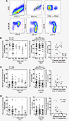 Figure 2
Monocyte subset analysis before and after ART. (A) Gating strategy used to identify monocyte subsets: CD14+CD16– (classical), CD14+CD16+ (intermediate), and CD14–CD16+ (inflammatory) monocytes. Top (left to right): selection gate for cells followed by live cell capture and lineage selection excluding CD3 and CD20 lymphocytes. Bottom (left to right): positive selection of monocytes by HLA-DR and CD14, and analysis of monocytes based on CD14 and CD16 staining. (B–D) Percentage of classical, intermediate, and inflammatory monocytes before and after ART in PLWH and PWoH. (B and C) Significant increase in CD14+CD16– monocytes after ART and significant decrease in CD14+CD16+ monocytes, approaching levels of PWoH by 12 months. Correlation of CD14+CD16– monocytes with plasma viral load (P = 0.03, r = 0.39). (D) Negative correlation of CD14–CD16+ monocytes with plasma viral load (P = 0.0001, r = –0.65). Statistical significance was determined using 1-way ANOVA with Tukey’s test for multigroup comparisons. **P < 0.01 compared with HIV– and pre-ART visits.
Before ART, the percentage of CD14+CD16– monocytes correlated directly with plasma viral load (r = 0.39, P = 0.03) (Figure 2B), whereas the percentage of CD14+CD16+ monocytes did not correlate. The percentage of CD14–CD16+ monocytes correlated indirectly with plasma viral load, while the percentage of CD14+CD16+ cells did not show a correlation (Figure 2C). The percentage of CD14–CD16+ monocytes indirectly correlated with plasma viral load (r = –0.65, P = 0.0001) (Figure 2D).
The classical monocytes and non-classical subpopulation are defined as CD14+CD16–CCR2+ and CD14–CD16+CX3CR1+, respectively. CCR2 is thought to promote monocyte extravasation into tissue, whereas CX3CR1 is associated with “patrolling” along blood vessels. CCR2 mean fluorescence intensity (MFI) on CD14+ monocytes was lower in HIV-infected individuals compared with uninfected controls. Before ART, CCR2 MFI did not distinguish between HAND categories. CCR2 MFI on classical monocytes increased significantly at 6 months of ART and then decreased from 6 to 12 months after ART initiation (Figure 3A). We observed a direct correlation between the MFI of CCR2 and the NPZglobal score (r = 0.39, P = 0.033) (Supplemental Figure 1). The MFI of CX3CR1 on CD14+CD16+ and CD14–CD16+ monocytes was lower in HIV+ individuals compared with HIV– individuals (Figure 3B). Notably, there was a significant decrease in the CX3CR1 MFIs in both the CD14+CD16+ and CD14–CD16– monocyte subsets in participants with HAD. CX3CR1 significantly increased 12 months after ART on CD14+CD16+ cells (Figure 3B), and CX3CR1 significantly increased on CD14–CD16+ cells 6 and 12 months after ART (Figure 3C).
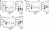 Figure 3
CCR2 and CX3CR1 expression on monocyte subsets before and after ART. (A) MFI of CCR2 on CD14+CD16– classical monocytes was lower in PLWH before ART and increased significantly 6 months after ART before declining at 12 months. (B and C) CX3CR1 MFI on CD14+CD16+ (B) and CD14–CD16+ monocytes (C) with reduced expression in HIV+ individuals compared with HIV–. PLWH with HAD displayed the lowest CX3CR1 expression, which significantly increased after ART. (C) Significant increase in CX3CR1 MFI on CD14loCD16+ monocytes at 6 and 12 months after ART. Statistical significance was determined using 1-way ANOVA with Tukey’s test for multigroup comparisons. *P < 0.05, **P < 0.01, ***P < 0.001 compared with PWoH and pre-ART visits.
Next, we asked whether monocyte-associated markers differentiate PLWH from people living without HIV (PWoH), and HAND categories before and after ART. Further analyses compared neurocognitively asymptomatic (NL and ANI) individuals with neurocognitively symptomatic participants (MND and HAD). CD4, CD86, and MAC387 MFIs were significantly decreased in all 3 monocyte subpopulations at 6 or 12 months of ART (Figure 4, A–C), and CD68 MFI was significantly decreased on CD14+CD16+ monocytes at 6 and 12 months of ART (Figure 4D). CD163 MFI significantly decreased on both the CD14+CD16+ and CD14loCD16+ monocytes with ART (Figure 4E), while CD44v6 and CCR5 levels did not change on any of the subsets with ART (Figure 4E and data not shown). None of the markers differentiated between HAND groups before ART.
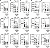 Figure 4
Cluster of differentiation marker expression on monocyte subsets before and after ART. (A–E) Decreased expression of CD4, CD86, MAC387, CD68 and CD163 on monocyte subsets following 6 and 12 months of ART. No significant changes were found in CD44v6 and CCR5 expression across any monocyte subsets before and after ART (data not shown). Statistical significance was determined using 1-way ANOVA with Tukey’s test for multigroup comparisons. *P < 0.05, **P < 0.01, ***P < 0.001 compared with pre-ART visits.
We previously demonstrated that sCD163 levels in plasma correlate with HIV replication in plasma, expansion of CD14+CD16+ monocytes, and neurocognitive deficits (HAND) in people living with chronic HIV infection on ART (28, 29, 31). In the current study, we found elevated sCD163 plasma levels in ART-naive individuals compared with HIV– controls, consistent with our earlier findings (Figure 5A). Elevated levels of sCD163 were significantly higher in neurocognitively symptomatic compared with asymptomatic participants. Moreover, we observed significantly increased sCD163 levels in neurocognitively symptomatic participants (MND and HAD). After 6 and 12 months of ART, plasma sCD163 significantly decreased (Figure 5B). In ART-naive participants, there was a correlation between plasma sCD163 and plasma viral load, consistent with our prior reports in chronically infected patients (Figure 5C).
 Figure 5
Plasma soluble CD163 levels before and after ART. (A) Elevated soluble CD163 (sCD163) levels in ART-naive PLWH compared with PWoH controls, especially in neurocognitively symptomatic individuals (MND and HAD) compared with asymptomatic (NL, ANI) individuals. (B) Significant reduction of sCD163 levels after 6 and 12 months of ART. (C) Correlation of sCD163 levels with plasma viral load before ART. Statistical significance was determined using 1-way ANOVA with Tukey’s test for multigroup comparisons. *P < 0.05, **P < 0.01, ***P < 0.001 compared with HIV– and pre-ART visits.
Among myeloid markers studied on monocytes and monocyte subsets, the sialoadhesin molecule CD169, like sCD163 described above, appears to be a reliable indicator of HIV activity including viral replication and plasma virus (20, 32). We did not find CD169 expression on monocytes in PWoH, but high levels were present in PLWH (Figure 6 and data not shown). The MFIs of CD169 on CD14+CD16– (r = 0.60, P < 0.001), CD14+CD16+ (r = 0.57, P = 0.001), and CD14loCD16+ (r = 0.66, P < 0.0001) monocytes correlated with plasma viral load prior to ART (Figure 6, A–C). The MFI of CD169 on monocyte subsets also negatively correlated with CD4 counts pre-ART (Figure 6, D–F) and NPZglobal scores (Figure 6, G–I).
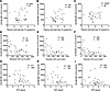 Figure 6
CD169 expression on monocyte subsets and correlations with plasma viral load and neurocognitive impairment. (A–C) MFI of CD169 on classical, intermediate, and inflammatory monocytes positively correlates with plasma viral load in ART-naive PLWH (P < 0.01 for all). (D–F) Negative correlation between monocyte CD169 MFI and CD4+ T cell counts. (G–I) Negative correlation between CD169 MFI on monocyte subsets and NPZglobal scores.
To further interrogate the relationship between CD169 expression on monocyte subsets and cognitive performance, we analyzed data from 7 individuals (P003, P004, P005, P013, P014, P015, and P016) who achieved viral suppression (plasma viral load < 50 copies/mL) but were diagnosed with ANI, MND, or HAD at 6 and 12 months after ART. These 7 individuals from the cohort were examined because we had frozen PBMCs collected from them at both data points, and they had viral suppression and ANI, MND, or HAD. Although no statistically significant correlation was observed between the CD169 MFI on monocyte subsets and NPZcomp scores in this cohort (Supplemental Figure 2), there were strong negative correlations between CD169 MFI on CD14+CD16– (r = –0.70, P = 0.034), CD14+CD16+ (r = –0.72, P = 0.03), and CD14–CD16+ (r = –0.70, P = 0.035) monocytes and CD4+ T cell count. NPZcomp scores did not correlate with CD4+ T cell counts (r = 0.12, P = 0.75) (Figure 7, A–D). Taken together, in this small group of individuals with suppressed plasma viral load but with cognitive impairment, CD169 levels did not show a direct linear correlation with NPZcomp scores at any of the time points examined after ART.
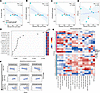 Figure 7
CD169 expression on monocyte subsets is associated with cognitive impairment compared with CD4+ T cell count, plasma viral load, and monocyte activation markers linked to HAND. (A–D) Spearman’s correlation analysis showing correlations between CD169 MFI on CD14+CD16–, CD14+CD16+, and CD14loCD16+ monocytes and CD4+ T cell count. Analyses included 7 patients (P003, P004, P005, P013, P014, P015, and P016) who achieved viral suppression (plasma viral load < 50 copies/mL) but were diagnosed with ANI, MND, or HAD at 6 and 12 months after ART. (E) Random forest model identifying key predictors of cognitive impairment. The top 15 features contributing most to classification accuracy in distinguishing individuals with MND or HAD from those with normal cognition (NL) or ANI are shown. A higher mean decrease in accuracy indicates greater variable importance. The model used data from 30 patients with CD169 MFI measurements at baseline (pre-ART). (F) Bayesian kernel machine regression (BKMR) analysis evaluating nonlinear associations between CD169 expression on monocyte subsets, CD4+ T cell count, plasma viral load, monocyte activation markers (sCD163, CCR2, CD86, CX3CR1, and MAC387), and NPZcomp score at baseline and 6 and 12 months after ART. Models were adjusted for age and sex using data from 30 patients with CD169 MFI measurements. Increased CD169 expression was associated with lower NPZcomp scores when other variables were held at median levels. (G) Heatmap showing CD169 expression on monocyte subsets, CD4+ T cell count, plasma viral load, and monocyte activation markers associated with HAND using baseline data from 30 patients with CD169 MFI measurements.
To further investigate potential nonlinear relationships among CD169 expression on monocyte subsets, CD4+ T cell count, plasma viral load, biomarkers previously associated with HAND in PLWH receiving ART (sCD163, CCR2, CD86, CX3CR1, and MAC387), and cognitive performance, we applied a Bayesian kernel machine regression (BKMR) model to data from 30 participants with CD169 measurements obtained at baseline (pre-ART) and at 6 and 12 months after ART initiation. BKMR allows for the identification of key predictors within a complex mixture model by estimating their individual and combined effects on NPZcomp scores. The BKMR model identified CD169 expression on CD14+CD16– (posterior inclusion probability [PIP] = 0.721), CD14+CD16+ (PIP = 0.654), and CD14loCD16+ (PIP = 0.660) monocytes among the most important predictors contributing to cognitive impairment within the mixture model (which included CD169 expression, CD4+ T cell count, plasma viral load, and monocyte activation markers associated with HAND [sCD163, CCR2, CD86, CX3CR1, MAC387]) in relation to NPZcomp score (Supplemental Table 2). For example, CD169 expression on classical, intermediate, and non-classical monocytes showed higher PIPs than plasma viral load (PIP = 0.339–0.492) and sCD163 (PIP = 0.409–0.620) and comparable or greater PIPs compared with CD4+ T cell counts (PIP = 0.463–0.738).
Furthermore, plasma viral load showed no association with cognitive performance, whereas CD4+ T cell count showed a weak linear association at baseline (pre-ART). Other markers (CD86, CXCR2, CX3CR1, and MAC387) showed a nonlinear relationship with NPZcomp scores (inverted U-shaped relationship). However, CD169 expression on monocyte subsets was significantly and negatively associated with cognitive performance. When all other factors were held at their median levels, higher CD169 expression on monocyte subsets was associated with a greater decline in NPZcomp scores at baseline (pre-ART) (Figure 7F, Supplemental Figure 3, and Supplemental Figure 4A). Consistent with these findings, heatmap analysis demonstrated distinct CD169 expression patterns between participants with MND/HAD and those with NL/ANI at baseline (pre-ART) (Figure 7G). To further validate our findings, we used random forest, a machine learning technique that combines multiple decision trees, to determine the key features that most strongly influence the model’s predictive accuracy, a concept known as feature importance. In this analysis, CD169 expression on monocyte subsets emerged as the most significant predictor of cognitive impairment, more so than sCD163, CCR2, and other markers shown to associate with HAND at baseline (Figure 7E and Supplemental Figure 5, A–E). We next evaluated biomarker importance at 6 and 12 months after ART among 30 participants. Although CD169 expression was no longer the highest-ranked variable in the BKMR models, it consistently remained among the top predictors based on PIPs and maintained an inverse association with cognitive function (Figure 7F). At 6 months after ART, CD169 expression remained highly ranked (PIP = 0.612–0.790), comparable to plasma viral load (PIP = 0.690–0.788) and CD4+ T cell count (PIP = 0.621–0.731), while exceeding sCD163 (PIP = 0.529–0.610). At 12 months after ART, CD169 expression continued to demonstrate importance similar to or greater than that of plasma viral load (PIP = 0.300–0.430), CD4+ T cell count (PIP = 0.248–0.413), and sCD163 (PIP = 0.201–0.411), supporting its persistent association with cognitive impairment throughout ART. Similarly, random forest analysis ranked CD169 expression among the top 15 predictors at 6 months and the top 7 predictors at 12 months after ART (Supplemental Figure 5, A–C and data not shown). Notably, at 6 months after ART, plasma viral load and CD4+ T cell count ranked sixth and seventh among the top 15 predictors, whereas CD169 ranked 15th. In contrast, at 12 months after ART, CD169 remained a prominent predictor of cognitive performance, ranking seventh overall, whereas CD4+ T cell count ranked 13th and plasma viral load was no longer among the top 15 predictors. These results suggest that CD169 expression remains associated with cognitive impairment despite sustained viral suppression.
Analyzing groups by categories of HAND, we found a stepwise increase in CD169 MFI across all 3 monocyte subpopulations, from NL to HAD, before ART. Comparing asymptomatic (NL and ANI) with symptomatic (MND and HAD) groups, significant differences in MFI were observed across all monocyte subsets (Figure 8, A–C). After ART was started, there were significant decreases in CD169 MFI across all 3 subsets, which dropped to levels similar to those in the HIV– cohort within 3 months after ART (Figure 9, A–C). In 2 individuals on ART whose plasma virus did not decrease, likely because of ART non-compliance, CD169 levels, while dipping initially, returned to HIV-infected levels thereafter and were significantly different from those in HIV-infected individuals who were virologically suppressed (Figure 9D and Supplemental Figure 6A). Interestingly, 5 individuals who were neurologically impaired were virologically suppressed (P04, P07, P012, P013, and P016) and had detectable CD169 on their monocytes, suggesting that CD169 may function as a biomarker of cognitive impairment with viral suppression.
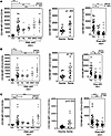 Figure 8
CD169 expression across HAND groups before ART. (A–C) Stepwise increase in MFI of CD169 on classical, intermediate, and inflammatory monocytes across HAND groups ranging from NL to HAD before ART. Significant differences were observed between asymptomatic (NL, ANI) and symptomatic (MND, HAD) patients in all monocyte subsets. Statistical significance was determined using Student’s t test and 1-way ANOVA with Tukey’s post hoc test for multigroup comparisons. *P < 0.05, **P < 0.01, ***P < 0.001 compared with HIV– and pre-ART visits.
 Figure 9
CD169 expression and viral load after ART. (A–C) Significant decreases in CD169 MFI on all monocyte subsets were observed at 6 and 12 months after ART on CD14–CD16+, CD14+CD16+, and CD14–CD16+ monocytes. V1, pre-ART; V2, 6 months post-ART; V5, 9 months post-ART. (D) Decreased plasma virus over time with ART. With ART, CD169 expression dropped to levels comparable to those of PWoH controls in most individuals examined. Non-virologically-suppressed individuals (P1, P2) had no significant changes in CD169 expression. The majority of virologically suppressed individuals exhibited a decrease in plasma viral load.
The percentage of CD14+CD16– monocytes in suppressed and non-virologically-suppressed individuals remained stable at 6 and 12 months after ART initiation (Supplemental Figure 6B). In contrast, the percentage of CD14+CD16+ monocytes in virologically suppressed and non-suppressed individuals varied at 6 months with both increases and decreases, before declining at 12 months (Supplemental Figure 6C). Meanwhile, the percentage of CD14–CD16+ monocytes in virologically suppressed individuals increased at 6 months after ART and then stabilized to pre-ART levels by 12 months (Supplemental Figure 6D).


