
Research ArticleNephrologyPublic Health
Open Access |  10.1172/jci.insight.203039
Kidney fibrosis is mediated by GARP-restricted TGF-β activation in fibroblasts
Yintong Chen,1 Weiwei Xu,1 Jieli Yu,1 Pei Deng,1 Nianping Liu,2 Yinyin Li,1 Hui Zhou,1 Hong Zhou,1 Jianchuan Wang,3 Bo Zhao,4 Florian Winau,5,6 Fan Fan Hou,1 and Yu Hu1
1Division of Nephrology, Nanfang Hospital, Southern Medical University, State Key Laboratory of Multi-organ Injury Prevention and Treatment; National Clinical Research Center for Kidney and Urological Diseases, Guangdong Provincial Institute of Nephrology, Guangdong Provincial Key Laboratory of Renal Failure Research, Guangzhou, Guangdong, China.
2The First Affiliated Hospital of USTC, School of Basic Medical Sciences, Division of Life Sciences and Medicine, University of Science and Technology of China, Hefei, China.
3School of Pharmaceutical Sciences, Sun Yat-sen University, Guangzhou, Guangdong, China.
4Molecular Cancer Research Center, School of Medicine, Shenzhen Campus of Sun Yat-sen University, Shenzhen, Guangdong, China.
5Program in Cellular and Molecular Medicine, Boston Children’s Hospital, Department of Pediatrics, Harvard Medical School, Boston, Massachusetts, USA.
6Singapore Immunology Network (SIgN), Agency for Science, Technology and Research (A*STAR), Singapore.
Address correspondence to: Yu Hu or Fan Fan Hou, Division of Nephrology, Nanfang Hospital, Southern Medical University, 1838 Guangzhou Avenue North, Guangzhou, Guangdong 510515, China. Email: yuhu@smu.edu.cn (YH); ffhouguangzhou@163.com (FFH). Or to: Florian Winau, Program in Cellular and Molecular Medicine, Boston Children’s Hospital, Department of Pediatrics, Harvard Medical School, 3 Blackfan Circle, Boston, Massachusetts, 02115, USA. Email: florian.winau@childrens.harvard.edu.
Authorship note: YC, WX, and JY contributed equally to this work.
Find articles by Chen, Y. in: PubMed | Google Scholar
1Division of Nephrology, Nanfang Hospital, Southern Medical University, State Key Laboratory of Multi-organ Injury Prevention and Treatment; National Clinical Research Center for Kidney and Urological Diseases, Guangdong Provincial Institute of Nephrology, Guangdong Provincial Key Laboratory of Renal Failure Research, Guangzhou, Guangdong, China.
2The First Affiliated Hospital of USTC, School of Basic Medical Sciences, Division of Life Sciences and Medicine, University of Science and Technology of China, Hefei, China.
3School of Pharmaceutical Sciences, Sun Yat-sen University, Guangzhou, Guangdong, China.
4Molecular Cancer Research Center, School of Medicine, Shenzhen Campus of Sun Yat-sen University, Shenzhen, Guangdong, China.
5Program in Cellular and Molecular Medicine, Boston Children’s Hospital, Department of Pediatrics, Harvard Medical School, Boston, Massachusetts, USA.
6Singapore Immunology Network (SIgN), Agency for Science, Technology and Research (A*STAR), Singapore.
Address correspondence to: Yu Hu or Fan Fan Hou, Division of Nephrology, Nanfang Hospital, Southern Medical University, 1838 Guangzhou Avenue North, Guangzhou, Guangdong 510515, China. Email: yuhu@smu.edu.cn (YH); ffhouguangzhou@163.com (FFH). Or to: Florian Winau, Program in Cellular and Molecular Medicine, Boston Children’s Hospital, Department of Pediatrics, Harvard Medical School, 3 Blackfan Circle, Boston, Massachusetts, 02115, USA. Email: florian.winau@childrens.harvard.edu.
Authorship note: YC, WX, and JY contributed equally to this work.
Find articles by Xu, W. in: PubMed | Google Scholar
1Division of Nephrology, Nanfang Hospital, Southern Medical University, State Key Laboratory of Multi-organ Injury Prevention and Treatment; National Clinical Research Center for Kidney and Urological Diseases, Guangdong Provincial Institute of Nephrology, Guangdong Provincial Key Laboratory of Renal Failure Research, Guangzhou, Guangdong, China.
2The First Affiliated Hospital of USTC, School of Basic Medical Sciences, Division of Life Sciences and Medicine, University of Science and Technology of China, Hefei, China.
3School of Pharmaceutical Sciences, Sun Yat-sen University, Guangzhou, Guangdong, China.
4Molecular Cancer Research Center, School of Medicine, Shenzhen Campus of Sun Yat-sen University, Shenzhen, Guangdong, China.
5Program in Cellular and Molecular Medicine, Boston Children’s Hospital, Department of Pediatrics, Harvard Medical School, Boston, Massachusetts, USA.
6Singapore Immunology Network (SIgN), Agency for Science, Technology and Research (A*STAR), Singapore.
Address correspondence to: Yu Hu or Fan Fan Hou, Division of Nephrology, Nanfang Hospital, Southern Medical University, 1838 Guangzhou Avenue North, Guangzhou, Guangdong 510515, China. Email: yuhu@smu.edu.cn (YH); ffhouguangzhou@163.com (FFH). Or to: Florian Winau, Program in Cellular and Molecular Medicine, Boston Children’s Hospital, Department of Pediatrics, Harvard Medical School, 3 Blackfan Circle, Boston, Massachusetts, 02115, USA. Email: florian.winau@childrens.harvard.edu.
Authorship note: YC, WX, and JY contributed equally to this work.
Find articles by Yu, J. in: PubMed | Google Scholar
1Division of Nephrology, Nanfang Hospital, Southern Medical University, State Key Laboratory of Multi-organ Injury Prevention and Treatment; National Clinical Research Center for Kidney and Urological Diseases, Guangdong Provincial Institute of Nephrology, Guangdong Provincial Key Laboratory of Renal Failure Research, Guangzhou, Guangdong, China.
2The First Affiliated Hospital of USTC, School of Basic Medical Sciences, Division of Life Sciences and Medicine, University of Science and Technology of China, Hefei, China.
3School of Pharmaceutical Sciences, Sun Yat-sen University, Guangzhou, Guangdong, China.
4Molecular Cancer Research Center, School of Medicine, Shenzhen Campus of Sun Yat-sen University, Shenzhen, Guangdong, China.
5Program in Cellular and Molecular Medicine, Boston Children’s Hospital, Department of Pediatrics, Harvard Medical School, Boston, Massachusetts, USA.
6Singapore Immunology Network (SIgN), Agency for Science, Technology and Research (A*STAR), Singapore.
Address correspondence to: Yu Hu or Fan Fan Hou, Division of Nephrology, Nanfang Hospital, Southern Medical University, 1838 Guangzhou Avenue North, Guangzhou, Guangdong 510515, China. Email: yuhu@smu.edu.cn (YH); ffhouguangzhou@163.com (FFH). Or to: Florian Winau, Program in Cellular and Molecular Medicine, Boston Children’s Hospital, Department of Pediatrics, Harvard Medical School, 3 Blackfan Circle, Boston, Massachusetts, 02115, USA. Email: florian.winau@childrens.harvard.edu.
Authorship note: YC, WX, and JY contributed equally to this work.
Find articles by Deng, P. in: PubMed | Google Scholar
1Division of Nephrology, Nanfang Hospital, Southern Medical University, State Key Laboratory of Multi-organ Injury Prevention and Treatment; National Clinical Research Center for Kidney and Urological Diseases, Guangdong Provincial Institute of Nephrology, Guangdong Provincial Key Laboratory of Renal Failure Research, Guangzhou, Guangdong, China.
2The First Affiliated Hospital of USTC, School of Basic Medical Sciences, Division of Life Sciences and Medicine, University of Science and Technology of China, Hefei, China.
3School of Pharmaceutical Sciences, Sun Yat-sen University, Guangzhou, Guangdong, China.
4Molecular Cancer Research Center, School of Medicine, Shenzhen Campus of Sun Yat-sen University, Shenzhen, Guangdong, China.
5Program in Cellular and Molecular Medicine, Boston Children’s Hospital, Department of Pediatrics, Harvard Medical School, Boston, Massachusetts, USA.
6Singapore Immunology Network (SIgN), Agency for Science, Technology and Research (A*STAR), Singapore.
Address correspondence to: Yu Hu or Fan Fan Hou, Division of Nephrology, Nanfang Hospital, Southern Medical University, 1838 Guangzhou Avenue North, Guangzhou, Guangdong 510515, China. Email: yuhu@smu.edu.cn (YH); ffhouguangzhou@163.com (FFH). Or to: Florian Winau, Program in Cellular and Molecular Medicine, Boston Children’s Hospital, Department of Pediatrics, Harvard Medical School, 3 Blackfan Circle, Boston, Massachusetts, 02115, USA. Email: florian.winau@childrens.harvard.edu.
Authorship note: YC, WX, and JY contributed equally to this work.
Find articles by Liu, N. in: PubMed | Google Scholar
1Division of Nephrology, Nanfang Hospital, Southern Medical University, State Key Laboratory of Multi-organ Injury Prevention and Treatment; National Clinical Research Center for Kidney and Urological Diseases, Guangdong Provincial Institute of Nephrology, Guangdong Provincial Key Laboratory of Renal Failure Research, Guangzhou, Guangdong, China.
2The First Affiliated Hospital of USTC, School of Basic Medical Sciences, Division of Life Sciences and Medicine, University of Science and Technology of China, Hefei, China.
3School of Pharmaceutical Sciences, Sun Yat-sen University, Guangzhou, Guangdong, China.
4Molecular Cancer Research Center, School of Medicine, Shenzhen Campus of Sun Yat-sen University, Shenzhen, Guangdong, China.
5Program in Cellular and Molecular Medicine, Boston Children’s Hospital, Department of Pediatrics, Harvard Medical School, Boston, Massachusetts, USA.
6Singapore Immunology Network (SIgN), Agency for Science, Technology and Research (A*STAR), Singapore.
Address correspondence to: Yu Hu or Fan Fan Hou, Division of Nephrology, Nanfang Hospital, Southern Medical University, 1838 Guangzhou Avenue North, Guangzhou, Guangdong 510515, China. Email: yuhu@smu.edu.cn (YH); ffhouguangzhou@163.com (FFH). Or to: Florian Winau, Program in Cellular and Molecular Medicine, Boston Children’s Hospital, Department of Pediatrics, Harvard Medical School, 3 Blackfan Circle, Boston, Massachusetts, 02115, USA. Email: florian.winau@childrens.harvard.edu.
Authorship note: YC, WX, and JY contributed equally to this work.
Find articles by Li, Y. in: PubMed | Google Scholar
1Division of Nephrology, Nanfang Hospital, Southern Medical University, State Key Laboratory of Multi-organ Injury Prevention and Treatment; National Clinical Research Center for Kidney and Urological Diseases, Guangdong Provincial Institute of Nephrology, Guangdong Provincial Key Laboratory of Renal Failure Research, Guangzhou, Guangdong, China.
2The First Affiliated Hospital of USTC, School of Basic Medical Sciences, Division of Life Sciences and Medicine, University of Science and Technology of China, Hefei, China.
3School of Pharmaceutical Sciences, Sun Yat-sen University, Guangzhou, Guangdong, China.
4Molecular Cancer Research Center, School of Medicine, Shenzhen Campus of Sun Yat-sen University, Shenzhen, Guangdong, China.
5Program in Cellular and Molecular Medicine, Boston Children’s Hospital, Department of Pediatrics, Harvard Medical School, Boston, Massachusetts, USA.
6Singapore Immunology Network (SIgN), Agency for Science, Technology and Research (A*STAR), Singapore.
Address correspondence to: Yu Hu or Fan Fan Hou, Division of Nephrology, Nanfang Hospital, Southern Medical University, 1838 Guangzhou Avenue North, Guangzhou, Guangdong 510515, China. Email: yuhu@smu.edu.cn (YH); ffhouguangzhou@163.com (FFH). Or to: Florian Winau, Program in Cellular and Molecular Medicine, Boston Children’s Hospital, Department of Pediatrics, Harvard Medical School, 3 Blackfan Circle, Boston, Massachusetts, 02115, USA. Email: florian.winau@childrens.harvard.edu.
Authorship note: YC, WX, and JY contributed equally to this work.
Find articles by Zhou, H. in: PubMed | Google Scholar
1Division of Nephrology, Nanfang Hospital, Southern Medical University, State Key Laboratory of Multi-organ Injury Prevention and Treatment; National Clinical Research Center for Kidney and Urological Diseases, Guangdong Provincial Institute of Nephrology, Guangdong Provincial Key Laboratory of Renal Failure Research, Guangzhou, Guangdong, China.
2The First Affiliated Hospital of USTC, School of Basic Medical Sciences, Division of Life Sciences and Medicine, University of Science and Technology of China, Hefei, China.
3School of Pharmaceutical Sciences, Sun Yat-sen University, Guangzhou, Guangdong, China.
4Molecular Cancer Research Center, School of Medicine, Shenzhen Campus of Sun Yat-sen University, Shenzhen, Guangdong, China.
5Program in Cellular and Molecular Medicine, Boston Children’s Hospital, Department of Pediatrics, Harvard Medical School, Boston, Massachusetts, USA.
6Singapore Immunology Network (SIgN), Agency for Science, Technology and Research (A*STAR), Singapore.
Address correspondence to: Yu Hu or Fan Fan Hou, Division of Nephrology, Nanfang Hospital, Southern Medical University, 1838 Guangzhou Avenue North, Guangzhou, Guangdong 510515, China. Email: yuhu@smu.edu.cn (YH); ffhouguangzhou@163.com (FFH). Or to: Florian Winau, Program in Cellular and Molecular Medicine, Boston Children’s Hospital, Department of Pediatrics, Harvard Medical School, 3 Blackfan Circle, Boston, Massachusetts, 02115, USA. Email: florian.winau@childrens.harvard.edu.
Authorship note: YC, WX, and JY contributed equally to this work.
Find articles by Zhou, H. in: PubMed | Google Scholar
1Division of Nephrology, Nanfang Hospital, Southern Medical University, State Key Laboratory of Multi-organ Injury Prevention and Treatment; National Clinical Research Center for Kidney and Urological Diseases, Guangdong Provincial Institute of Nephrology, Guangdong Provincial Key Laboratory of Renal Failure Research, Guangzhou, Guangdong, China.
2The First Affiliated Hospital of USTC, School of Basic Medical Sciences, Division of Life Sciences and Medicine, University of Science and Technology of China, Hefei, China.
3School of Pharmaceutical Sciences, Sun Yat-sen University, Guangzhou, Guangdong, China.
4Molecular Cancer Research Center, School of Medicine, Shenzhen Campus of Sun Yat-sen University, Shenzhen, Guangdong, China.
5Program in Cellular and Molecular Medicine, Boston Children’s Hospital, Department of Pediatrics, Harvard Medical School, Boston, Massachusetts, USA.
6Singapore Immunology Network (SIgN), Agency for Science, Technology and Research (A*STAR), Singapore.
Address correspondence to: Yu Hu or Fan Fan Hou, Division of Nephrology, Nanfang Hospital, Southern Medical University, 1838 Guangzhou Avenue North, Guangzhou, Guangdong 510515, China. Email: yuhu@smu.edu.cn (YH); ffhouguangzhou@163.com (FFH). Or to: Florian Winau, Program in Cellular and Molecular Medicine, Boston Children’s Hospital, Department of Pediatrics, Harvard Medical School, 3 Blackfan Circle, Boston, Massachusetts, 02115, USA. Email: florian.winau@childrens.harvard.edu.
Authorship note: YC, WX, and JY contributed equally to this work.
Find articles by Wang, J. in: PubMed | Google Scholar
1Division of Nephrology, Nanfang Hospital, Southern Medical University, State Key Laboratory of Multi-organ Injury Prevention and Treatment; National Clinical Research Center for Kidney and Urological Diseases, Guangdong Provincial Institute of Nephrology, Guangdong Provincial Key Laboratory of Renal Failure Research, Guangzhou, Guangdong, China.
2The First Affiliated Hospital of USTC, School of Basic Medical Sciences, Division of Life Sciences and Medicine, University of Science and Technology of China, Hefei, China.
3School of Pharmaceutical Sciences, Sun Yat-sen University, Guangzhou, Guangdong, China.
4Molecular Cancer Research Center, School of Medicine, Shenzhen Campus of Sun Yat-sen University, Shenzhen, Guangdong, China.
5Program in Cellular and Molecular Medicine, Boston Children’s Hospital, Department of Pediatrics, Harvard Medical School, Boston, Massachusetts, USA.
6Singapore Immunology Network (SIgN), Agency for Science, Technology and Research (A*STAR), Singapore.
Address correspondence to: Yu Hu or Fan Fan Hou, Division of Nephrology, Nanfang Hospital, Southern Medical University, 1838 Guangzhou Avenue North, Guangzhou, Guangdong 510515, China. Email: yuhu@smu.edu.cn (YH); ffhouguangzhou@163.com (FFH). Or to: Florian Winau, Program in Cellular and Molecular Medicine, Boston Children’s Hospital, Department of Pediatrics, Harvard Medical School, 3 Blackfan Circle, Boston, Massachusetts, 02115, USA. Email: florian.winau@childrens.harvard.edu.
Authorship note: YC, WX, and JY contributed equally to this work.
Find articles by Zhao, B. in: PubMed | Google Scholar
1Division of Nephrology, Nanfang Hospital, Southern Medical University, State Key Laboratory of Multi-organ Injury Prevention and Treatment; National Clinical Research Center for Kidney and Urological Diseases, Guangdong Provincial Institute of Nephrology, Guangdong Provincial Key Laboratory of Renal Failure Research, Guangzhou, Guangdong, China.
2The First Affiliated Hospital of USTC, School of Basic Medical Sciences, Division of Life Sciences and Medicine, University of Science and Technology of China, Hefei, China.
3School of Pharmaceutical Sciences, Sun Yat-sen University, Guangzhou, Guangdong, China.
4Molecular Cancer Research Center, School of Medicine, Shenzhen Campus of Sun Yat-sen University, Shenzhen, Guangdong, China.
5Program in Cellular and Molecular Medicine, Boston Children’s Hospital, Department of Pediatrics, Harvard Medical School, Boston, Massachusetts, USA.
6Singapore Immunology Network (SIgN), Agency for Science, Technology and Research (A*STAR), Singapore.
Address correspondence to: Yu Hu or Fan Fan Hou, Division of Nephrology, Nanfang Hospital, Southern Medical University, 1838 Guangzhou Avenue North, Guangzhou, Guangdong 510515, China. Email: yuhu@smu.edu.cn (YH); ffhouguangzhou@163.com (FFH). Or to: Florian Winau, Program in Cellular and Molecular Medicine, Boston Children’s Hospital, Department of Pediatrics, Harvard Medical School, 3 Blackfan Circle, Boston, Massachusetts, 02115, USA. Email: florian.winau@childrens.harvard.edu.
Authorship note: YC, WX, and JY contributed equally to this work.
Find articles by Winau, F. in: PubMed | Google Scholar
1Division of Nephrology, Nanfang Hospital, Southern Medical University, State Key Laboratory of Multi-organ Injury Prevention and Treatment; National Clinical Research Center for Kidney and Urological Diseases, Guangdong Provincial Institute of Nephrology, Guangdong Provincial Key Laboratory of Renal Failure Research, Guangzhou, Guangdong, China.
2The First Affiliated Hospital of USTC, School of Basic Medical Sciences, Division of Life Sciences and Medicine, University of Science and Technology of China, Hefei, China.
3School of Pharmaceutical Sciences, Sun Yat-sen University, Guangzhou, Guangdong, China.
4Molecular Cancer Research Center, School of Medicine, Shenzhen Campus of Sun Yat-sen University, Shenzhen, Guangdong, China.
5Program in Cellular and Molecular Medicine, Boston Children’s Hospital, Department of Pediatrics, Harvard Medical School, Boston, Massachusetts, USA.
6Singapore Immunology Network (SIgN), Agency for Science, Technology and Research (A*STAR), Singapore.
Address correspondence to: Yu Hu or Fan Fan Hou, Division of Nephrology, Nanfang Hospital, Southern Medical University, 1838 Guangzhou Avenue North, Guangzhou, Guangdong 510515, China. Email: yuhu@smu.edu.cn (YH); ffhouguangzhou@163.com (FFH). Or to: Florian Winau, Program in Cellular and Molecular Medicine, Boston Children’s Hospital, Department of Pediatrics, Harvard Medical School, 3 Blackfan Circle, Boston, Massachusetts, 02115, USA. Email: florian.winau@childrens.harvard.edu.
Authorship note: YC, WX, and JY contributed equally to this work.
Find articles by
Hou, F.
in:
PubMed
|
Google Scholar
|

1Division of Nephrology, Nanfang Hospital, Southern Medical University, State Key Laboratory of Multi-organ Injury Prevention and Treatment; National Clinical Research Center for Kidney and Urological Diseases, Guangdong Provincial Institute of Nephrology, Guangdong Provincial Key Laboratory of Renal Failure Research, Guangzhou, Guangdong, China.
2The First Affiliated Hospital of USTC, School of Basic Medical Sciences, Division of Life Sciences and Medicine, University of Science and Technology of China, Hefei, China.
3School of Pharmaceutical Sciences, Sun Yat-sen University, Guangzhou, Guangdong, China.
4Molecular Cancer Research Center, School of Medicine, Shenzhen Campus of Sun Yat-sen University, Shenzhen, Guangdong, China.
5Program in Cellular and Molecular Medicine, Boston Children’s Hospital, Department of Pediatrics, Harvard Medical School, Boston, Massachusetts, USA.
6Singapore Immunology Network (SIgN), Agency for Science, Technology and Research (A*STAR), Singapore.
Address correspondence to: Yu Hu or Fan Fan Hou, Division of Nephrology, Nanfang Hospital, Southern Medical University, 1838 Guangzhou Avenue North, Guangzhou, Guangdong 510515, China. Email: yuhu@smu.edu.cn (YH); ffhouguangzhou@163.com (FFH). Or to: Florian Winau, Program in Cellular and Molecular Medicine, Boston Children’s Hospital, Department of Pediatrics, Harvard Medical School, 3 Blackfan Circle, Boston, Massachusetts, 02115, USA. Email: florian.winau@childrens.harvard.edu.
Authorship note: YC, WX, and JY contributed equally to this work.
Find articles by Hu, Y. in: PubMed | Google Scholar
Published September 17, 2026 - More info
JCI Insight. 2026;11(20):e203039. https://doi.org/10.1172/jci.insight.203039.
© 2026 Chen et al. This work is licensed under the Creative Commons Attribution 4.0 International License. To view a copy of this license, visit http://creativecommons.org/licenses/by/4.0/.
Received: December 2, 2025; Accepted: September 2, 2026


-
Results
Upregulation of GARP in human and mouse fibrotic kidneys. To investigate the potential role of GARP-mediated TGF-β activation in kidney fibrosis, we first assessed its expression by analyzing publicly available single-cell RNA sequencing (scRNA-seq) data from murine fibrotic kidneys (GSE190887) (27). At the whole-kidney sample level, analysis of unilateral ureteral obstruction (UUO) kidneys across multiple time points revealed that Lrrc32 expression progressively increased along with fibrosis progression (Figure 1A). To validate these scRNA-seq findings, we next examined GARP expression by quantitative real-time PCR (RT-PCR) in 3 established rodent models of kidney fibrosis: UUO, unilateral renal ischemia-reperfusion injury (UIRI), and 5/6 nephrectomy (5/6Nx). In the UUO mouse model, renal Lrrc32 mRNA levels were upregulated by day 7 and further increased by day 14 (Figure 1B) and correlated positively with fibrosis markers Col1a1 and Acta2 (Figure 1C). Immunoblot confirmed elevated protein levels of GARP and fibronectin (FN) in UUO kidneys (Figure 1, D and E). Similarly, increased expression of GARP was observed in 5/6Nx rats (Supplemental Figure 1, A–C; supplemental material available online with this article; https://doi.org/10.1172/jci.insight.203039DS1) and UIRI mice (Supplemental Figure 1, D–F). Immunohistochemistry revealed robust GARP staining in the interstitial regions of fibrotic kidneys, whereas expression was minimal in normal kidneys (Figure 1, F and G, and Supplemental Figure 1G). We next examined GARP expression in human kidneys. Microarray analysis of renal biopsy specimens from CKD patients (GSE116626) showed significantly elevated LRRC32, along with fibrosis markers FN1 and COL3A1, compared with healthy controls (Figure 1H). To define the cellular sources of GARP in fibrotic kidneys, we re-analyzed the scRNA-seq datasets using the original cell-type annotations provided in the published study (27). Among all kidney cell populations, Lrrc32 was predominantly expressed in myofibroblasts in the fibrotic kidney, with relatively low levels in fibroblasts and endothelial cells (Figure 1I). We next performed flow cytometry of kidney single-cell suspensions from sham and UUO mice to characterize GARP-expressing cells in fibrotic kidneys. The gating strategy used to identify fibroblasts, endothelial cells, and immune cells is shown in Supplemental Figure 1H. Briefly, fibroblasts were defined as PDGFRα+CD31–CD45– cells, endothelial cells as CD31+CD45–PDGFRα– cells, and immune cells as CD45+CD31–PDGFRα– cells. Isotype control was used to exclude non-specific binding (Supplemental Figure 1I). Flow cytometric analysis showed that UUO markedly increased the proportion of GARP+LAP (TGF-β1)+ cells in the fibroblast population, whereas only modest increases were observed in endothelial and immune cells (Figure 1, J and K). Collectively, these data demonstrate an upregulation of GARP in human and murine fibrotic kidneys and identify fibroblasts as a major source of GARP-mediated latent TGF-β1 presentation in fibrotic kidney, suggesting a potential role of GARP-dependent TGF-β activation in the pathogenesis of kidney fibrosis.
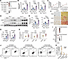 Figure 1
Increased expression of GARP in kidneys of fibrotic mice and CKD patients. (A) scRNA-seq analysis for Lrrc32 expression in the kidneys of mice at different days after UUO. (B) mRNA levels of Lrrc32 and fibrotic markers in mouse kidneys (day 7 or day 14 after surgery) (n = 4–5). Sham-operated mice were used as negative controls. (C) Pearson’s correlation analysis between Lrrc32 and fibrotic marker expression levels. (D and E) Representative (D) and quantification (E) of immunoblot for GARP and FN expression in UUO kidneys. Each lane represents 1 mouse. FN, fibronectin. (F and G) Representative (F) and quantification (G) of immunohistochemical staining for GARP (left) and Sirius red staining (right) of mouse kidney sections. Scale bars: 50 μm. (H) Microarray analysis of LRRC32, FN, and COL3A1 in renal biopsy specimens from control and CKD patients. (I) Bubble plot comparing expression of Lrrc32 in various cell types in mouse UUO kidneys. (J and K) Representative flow cytometry plots (J) and quantification (K) of GARP+LAP (TGF-β1)+ cell frequencies among fibroblasts, endothelial cells, and immune cells from sham and UUO kidneys. Uro, urothelium; TAL, thick ascending limb of LoH; PT-R, repairing proximal tubule; PT-Inj, injured PT; PT-FR, failed repair PT cells; PT-AcInj, acute injury PT; Pod, podocyte; PC, principal cell of collecting duct; Mφ, macrophage; Myofib, myofibroblast; ICB, type B intercalated cell of collecting duct; ICA, type A intercalated cell of collecting duct; Fib, fibroblast; EC, endothelial cell; DTL, descending limb of loop of Henle (LoH); ATL, thin ascending limb of LoH; DCT, distal convoluted tubule; CNT, connecting tubule; B/T, B and T cells. Data represent mean ± SEM. *P < 0.05; **P < 0.01; ***P < 0.001; ****P < 0.0001. Statistical significance was determined by 2-way ANOVA followed by Tukey’s multiple-comparison test (B and E) and unpaired 2-tailed Student’s t test (G, H, and K).
Global GARP deletion attenuates kidney fibrosis. Given the critical role of TGF-β in fibrosis, we next evaluated the in vivo function of GARP using GARP-knockout (GARP-KO) mice. Because constitutive GARP deletion causes perinatal lethality due to defective palatogenesis (28), we crossed Lrrc32fl/fl mice (wild-type, WT) (29) with UBCCreERT2 mice to establish inducible global GARP-KO mice (UBCCreERT2 Lrrc32fl/fl). To this end, both WT and GARP-KO mice were administered tamoxifen before surgery and were subsequently subjected to UUO (Figure 2A). As expected, GARP expression was drastically reduced in both sham and UUO kidneys of GARP-KO compared with WT littermates (Figure 2, B–D), confirming efficient gene deletion. RT-PCR and immunoblot analysis demonstrated that ECM components including Fn1 (FN) and Col1a1 (COL1A1), as well as the myofibroblast marker Acta2 (α-SMA), (Figure 2, B, C, and E) and Vimentin (Figure 2C), were markedly upregulated in fibrotic kidneys, whereas the upregulation was largely abrogated by GARP deletion (Figure 2, B–F). Consistently, Sirius red staining revealed extensive kidney fibrosis in WT-UUO mice, whereas fibrosis was substantially reduced in GARP-KO kidneys (Figure 2, E and F).
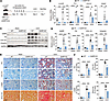 Figure 2
Global deletion of GARP mitigates UUO-induced kidney fibrosis. (A) Experimental design. (B) mRNA levels of Lrrc32 and fibrotic markers (Fn1, Col1a1, and Acta2) in kidneys of WT and GARP-KO mice (n = 5 per group). (C and D) Representative (C) and quantification (D) of immunoblot for GARP, FN, COL1A1, and Vimentin in sham or UUO-treated WT and GARP-KO mice (n = 5 per group). Each lane represents 1 mouse. GAPDH was used as a loading control. (E and F) Representative staining (E) and quantitation (F) for FN, COL1A1 and α-SMA and Sirius red in kidneys of WT and GARP-KO mice. Scale bars: 50 μm. Data represent mean ± SEM. **P < 0.01; ***P < 0.001; ****P < 0.0001. Statistical significance was determined by 2-way ANOVA followed by Tukey’s multiple-comparison test (B, D, and F).
To corroborate these findings in another animal model, WT and GARP-KO mice were treated with tamoxifen and subjected to UIRI combined with contralateral uninephrectomy (Supplemental Figure 2A). Compared with WT controls, GARP-KO mice exhibited reduced ECM deposition, decreased expression of Vimentin and α-SMA, and significantly attenuated tubulointerstitial fibrosis, as assessed by Sirius red staining (Supplemental Figure 2, B–E). Notably, serum creatinine and blood urea nitrogen (BUN) levels were significantly lower in GARP-KO mice following UIRI (Supplemental Figure 2F). Together, these results demonstrate that global deletion of GARP markedly mitigates kidney fibrosis.
Fibroblast-specific GARP deficiency ameliorates kidney fibrosis. Given that GARP expression was predominantly enriched in myofibroblasts and, to a lesser degree, in fibroblasts of fibrotic kidneys (Figure 1I), we generated inducible, fibroblast-specific GARP conditional KO mice (Col1a2CreERT × Lrrc32fl/fl, GARP-CKO) to assess the fibroblast-restricted role of GARP in kidney fibrosis. Accordingly, both genotypes (GARP-CKO and WT) were administered tamoxifen before UUO surgery, with administration continued until sacrifice to ensure sustained GARP deletion in activated fibroblasts (Figure 3A). Immunoblotting confirmed efficient GARP deletion, with reduced UUO-induced expression in GARP-CKO kidneys compared with WT controls (Figure 3, B and C), indicating fibroblasts as the major source of elevated GARP during fibrosis. Consistently, GARP-CKO mice exhibited reduced expression of FN, COL1A1, and Vimentin relative to WT controls (Figure 3, B and C). Sirius red staining revealed extensive tubulointerstitial fibrosis in WT UUO kidneys, whereas fibrosis was attenuated in GARP-CKO kidneys (Figure 3, D and E). In addition, immunohistochemical staining validated that fibroblast-specific GARP deletion weakened ECM deposition and α-SMA expression (Figure 3, D and E). Together, these findings suggest that fibroblast-expressed GARP is required for enhanced activation of fibroblasts and ECM accumulation during kidney fibrosis.
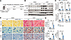 Figure 3
Fibroblast-specific deletion of GARP ameliorates UUO-induced kidney fibrosis in mice. (A) Schematic diagram of the experimental design. (B and C) Representative (B) and quantification (C) of immunoblot for GARP, FN, COL1A1, and Vimentin in WT and GARP-CKO mice (n = 5–6 per group). GAPDH was used as a loading control. (D and E) Representative staining (D) and quantification (E) for Sirius red, COL1A1, and α-SMA in WT and GARP-CKO mice (n = 6 per group). Scale bars: 50 μm. Data represent mean ± SEM. **P < 0.01; ***P < 0.001; ****P < 0.0001. Statistical significance was determined by 2-way ANOVA followed by Tukey’s multiple-comparison test (C and E).
TGF-β signaling as a major pathway driving fibroblast profibrotic activity. Kidney fibrosis develops through complex crosstalk between injured parenchymal cells, such as tubular epithelial cells, and non-parenchymal populations, including immune-inflammatory and mesenchymal cells (30, 31). To clarify the contributions of different cell types during kidney fibrosis, we re-analyzed a published integrated single-cell and single-nucleus RNA-seq (sc/snRNA-seq) dataset comprising 219,906 single cells/nuclei from human kidneys (GSE183279) (31). This dataset included specimens from healthy individuals (Ref, n = 38) and patients with various forms of CKD (n = 28), encompassing 16 distinct cell clusters (Figure 4A). To quantify ECM-related activity, we computed an ECM score using the MSigDB Matrisome (NABA) core matrisome gene sets (collagens, ECM glycoproteins and proteoglycans) (31). Fibroblasts exhibited the highest ECM score among all cell types (Figure 4B), supporting their central role in ECM production and deposition (32–34). The above finding was further validated in the scRNA-seq dataset from UUO-kidneys (27) (Supplemental Figure 3A). Given the pivotal role of TGF-β in fibrogenesis (7), we next assessed the expression of TGF-β pathway–related genes across kidney cell types. Similar to the ECM score, fibroblasts demonstrated the highest TGF-β signaling score in both human and mouse datasets (Figure 4C and Supplemental Figure 3B). Uniform Manifold Approximation and Projection (UMAP) visualization and feature plotting showed that LRRC32 signal was enriched in the fibroblast compartment (Supplemental Figure 3C), and violin plots further confirmed increased LRRC32 expression in CKD compared with Ref kidneys (Supplemental Figure 3D). Gene Ontology (GO) enrichment analysis of fibroblast-specific differentially expressed genes highlighted ECM organization and TGF-β signaling as key biological processes (Figure 4D). Gene set enrichment analysis (GSEA) of the top 5 ECM score–enriched cell clusters further confirmed significant activation of TGF-β signaling in fibroblasts (Figure 4E and Supplemental Figure 3E). To spatially map the relationship among fibroblasts, ECM production, and TGF-β signaling, we analyzed a spatial transcriptomics dataset of CKD and control kidneys (31). Spatial analysis revealed colocalization of fibroblast markers with high ECM and TGF-β signaling scores (Figure 4F and Supplemental Figure 3, F–H), and LRRC32 showed spatial enrichment in fibrotic regions (Figure 4F). Moreover, ordinary least-squares regression analysis demonstrated a positive correlation among fibroblast abundance, ECM score, and TGF-β signaling score (Figure 4G). Within fibroblasts, LRRC32 expression was increased in CKD and positively correlated with ECM, TGF-β signaling, and fibrotic program scores (Supplemental Figure 3, I and J). Together, these analyses identify fibroblasts as key mediators of ECM accumulation in kidney fibrosis and highlight TGF-β signaling as a major pathway driving their profibrotic activity, with LRRC32 showing fibroblast enrichment and positive association with fibrotic programs.
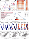 Figure 4
Fibroblasts and TGF-β signaling orchestrate fibrosis progression. (A) Uniform Manifold Approximation and Projection (UMAP) embeddings of the integrated sc/snRNA-seq dataset from CKD patients and control samples (GSE183279), colored by ECM score (left), group information (right top) and cell types (right bottom). FIB, fibroblast; NEU, neural cell; VSM/P, vascular smooth muscle cell or pericyte; DTL, descending thin limb cell; PEC, parietal epithelial cells; POD, podocyte; ATL, ascending thin limbs; TAL, thick ascending limb; PT, proximal tubule; DCT, distal convoluted tubule; PC, principal cell; CNT, connecting tubule; EC, endothelial cells; IC, intercalated cells; PapE, papillary tip epithelial cells abutting the calyx; IMM, immune cells. (B) Heatmap showing the collagen score, glycoprotein score, and proteoglycan score across cell types, which collectively form the ECM score. (C) Heatmap showing the TGF-β score across the cell types. (D) Bar plots of representative biological pathways enriched in fibroblasts defined by differentially expressed genes (DEGs). (E) Gene set enrichment analysis (GSEA) of fibroblasts for KEGG TGF-β signaling (left). The table (right) lists the normalized enrichment scores (NES) for the top 5 cell types ranked by ECM scores. (F) The TGF-β score, ECM score, Fibroblast score, and LRRC32 expression in the 10× Visium data of control and CKD samples. Each score shares the same threshold in the color bar for comparison. (G) Pairwise correlations of TGF-β score, ECM score, and fibroblast score in CKD Visium data. P values were calculated using F-statistics from ordinary least-squares models, with the error band representing the 95% confidence interval.
Because perturbation of TGF-β–related pathways in fibroblasts may affect not only matrix production but also matrix remodeling, we next performed an unbiased matrisome-focused analysis to further characterize the ECM program associated with LRRC32-expressing fibroblasts. Fibroblasts from the human kidney sc/snRNA-seq dataset were stratified into LRRC32-positive and LRRC32-negative populations, and differentially expressed genes annotated by the Matrisome Project were analyzed (Supplemental Figure 4A). Among detected matrisome genes, 158 were upregulated and only 1 was downregulated in LRRC32-positive fibroblasts (Supplemental Figure 4B and Supplemental Table 1). LRRC32-positive fibroblasts exhibited a broad increase in matrisome genes spanning multiple categories, including collagens, ECM glycoproteins, ECM-affiliated proteins, secreted factors, proteoglycans, and ECM regulators (Supplemental Figure 4C). Within the MMP/TIMP family of ECM regulators, MMP2 was modestly increased, whereas TIMP1, TIMP2, and TIMP3 were also significantly elevated in LRRC32-positive fibroblasts (Supplemental Figure 4D). These findings suggest that LRRC32-positive fibroblasts are associated with a coordinated matrisome program involving both matrix-producing and matrix-regulatory components.
We further examined matrisome features in the mouse kidney fibrosis scRNA-seq dataset (GSE190887). Because the sci-RNA-seq3 platform showed sparse detection of low-abundance matrisome transcripts in fibroblasts, we first evaluated matrisome gene detection rates and found that many matrisome genes were detected in only a small fraction of fibroblasts, with overall lower detection rates in the mouse dataset than in the human sc/snRNA-seq dataset (Supplemental Figure 5, A and B). Therefore, the mouse analysis was used as supportive evidence rather than as a direct one-to-one validation of all human matrisome genes. In mouse fibroblasts, Lrrc32-positive cells were identified and compared with Lrrc32-negative fibroblasts (Supplemental Figure 5C). Differential analysis showed a broad upward shift of matrisome genes in Lrrc32-positive fibroblasts, with 47 upregulated and no downregulated matrisome genes at the nominal significance level (Supplemental Figure 5D). To examine whether GARP loss affects fibroblast matrisome-related programs in vivo, we FACS-isolated PDGFRα-positive fibroblasts from WT and fibroblast-specific GARP-CKO mice after UUO for RT-PCR analysis (Supplemental Figure 5E). GARP-deficient fibroblasts showed reduced expression of selected fibrotic ECM genes, including Col1a1 and Col3a1, whereas Col1a2 showed no significant change (Supplemental Figure 5F). Among matrix-remodeling genes, Mmp2 showed a modest, nonsignificant decrease (P = 0.1504), whereas Timp2 was reduced to a greater extent (P < 0.01) in GARP-deficient fibroblasts (Supplemental Figure 5F). Together, these data suggest that LRRC32/GARP is associated with a coordinated fibroblast matrisome program and that GARP deletion attenuates selected matrix-producing and TIMP2-associated matrix-stabilizing responses.
GARP-dependent TGF-β activation sustains Smad signaling and induces fibroblast stimulation. Because global or fibroblast-specific deletion of GARP attenuated kidney fibrosis, we next examined how GARP regulates TGF-β signaling in fibroblasts and contributes to their activation. GARP is known to tether latent TGF-β on the plasma membrane of blood cells and hepatic stellate cells (18, 35), thereby providing a platform for TGF-β activation. In agreement with this mechanism, flow cytometric analysis showed that fibroblast-specific deletion of GARP markedly reduced the proportion of GARP+LAP (TGF-β1)+ fibroblasts following UUO (Figure 5, A and B). Moreover, immunoblot analysis revealed robust induction of p-Smad2/3 after UUO (Figure 5, C and D) and UIRI (Supplemental Figure 6, A and B), which was significantly reduced in GARP-KO kidneys compared with WT controls. These results led us to hypothesize that GARP promotes the local activation of latent TGF-β1, thereby sustaining TGF-β signaling during kidney fibrosis. Indeed, a TGF-β reporter assay (36) showed impaired generation of active TGF-β in GARP-deficient primary kidney fibroblasts (PKFs) (Figure 5E and Supplemental Figure 7, A and B). Upon TGF-β1 stimulation, PKFs from WT mice exhibited elevated p-Smad2/3 levels that persisted for at least 48 hours, whereas GARP deletion clearly blunted Smad2/3 phosphorylation (Figure 5, F and G). Because GARP expression is relatively low at baseline, we reasoned that newly induced GARP may reinforce TGF-β activation upon stimulation. To test whether GARP upregulation promotes TGF-β activation, we overexpressed GARP in normal rat kidney fibroblasts (NRK-49F) using lentivirus (LV) (Figure 5H). Immunoblot analysis showed that, compared with control cells (LV-vector), GARP-overexpressing NRK-49F cells (LV-GARP) exhibited increased levels of mature TGF-β1 (Figure 5I), regardless of exogenous TGF-β1 stimulation. Because no antibody is available to detect rat latent TGF-β1, we were unable to directly assess whether GARP overexpression increased the surface docking of latent TGF-β1. To address this, we performed a TGF-β reporter assay, which showed that GARP overexpression significantly increased active TGF-β levels in the supernatant compared with the control, an effect abolished by incubation with the αv inhibitor CWHM-12 (Figure 5J). Consistently, Smad2/3 phosphorylation was enhanced in GARP-overexpressing cells (Figure 5, K and L), which could be also blocked by CWHM-12 (Supplemental Figure 7C) or pharmacological inhibition of TGF-β receptor I with SB431542 (Supplemental Figure 7D). Together, these findings demonstrate that upregulation of GARP enables integrin-dependent activation of latent TGF-β by fibroblasts, thereby sustaining TGF-β signaling.
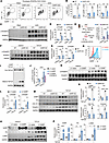 Figure 5
GARP-dependent TGF-β activation sustains Smad signaling and induces fibroblast stimulation. (A and B) Representative flow cytometry plots (A) and quantification (B) of GARP and LAP (TGF-β1) expression in kidney fibroblasts from sham or UUO-treated WT and GARP-KO mice (n = 3–4). MFI, mean fluorescence intensity. (C and D) Representative (C) and quantification (D) of immunoblot for p-Smad2/3 in sham or UUO-treated WT and GARP-KO mice (n = 5). (E) TGF-β reporter assay detecting active TGF-β released by PKFs with or without TGF-β1 priming. (F and G) Representative (F) and quantification (G) of immunoblot for p-Smad2/3 in medium only or TGF-β1–primed PKFs from WT and GARP-KO mice. (H) Flow cytometry of GARP expression in NRK-49F cells transduced with empty lentivirus (LV-vector) or GARP-overexpression lentivirus (LV-GARP). (I) Representative immunoblots showing TGF-β1 levels in cell lysates from medium (control) or TGF-β1–primed NRK-49F cells transduced with LV-vector or LV-GARP. (J) TGF-β reporter cell assay detecting active TGF-β release in NRK49F cells following TGF-β1 treatment in the absence or presence of CWHM-12 (an integrin αv inhibitor). (K and L) Representative (K) and quantification (L) of immunoblot for p-Smad2/3 in medium only or TGF-β1–primed NRK-49F cells. (M and N) Representative (M) and quantification (N) of immunoblot for GARP, FN, COL1A1, and Vimentin in medium only or TGF-β1–primed PKF cells from WT and GARP-KO mice. (O and P) Representative (O) and quantification (P) of immunoblot for GARP, FN, and α-SMA in medium only or TGF-β1–primed NRK-49F cells transduced with LV-vector or LV-GARP. Each lane in C, F, I, K, M, and O represents 1 well of cells. All in vitro experiments were independently repeated 3 times with similar results. Data represent mean ± SEM. *P < 0.05; **P < 0.01; ***P < 0.001; ****P < 0.0001. Statistical significance was determined by 2-way ANOVA followed by Tukey’s multiple-comparison test (B, D, E, G, J, L, N, and P).
We next assessed the impact of GARP-dependent TGF-β activation on fibroblast phenotype. Activated fibroblasts are characterized by increased ECM production and elevated expression of filament proteins such as α-SMA and Vimentin (6). In PKFs, TGF-β1 stimulation induced upregulation of GARP, ECM proteins (FN and COL1A1), and Vimentin. Deletion of GARP prevented the TGF-β1–induced upregulation of ECM proteins and Vimentin (Figure 5, M and N). Conversely, GARP overexpression in NRK-49F cells enhanced FN and α-SMA expression compared with the vector control (Figure 5, O and P). Thus, these findings indicate that increased expression of GARP amplifies TGF-β signaling to promote renal fibroblast activation.
TNF upregulates GARP in an NF-κB–dependent fashion and synergizes with TGF-β1. Since GARP expression was upregulated in both fibrotic kidneys and activated fibroblasts, we next sought to elucidate the mechanisms underlying its induction. Using Harmonizome 3.0 (37), we searched for putative transcription factors regulating GARP expression (https://maayanlab.cloud/Harmonizome/gene/LRRC32). After cross-referencing results from ChEA Transcription Factor Targets 2022 and JASPAR Predicted Transcription Factor Targets (https://maayanlab.cloud/Harmonizome/gene/LRRC32), NFKB1 emerged as the remaining candidate (Figure 6A), in line with prior studies showing NF-κB binding to the Lrrc32 promoter (38) and enhancer (26). Furthermore, in human kidney single-cell transcriptomic data (GSE183279) (31), fibroblast-restricted comparison between CKD and Ref further showed significant enrichment of the Hallmark TNFA signaling via NF-κB gene set, indicating activation of NF-κB–related transcriptional programs in fibroblasts under CKD condition (Figure 6B). Given that NF-κB signaling is tightly linked to kidney inflammation and fibrosis (39) and can be activated by cytokines such as TNF (40), we examined whether TNF regulates GARP expression through NF-κB. Stimulation of NRK-49F fibroblasts with TNF induced a time-dependent increase in GARP expression (Figure 6, C and D). During UUO-induced kidney fibrosis, upregulation of Nfkb1 preceded that of Lrrc32 (Supplemental Figure 8A). Similarly, TNF rapidly activated NF-κB signaling in NRK-49F cells, as evidenced by increased phosphorylation of P105 (NF-κB1) and P65 and reduced IκBα expression, preceding the subsequent induction of GARP protein expression (Supplemental Figure 8B). Finally, pharmacological inhibition of NF-κB with pyrrolidinedithiocarbamate (PDTC) blocked TNF-induced GARP expression (Figure 6, E–G). Guided by the consensus NF-κB binding sequence 5′-GGGRNNNYCC-3′ (where R = A or G, Y = C or T, and N = any nucleotide) (41), we identified 2 potential NF-κB binding sites (NBSs) in the rat Lrrc32 promoter. Cross-species sequence alignment showed that the promoter NBS (NBS1) is highly conserved across rat, human, mouse, macaque, and dog, whereas NBS2 is less well conserved (Figure 6H). Chromatin immunoprecipitation followed by quantitative PCR (ChIP-qPCR) confirmed that TNF increased P65 occupancy at NBS1 and, to a lesser extent, at NBS2 (Figure 6I). Interestingly, TNF increased P65 occupancy not only at the Lrrc32 promoter but also at the previously reported distal enhancer (26) (Figure 6I). These data indicate that TNF-induced NF-κB directly binds both proximal and distal regulatory elements of the Lrrc32 locus in fibroblasts.
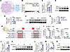 Figure 6
TNF upregulates GARP in an NF-κB–dependent fashion and synergizes with TGF-β1. (A) Venn diagram of transcription factors predicted to bind to the Lrrc32 promoter region using ChEA and JASPAR tools. (B) Gene set enrichment analysis (GSEA) of fibroblasts from human kidney single-cell transcriptomic data (GSE183279) comparing CKD and reference samples, showing significant positive enrichment of the Hallmark TNFA signaling via NF-κB gene set in CKD fibroblasts. (C and D) mRNA (C) and protein (D) levels of GARP in NRK-49F cells treated with TNF (10 ng/mL) for the indicated durations. (E–G) mRNA (E) and protein (F and G) levels of GARP in TNF-stimulated NRK-49F cells in the absence or presence of PDTC (an NF-κB inhibitor). Representative images and quantitative analysis are shown in E and F, respectively. (H) The NF-κB consensus motif is shown at the top, and the gray regions mark 2 potential NF-κB binding sites (NBS1 and NBS2) in the rat Lrrc32 promoter. (I) ChIP analysis of NF-κB p65 binding to NBS1 and NBS2 in the rat Lrrc32 promoter and to the distal Lrrc32 enhancer in NRK-49F cells under basal or TNF-stimulated conditions. Normal IgG served as a control. (J–L) mRNA (J) and protein (K and L) levels of GARP in NRK-49F cells treated with TGF-β1 (2 ng/mL) for the indicated durations. Representative images and quantitative analysis are shown in J and K, respectively. (M and N) mRNA (M) and protein (N) levels of GARP in NRK-49F cells treated with TNF, TGF-β1, or the combination of TGF-β1 and TNF. All in vitro experiments were independently repeated 3 times with similar results. Data represent mean ± SEM. *P < 0.05; **P < 0.01; ***P < 0.001; ****P < 0.0001. Statistical significance was determined by 1-way ANOVA (C, E, G, J, L, M) or 2-way ANOVA (I) with Tukey’s test for multiple comparisons.
Given that GARP expression was elevated in activated fibroblasts and TGF-β1 is a central driver of fibroblast activation during kidney fibrosis, we next examined whether TGF-β1 also regulates GARP expression. TGF-β1 induced a time-dependent upregulation of GARP (Figure 6, J–L). Interestingly, co-stimulation with TNF and TGF-β1 synergistically enhanced Lrrc32 mRNA and GARP protein expression, exhibiting a greater effect than either cytokine alone (Figure 6, M and N). Together, these findings demonstrate that TNF/NF-κB signaling engages evolutionarily conserved regulatory elements of Lrrc32 in renal fibroblasts and that TNF synergizes with TGF-β1 to reinforce GARP upregulation during fibroblast activation.
Therapeutic deletion of GARP attenuates established kidney fibrosis. Lastly, we asked whether therapeutic deletion of GARP could mitigate established fibrosis. To this end, kidney fibrosis was first allowed to develop for 7 days in inducible GARP-CKO UUO mice, after which tamoxifen was administered for 5 consecutive days to induce GARP deletion (Figure 7A). Immunoblots confirmed reduced renal GARP expression, along with attenuated induction of FN, COL1A1, and Vimentin on day 14 compared with controls (Figure 7, B and C). Sirius red staining showed comparable fibrosis between the 2 groups on day 7. However, following tamoxifen-induced GARP deletion, fibrosis progressed to a much greater extent in control than in GARP-CKO mice (Figure 7, D and E). Immunohistochemistry also confirmed reduced α-SMA expression after GARP deletion (Figure 7F). To extend these findings, we next induced global GARP deletion after fibrosis onset, which similarly reduced collagen deposition, ECM accumulation, and Smad2/3 phosphorylation, yielding a more pronounced antifibrotic response than fibroblast-specific deletion (Supplemental Figure 9). Collectively, these results demonstrate that therapeutic deletion of GARP effectively attenuates ongoing kidney fibrosis, highlighting GARP as a promising therapeutic target.
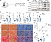 Figure 7
Therapeutic deletion of GARP in fibroblasts reduces established kidney fibrosis. (A) Experimental design illustrating the therapeutic targeting of GARP in fibroblasts after fibrosis establishment. (B and C) Representative immunoblots (B) and quantification (C) of GARP, fibronectin (FN), COL1A1, and Vimentin in kidneys from WT and GARP-CKO mice treated as in A (n = 5–6 per group). (D–F) Representative images (D) and quantitative analyses of Sirius red–stained fibrotic area (E) and α-SMA–positive area (F) in kidney sections from WT and GARP-CKO mice. Scale bars: 20 μm. Data represent mean ± SEM. ****P < 0.0001. NS, not significant (P > 0.05). Statistical significance was determined by 2-way ANOVA followed by Tukey’s multiple-comparison test (C, E, and F).


