
Go to The Journal of Clinical Investigation
  • About
  • Editors
  • Consulting Editors
  • For authors
  • Journal stats
  • Publication ethics
  • Publication alerts by email
  • Transfers
  • Advertising
  • Job board
  • Contact
  • Physician-Scientist Development
  • Current issue
  • Past issues
  • By specialty
    • COVID-19
    • Cardiology
    • Immunology
    • Metabolism
    • Nephrology
    • Oncology
    • Pulmonology
    • All ...
  • Videos
  • Collections
    • In-Press Preview
    • Resource and Technical Advances
    • Clinical Research and Public Health
    • Research Letters
    • Editorials
    • Perspectives
    • Physician-Scientist Development
    • Reviews
    • Top read articles

  • Current issue
  • Past issues
  • Specialties
  • In-Press Preview
  • Resource and Technical Advances
  • Clinical Research and Public Health
  • Research Letters
  • Editorials
  • Perspectives
  • Physician-Scientist Development
  • Reviews
  • Top read articles
  • About
  • Editors
  • Consulting Editors
  • For authors
  • Journal stats
  • Publication ethics
  • Publication alerts by email
  • Transfers
  • Advertising
  • Job board
  • Contact
Top
  • View PDF
  • Download citation information
  • Send a comment
  • Terms of use
  • Standard abbreviations
  • Need help? Email the journal
  • Top
  • Abstract
  • Introduction
  • Results
  • Discussion
  • Methods
  • Manuscript information
  • Supplemental material
  • Acknowledgments
  • Footnotes
  • References
  • Version history
  • Article usage
  • Citations to this article
Advertisement

Research ArticleInfectious diseaseInflammationNeuroscience Open Access | 10.1172/jci.insight.203088

Disrupted neuronal expression of autism risk genes and white matter micro-organization after infant Zika infection

Venkata-Viswanadh Edara,1 Sienna Freeman,2 Maureen Sampson,3 Kathryn M. Moore,2 Rebecca Richardson,2 Nils Schoof,1 Divine Burgess,1 Michelle J. Lee,2,4 Gregory K. Tharp,2,4 Roza Vlasova,5 Shane Taylor,6 Ariana Hodjatzadeh,6 Yihana P. Melendez-Alejandro,6 Samia M. Abdallah,6 Esther W. Ndungu,6 Ava K. Mascarenhas,6 Chao-Hsiung Hsu,7 Tsang-Wei Tu,7,8 Martin Styner,5 Mehul S. Suthar,1,2,4,9 Mar M. Sanchez,2,10 Steven E. Bosinger,2,4 Mark W. Burke,6 Steven A. Sloan,3,11 Jessica Raper,1,2 and Ann Chahroudi1,2,4,9

1Department of Pediatrics, Emory University School of Medicine, Atlanta, Georgia, USA.

2Emory National Primate Research Center, Atlanta, Georgia, USA.

3Department of Human Genetics, Emory University School of Medicine, Atlanta, Georgia, USA.

4Emory Vaccine Center, Atlanta, Georgia, USA.

5Department of Psychiatry, University of North Carolina, Chapel Hill, North Carolina, USA.

6Department of Physiology and Biophysics and

7Molecular Imaging Laboratory, Department of Radiology, Howard University, Washington, DC, USA.

8Department of Pediatrics, George Washington University School of Medicine and Health Sciences, Washington, DC, USA.

9Center for Childhood Infections and Vaccines of Children’s Healthcare of Atlanta and Emory University, Atlanta, Georgia, USA.

10Department of Psychiatry and Behavioral Sciences and

11Emory Center for Neurodegenerative Disease, Emory University School of Medicine, Atlanta, Georgia, USA.

Address for correspondence. Ann Chahroudi, Health Science Research Building, Suite N447, 1750 Haygood Drive NE, Atlanta, Georgia 30322, USA. Email: ann.m.chahroudi@emory.edu.

Find articles by Edara, V. in: PubMed | Google Scholar

1Department of Pediatrics, Emory University School of Medicine, Atlanta, Georgia, USA.

2Emory National Primate Research Center, Atlanta, Georgia, USA.

3Department of Human Genetics, Emory University School of Medicine, Atlanta, Georgia, USA.

4Emory Vaccine Center, Atlanta, Georgia, USA.

5Department of Psychiatry, University of North Carolina, Chapel Hill, North Carolina, USA.

6Department of Physiology and Biophysics and

7Molecular Imaging Laboratory, Department of Radiology, Howard University, Washington, DC, USA.

8Department of Pediatrics, George Washington University School of Medicine and Health Sciences, Washington, DC, USA.

9Center for Childhood Infections and Vaccines of Children’s Healthcare of Atlanta and Emory University, Atlanta, Georgia, USA.

10Department of Psychiatry and Behavioral Sciences and

11Emory Center for Neurodegenerative Disease, Emory University School of Medicine, Atlanta, Georgia, USA.

Address for correspondence. Ann Chahroudi, Health Science Research Building, Suite N447, 1750 Haygood Drive NE, Atlanta, Georgia 30322, USA. Email: ann.m.chahroudi@emory.edu.

Find articles by Freeman, S. in: PubMed | Google Scholar

1Department of Pediatrics, Emory University School of Medicine, Atlanta, Georgia, USA.

2Emory National Primate Research Center, Atlanta, Georgia, USA.

3Department of Human Genetics, Emory University School of Medicine, Atlanta, Georgia, USA.

4Emory Vaccine Center, Atlanta, Georgia, USA.

5Department of Psychiatry, University of North Carolina, Chapel Hill, North Carolina, USA.

6Department of Physiology and Biophysics and

7Molecular Imaging Laboratory, Department of Radiology, Howard University, Washington, DC, USA.

8Department of Pediatrics, George Washington University School of Medicine and Health Sciences, Washington, DC, USA.

9Center for Childhood Infections and Vaccines of Children’s Healthcare of Atlanta and Emory University, Atlanta, Georgia, USA.

10Department of Psychiatry and Behavioral Sciences and

11Emory Center for Neurodegenerative Disease, Emory University School of Medicine, Atlanta, Georgia, USA.

Address for correspondence. Ann Chahroudi, Health Science Research Building, Suite N447, 1750 Haygood Drive NE, Atlanta, Georgia 30322, USA. Email: ann.m.chahroudi@emory.edu.

Find articles by Sampson, M. in: PubMed | Google Scholar

1Department of Pediatrics, Emory University School of Medicine, Atlanta, Georgia, USA.

2Emory National Primate Research Center, Atlanta, Georgia, USA.

3Department of Human Genetics, Emory University School of Medicine, Atlanta, Georgia, USA.

4Emory Vaccine Center, Atlanta, Georgia, USA.

5Department of Psychiatry, University of North Carolina, Chapel Hill, North Carolina, USA.

6Department of Physiology and Biophysics and

7Molecular Imaging Laboratory, Department of Radiology, Howard University, Washington, DC, USA.

8Department of Pediatrics, George Washington University School of Medicine and Health Sciences, Washington, DC, USA.

9Center for Childhood Infections and Vaccines of Children’s Healthcare of Atlanta and Emory University, Atlanta, Georgia, USA.

10Department of Psychiatry and Behavioral Sciences and

11Emory Center for Neurodegenerative Disease, Emory University School of Medicine, Atlanta, Georgia, USA.

Address for correspondence. Ann Chahroudi, Health Science Research Building, Suite N447, 1750 Haygood Drive NE, Atlanta, Georgia 30322, USA. Email: ann.m.chahroudi@emory.edu.

Find articles by Moore, K. in: PubMed | Google Scholar

1Department of Pediatrics, Emory University School of Medicine, Atlanta, Georgia, USA.

2Emory National Primate Research Center, Atlanta, Georgia, USA.

3Department of Human Genetics, Emory University School of Medicine, Atlanta, Georgia, USA.

4Emory Vaccine Center, Atlanta, Georgia, USA.

5Department of Psychiatry, University of North Carolina, Chapel Hill, North Carolina, USA.

6Department of Physiology and Biophysics and

7Molecular Imaging Laboratory, Department of Radiology, Howard University, Washington, DC, USA.

8Department of Pediatrics, George Washington University School of Medicine and Health Sciences, Washington, DC, USA.

9Center for Childhood Infections and Vaccines of Children’s Healthcare of Atlanta and Emory University, Atlanta, Georgia, USA.

10Department of Psychiatry and Behavioral Sciences and

11Emory Center for Neurodegenerative Disease, Emory University School of Medicine, Atlanta, Georgia, USA.

Address for correspondence. Ann Chahroudi, Health Science Research Building, Suite N447, 1750 Haygood Drive NE, Atlanta, Georgia 30322, USA. Email: ann.m.chahroudi@emory.edu.

Find articles by Richardson, R. in: PubMed | Google Scholar

1Department of Pediatrics, Emory University School of Medicine, Atlanta, Georgia, USA.

2Emory National Primate Research Center, Atlanta, Georgia, USA.

3Department of Human Genetics, Emory University School of Medicine, Atlanta, Georgia, USA.

4Emory Vaccine Center, Atlanta, Georgia, USA.

5Department of Psychiatry, University of North Carolina, Chapel Hill, North Carolina, USA.

6Department of Physiology and Biophysics and

7Molecular Imaging Laboratory, Department of Radiology, Howard University, Washington, DC, USA.

8Department of Pediatrics, George Washington University School of Medicine and Health Sciences, Washington, DC, USA.

9Center for Childhood Infections and Vaccines of Children’s Healthcare of Atlanta and Emory University, Atlanta, Georgia, USA.

10Department of Psychiatry and Behavioral Sciences and

11Emory Center for Neurodegenerative Disease, Emory University School of Medicine, Atlanta, Georgia, USA.

Address for correspondence. Ann Chahroudi, Health Science Research Building, Suite N447, 1750 Haygood Drive NE, Atlanta, Georgia 30322, USA. Email: ann.m.chahroudi@emory.edu.

Find articles by Schoof, N. in: PubMed | Google Scholar

1Department of Pediatrics, Emory University School of Medicine, Atlanta, Georgia, USA.

2Emory National Primate Research Center, Atlanta, Georgia, USA.

3Department of Human Genetics, Emory University School of Medicine, Atlanta, Georgia, USA.

4Emory Vaccine Center, Atlanta, Georgia, USA.

5Department of Psychiatry, University of North Carolina, Chapel Hill, North Carolina, USA.

6Department of Physiology and Biophysics and

7Molecular Imaging Laboratory, Department of Radiology, Howard University, Washington, DC, USA.

8Department of Pediatrics, George Washington University School of Medicine and Health Sciences, Washington, DC, USA.

9Center for Childhood Infections and Vaccines of Children’s Healthcare of Atlanta and Emory University, Atlanta, Georgia, USA.

10Department of Psychiatry and Behavioral Sciences and

11Emory Center for Neurodegenerative Disease, Emory University School of Medicine, Atlanta, Georgia, USA.

Address for correspondence. Ann Chahroudi, Health Science Research Building, Suite N447, 1750 Haygood Drive NE, Atlanta, Georgia 30322, USA. Email: ann.m.chahroudi@emory.edu.

Find articles by Burgess, D. in: PubMed | Google Scholar

1Department of Pediatrics, Emory University School of Medicine, Atlanta, Georgia, USA.

2Emory National Primate Research Center, Atlanta, Georgia, USA.

3Department of Human Genetics, Emory University School of Medicine, Atlanta, Georgia, USA.

4Emory Vaccine Center, Atlanta, Georgia, USA.

5Department of Psychiatry, University of North Carolina, Chapel Hill, North Carolina, USA.

6Department of Physiology and Biophysics and

7Molecular Imaging Laboratory, Department of Radiology, Howard University, Washington, DC, USA.

8Department of Pediatrics, George Washington University School of Medicine and Health Sciences, Washington, DC, USA.

9Center for Childhood Infections and Vaccines of Children’s Healthcare of Atlanta and Emory University, Atlanta, Georgia, USA.

10Department of Psychiatry and Behavioral Sciences and

11Emory Center for Neurodegenerative Disease, Emory University School of Medicine, Atlanta, Georgia, USA.

Address for correspondence. Ann Chahroudi, Health Science Research Building, Suite N447, 1750 Haygood Drive NE, Atlanta, Georgia 30322, USA. Email: ann.m.chahroudi@emory.edu.

Find articles by Lee, M. in: PubMed | Google Scholar

1Department of Pediatrics, Emory University School of Medicine, Atlanta, Georgia, USA.

2Emory National Primate Research Center, Atlanta, Georgia, USA.

3Department of Human Genetics, Emory University School of Medicine, Atlanta, Georgia, USA.

4Emory Vaccine Center, Atlanta, Georgia, USA.

5Department of Psychiatry, University of North Carolina, Chapel Hill, North Carolina, USA.

6Department of Physiology and Biophysics and

7Molecular Imaging Laboratory, Department of Radiology, Howard University, Washington, DC, USA.

8Department of Pediatrics, George Washington University School of Medicine and Health Sciences, Washington, DC, USA.

9Center for Childhood Infections and Vaccines of Children’s Healthcare of Atlanta and Emory University, Atlanta, Georgia, USA.

10Department of Psychiatry and Behavioral Sciences and

11Emory Center for Neurodegenerative Disease, Emory University School of Medicine, Atlanta, Georgia, USA.

Address for correspondence. Ann Chahroudi, Health Science Research Building, Suite N447, 1750 Haygood Drive NE, Atlanta, Georgia 30322, USA. Email: ann.m.chahroudi@emory.edu.

Find articles by Tharp, G. in: PubMed | Google Scholar |

1Department of Pediatrics, Emory University School of Medicine, Atlanta, Georgia, USA.

2Emory National Primate Research Center, Atlanta, Georgia, USA.

3Department of Human Genetics, Emory University School of Medicine, Atlanta, Georgia, USA.

4Emory Vaccine Center, Atlanta, Georgia, USA.

5Department of Psychiatry, University of North Carolina, Chapel Hill, North Carolina, USA.

6Department of Physiology and Biophysics and

7Molecular Imaging Laboratory, Department of Radiology, Howard University, Washington, DC, USA.

8Department of Pediatrics, George Washington University School of Medicine and Health Sciences, Washington, DC, USA.

9Center for Childhood Infections and Vaccines of Children’s Healthcare of Atlanta and Emory University, Atlanta, Georgia, USA.

10Department of Psychiatry and Behavioral Sciences and

11Emory Center for Neurodegenerative Disease, Emory University School of Medicine, Atlanta, Georgia, USA.

Address for correspondence. Ann Chahroudi, Health Science Research Building, Suite N447, 1750 Haygood Drive NE, Atlanta, Georgia 30322, USA. Email: ann.m.chahroudi@emory.edu.

Find articles by Vlasova, R. in: PubMed | Google Scholar

1Department of Pediatrics, Emory University School of Medicine, Atlanta, Georgia, USA.

2Emory National Primate Research Center, Atlanta, Georgia, USA.

3Department of Human Genetics, Emory University School of Medicine, Atlanta, Georgia, USA.

4Emory Vaccine Center, Atlanta, Georgia, USA.

5Department of Psychiatry, University of North Carolina, Chapel Hill, North Carolina, USA.

6Department of Physiology and Biophysics and

7Molecular Imaging Laboratory, Department of Radiology, Howard University, Washington, DC, USA.

8Department of Pediatrics, George Washington University School of Medicine and Health Sciences, Washington, DC, USA.

9Center for Childhood Infections and Vaccines of Children’s Healthcare of Atlanta and Emory University, Atlanta, Georgia, USA.

10Department of Psychiatry and Behavioral Sciences and

11Emory Center for Neurodegenerative Disease, Emory University School of Medicine, Atlanta, Georgia, USA.

Address for correspondence. Ann Chahroudi, Health Science Research Building, Suite N447, 1750 Haygood Drive NE, Atlanta, Georgia 30322, USA. Email: ann.m.chahroudi@emory.edu.

Find articles by Taylor, S. in: PubMed | Google Scholar

1Department of Pediatrics, Emory University School of Medicine, Atlanta, Georgia, USA.

2Emory National Primate Research Center, Atlanta, Georgia, USA.

3Department of Human Genetics, Emory University School of Medicine, Atlanta, Georgia, USA.

4Emory Vaccine Center, Atlanta, Georgia, USA.

5Department of Psychiatry, University of North Carolina, Chapel Hill, North Carolina, USA.

6Department of Physiology and Biophysics and

7Molecular Imaging Laboratory, Department of Radiology, Howard University, Washington, DC, USA.

8Department of Pediatrics, George Washington University School of Medicine and Health Sciences, Washington, DC, USA.

9Center for Childhood Infections and Vaccines of Children’s Healthcare of Atlanta and Emory University, Atlanta, Georgia, USA.

10Department of Psychiatry and Behavioral Sciences and

11Emory Center for Neurodegenerative Disease, Emory University School of Medicine, Atlanta, Georgia, USA.

Address for correspondence. Ann Chahroudi, Health Science Research Building, Suite N447, 1750 Haygood Drive NE, Atlanta, Georgia 30322, USA. Email: ann.m.chahroudi@emory.edu.

Find articles by Hodjatzadeh, A. in: PubMed | Google Scholar

1Department of Pediatrics, Emory University School of Medicine, Atlanta, Georgia, USA.

2Emory National Primate Research Center, Atlanta, Georgia, USA.

3Department of Human Genetics, Emory University School of Medicine, Atlanta, Georgia, USA.

4Emory Vaccine Center, Atlanta, Georgia, USA.

5Department of Psychiatry, University of North Carolina, Chapel Hill, North Carolina, USA.

6Department of Physiology and Biophysics and

7Molecular Imaging Laboratory, Department of Radiology, Howard University, Washington, DC, USA.

8Department of Pediatrics, George Washington University School of Medicine and Health Sciences, Washington, DC, USA.

9Center for Childhood Infections and Vaccines of Children’s Healthcare of Atlanta and Emory University, Atlanta, Georgia, USA.

10Department of Psychiatry and Behavioral Sciences and

11Emory Center for Neurodegenerative Disease, Emory University School of Medicine, Atlanta, Georgia, USA.

Address for correspondence. Ann Chahroudi, Health Science Research Building, Suite N447, 1750 Haygood Drive NE, Atlanta, Georgia 30322, USA. Email: ann.m.chahroudi@emory.edu.

Find articles by Melendez-Alejandro, Y. in: PubMed | Google Scholar

1Department of Pediatrics, Emory University School of Medicine, Atlanta, Georgia, USA.

2Emory National Primate Research Center, Atlanta, Georgia, USA.

3Department of Human Genetics, Emory University School of Medicine, Atlanta, Georgia, USA.

4Emory Vaccine Center, Atlanta, Georgia, USA.

5Department of Psychiatry, University of North Carolina, Chapel Hill, North Carolina, USA.

6Department of Physiology and Biophysics and

7Molecular Imaging Laboratory, Department of Radiology, Howard University, Washington, DC, USA.

8Department of Pediatrics, George Washington University School of Medicine and Health Sciences, Washington, DC, USA.

9Center for Childhood Infections and Vaccines of Children’s Healthcare of Atlanta and Emory University, Atlanta, Georgia, USA.

10Department of Psychiatry and Behavioral Sciences and

11Emory Center for Neurodegenerative Disease, Emory University School of Medicine, Atlanta, Georgia, USA.

Address for correspondence. Ann Chahroudi, Health Science Research Building, Suite N447, 1750 Haygood Drive NE, Atlanta, Georgia 30322, USA. Email: ann.m.chahroudi@emory.edu.

Find articles by Abdallah, S. in: PubMed | Google Scholar

1Department of Pediatrics, Emory University School of Medicine, Atlanta, Georgia, USA.

2Emory National Primate Research Center, Atlanta, Georgia, USA.

3Department of Human Genetics, Emory University School of Medicine, Atlanta, Georgia, USA.

4Emory Vaccine Center, Atlanta, Georgia, USA.

5Department of Psychiatry, University of North Carolina, Chapel Hill, North Carolina, USA.

6Department of Physiology and Biophysics and

7Molecular Imaging Laboratory, Department of Radiology, Howard University, Washington, DC, USA.

8Department of Pediatrics, George Washington University School of Medicine and Health Sciences, Washington, DC, USA.

9Center for Childhood Infections and Vaccines of Children’s Healthcare of Atlanta and Emory University, Atlanta, Georgia, USA.

10Department of Psychiatry and Behavioral Sciences and

11Emory Center for Neurodegenerative Disease, Emory University School of Medicine, Atlanta, Georgia, USA.

Address for correspondence. Ann Chahroudi, Health Science Research Building, Suite N447, 1750 Haygood Drive NE, Atlanta, Georgia 30322, USA. Email: ann.m.chahroudi@emory.edu.

Find articles by Ndungu, E. in: PubMed | Google Scholar

1Department of Pediatrics, Emory University School of Medicine, Atlanta, Georgia, USA.

2Emory National Primate Research Center, Atlanta, Georgia, USA.

3Department of Human Genetics, Emory University School of Medicine, Atlanta, Georgia, USA.

4Emory Vaccine Center, Atlanta, Georgia, USA.

5Department of Psychiatry, University of North Carolina, Chapel Hill, North Carolina, USA.

6Department of Physiology and Biophysics and

7Molecular Imaging Laboratory, Department of Radiology, Howard University, Washington, DC, USA.

8Department of Pediatrics, George Washington University School of Medicine and Health Sciences, Washington, DC, USA.

9Center for Childhood Infections and Vaccines of Children’s Healthcare of Atlanta and Emory University, Atlanta, Georgia, USA.

10Department of Psychiatry and Behavioral Sciences and

11Emory Center for Neurodegenerative Disease, Emory University School of Medicine, Atlanta, Georgia, USA.

Address for correspondence. Ann Chahroudi, Health Science Research Building, Suite N447, 1750 Haygood Drive NE, Atlanta, Georgia 30322, USA. Email: ann.m.chahroudi@emory.edu.

Find articles by Mascarenhas, A. in: PubMed | Google Scholar

1Department of Pediatrics, Emory University School of Medicine, Atlanta, Georgia, USA.

2Emory National Primate Research Center, Atlanta, Georgia, USA.

3Department of Human Genetics, Emory University School of Medicine, Atlanta, Georgia, USA.

4Emory Vaccine Center, Atlanta, Georgia, USA.

5Department of Psychiatry, University of North Carolina, Chapel Hill, North Carolina, USA.

6Department of Physiology and Biophysics and

7Molecular Imaging Laboratory, Department of Radiology, Howard University, Washington, DC, USA.

8Department of Pediatrics, George Washington University School of Medicine and Health Sciences, Washington, DC, USA.

9Center for Childhood Infections and Vaccines of Children’s Healthcare of Atlanta and Emory University, Atlanta, Georgia, USA.

10Department of Psychiatry and Behavioral Sciences and

11Emory Center for Neurodegenerative Disease, Emory University School of Medicine, Atlanta, Georgia, USA.

Address for correspondence. Ann Chahroudi, Health Science Research Building, Suite N447, 1750 Haygood Drive NE, Atlanta, Georgia 30322, USA. Email: ann.m.chahroudi@emory.edu.

Find articles by Hsu, C. in: PubMed | Google Scholar |

1Department of Pediatrics, Emory University School of Medicine, Atlanta, Georgia, USA.

2Emory National Primate Research Center, Atlanta, Georgia, USA.

3Department of Human Genetics, Emory University School of Medicine, Atlanta, Georgia, USA.

4Emory Vaccine Center, Atlanta, Georgia, USA.

5Department of Psychiatry, University of North Carolina, Chapel Hill, North Carolina, USA.

6Department of Physiology and Biophysics and

7Molecular Imaging Laboratory, Department of Radiology, Howard University, Washington, DC, USA.

8Department of Pediatrics, George Washington University School of Medicine and Health Sciences, Washington, DC, USA.

9Center for Childhood Infections and Vaccines of Children’s Healthcare of Atlanta and Emory University, Atlanta, Georgia, USA.

10Department of Psychiatry and Behavioral Sciences and

11Emory Center for Neurodegenerative Disease, Emory University School of Medicine, Atlanta, Georgia, USA.

Address for correspondence. Ann Chahroudi, Health Science Research Building, Suite N447, 1750 Haygood Drive NE, Atlanta, Georgia 30322, USA. Email: ann.m.chahroudi@emory.edu.

Find articles by Tu, T. in: PubMed | Google Scholar |

1Department of Pediatrics, Emory University School of Medicine, Atlanta, Georgia, USA.

2Emory National Primate Research Center, Atlanta, Georgia, USA.

3Department of Human Genetics, Emory University School of Medicine, Atlanta, Georgia, USA.

4Emory Vaccine Center, Atlanta, Georgia, USA.

5Department of Psychiatry, University of North Carolina, Chapel Hill, North Carolina, USA.

6Department of Physiology and Biophysics and

7Molecular Imaging Laboratory, Department of Radiology, Howard University, Washington, DC, USA.

8Department of Pediatrics, George Washington University School of Medicine and Health Sciences, Washington, DC, USA.

9Center for Childhood Infections and Vaccines of Children’s Healthcare of Atlanta and Emory University, Atlanta, Georgia, USA.

10Department of Psychiatry and Behavioral Sciences and

11Emory Center for Neurodegenerative Disease, Emory University School of Medicine, Atlanta, Georgia, USA.

Address for correspondence. Ann Chahroudi, Health Science Research Building, Suite N447, 1750 Haygood Drive NE, Atlanta, Georgia 30322, USA. Email: ann.m.chahroudi@emory.edu.

Find articles by Styner, M. in: PubMed | Google Scholar

1Department of Pediatrics, Emory University School of Medicine, Atlanta, Georgia, USA.

2Emory National Primate Research Center, Atlanta, Georgia, USA.

3Department of Human Genetics, Emory University School of Medicine, Atlanta, Georgia, USA.

4Emory Vaccine Center, Atlanta, Georgia, USA.

5Department of Psychiatry, University of North Carolina, Chapel Hill, North Carolina, USA.

6Department of Physiology and Biophysics and

7Molecular Imaging Laboratory, Department of Radiology, Howard University, Washington, DC, USA.

8Department of Pediatrics, George Washington University School of Medicine and Health Sciences, Washington, DC, USA.

9Center for Childhood Infections and Vaccines of Children’s Healthcare of Atlanta and Emory University, Atlanta, Georgia, USA.

10Department of Psychiatry and Behavioral Sciences and

11Emory Center for Neurodegenerative Disease, Emory University School of Medicine, Atlanta, Georgia, USA.

Address for correspondence. Ann Chahroudi, Health Science Research Building, Suite N447, 1750 Haygood Drive NE, Atlanta, Georgia 30322, USA. Email: ann.m.chahroudi@emory.edu.

Find articles by Suthar, M. in: PubMed | Google Scholar |

1Department of Pediatrics, Emory University School of Medicine, Atlanta, Georgia, USA.

2Emory National Primate Research Center, Atlanta, Georgia, USA.

3Department of Human Genetics, Emory University School of Medicine, Atlanta, Georgia, USA.

4Emory Vaccine Center, Atlanta, Georgia, USA.

5Department of Psychiatry, University of North Carolina, Chapel Hill, North Carolina, USA.

6Department of Physiology and Biophysics and

7Molecular Imaging Laboratory, Department of Radiology, Howard University, Washington, DC, USA.

8Department of Pediatrics, George Washington University School of Medicine and Health Sciences, Washington, DC, USA.

9Center for Childhood Infections and Vaccines of Children’s Healthcare of Atlanta and Emory University, Atlanta, Georgia, USA.

10Department of Psychiatry and Behavioral Sciences and

11Emory Center for Neurodegenerative Disease, Emory University School of Medicine, Atlanta, Georgia, USA.

Address for correspondence. Ann Chahroudi, Health Science Research Building, Suite N447, 1750 Haygood Drive NE, Atlanta, Georgia 30322, USA. Email: ann.m.chahroudi@emory.edu.

Find articles by Sanchez, M. in: PubMed | Google Scholar

1Department of Pediatrics, Emory University School of Medicine, Atlanta, Georgia, USA.

2Emory National Primate Research Center, Atlanta, Georgia, USA.

3Department of Human Genetics, Emory University School of Medicine, Atlanta, Georgia, USA.

4Emory Vaccine Center, Atlanta, Georgia, USA.

5Department of Psychiatry, University of North Carolina, Chapel Hill, North Carolina, USA.

6Department of Physiology and Biophysics and

7Molecular Imaging Laboratory, Department of Radiology, Howard University, Washington, DC, USA.

8Department of Pediatrics, George Washington University School of Medicine and Health Sciences, Washington, DC, USA.

9Center for Childhood Infections and Vaccines of Children’s Healthcare of Atlanta and Emory University, Atlanta, Georgia, USA.

10Department of Psychiatry and Behavioral Sciences and

11Emory Center for Neurodegenerative Disease, Emory University School of Medicine, Atlanta, Georgia, USA.

Address for correspondence. Ann Chahroudi, Health Science Research Building, Suite N447, 1750 Haygood Drive NE, Atlanta, Georgia 30322, USA. Email: ann.m.chahroudi@emory.edu.

Find articles by Bosinger, S. in: PubMed | Google Scholar |

1Department of Pediatrics, Emory University School of Medicine, Atlanta, Georgia, USA.

2Emory National Primate Research Center, Atlanta, Georgia, USA.

3Department of Human Genetics, Emory University School of Medicine, Atlanta, Georgia, USA.

4Emory Vaccine Center, Atlanta, Georgia, USA.

5Department of Psychiatry, University of North Carolina, Chapel Hill, North Carolina, USA.

6Department of Physiology and Biophysics and

7Molecular Imaging Laboratory, Department of Radiology, Howard University, Washington, DC, USA.

8Department of Pediatrics, George Washington University School of Medicine and Health Sciences, Washington, DC, USA.

9Center for Childhood Infections and Vaccines of Children’s Healthcare of Atlanta and Emory University, Atlanta, Georgia, USA.

10Department of Psychiatry and Behavioral Sciences and

11Emory Center for Neurodegenerative Disease, Emory University School of Medicine, Atlanta, Georgia, USA.

Address for correspondence. Ann Chahroudi, Health Science Research Building, Suite N447, 1750 Haygood Drive NE, Atlanta, Georgia 30322, USA. Email: ann.m.chahroudi@emory.edu.

Find articles by Burke, M. in: PubMed | Google Scholar

1Department of Pediatrics, Emory University School of Medicine, Atlanta, Georgia, USA.

2Emory National Primate Research Center, Atlanta, Georgia, USA.

3Department of Human Genetics, Emory University School of Medicine, Atlanta, Georgia, USA.

4Emory Vaccine Center, Atlanta, Georgia, USA.

5Department of Psychiatry, University of North Carolina, Chapel Hill, North Carolina, USA.

6Department of Physiology and Biophysics and

7Molecular Imaging Laboratory, Department of Radiology, Howard University, Washington, DC, USA.

8Department of Pediatrics, George Washington University School of Medicine and Health Sciences, Washington, DC, USA.

9Center for Childhood Infections and Vaccines of Children’s Healthcare of Atlanta and Emory University, Atlanta, Georgia, USA.

10Department of Psychiatry and Behavioral Sciences and

11Emory Center for Neurodegenerative Disease, Emory University School of Medicine, Atlanta, Georgia, USA.

Address for correspondence. Ann Chahroudi, Health Science Research Building, Suite N447, 1750 Haygood Drive NE, Atlanta, Georgia 30322, USA. Email: ann.m.chahroudi@emory.edu.

Find articles by Sloan, S. in: PubMed | Google Scholar

1Department of Pediatrics, Emory University School of Medicine, Atlanta, Georgia, USA.

2Emory National Primate Research Center, Atlanta, Georgia, USA.

3Department of Human Genetics, Emory University School of Medicine, Atlanta, Georgia, USA.

4Emory Vaccine Center, Atlanta, Georgia, USA.

5Department of Psychiatry, University of North Carolina, Chapel Hill, North Carolina, USA.

6Department of Physiology and Biophysics and

7Molecular Imaging Laboratory, Department of Radiology, Howard University, Washington, DC, USA.

8Department of Pediatrics, George Washington University School of Medicine and Health Sciences, Washington, DC, USA.

9Center for Childhood Infections and Vaccines of Children’s Healthcare of Atlanta and Emory University, Atlanta, Georgia, USA.

10Department of Psychiatry and Behavioral Sciences and

11Emory Center for Neurodegenerative Disease, Emory University School of Medicine, Atlanta, Georgia, USA.

Address for correspondence. Ann Chahroudi, Health Science Research Building, Suite N447, 1750 Haygood Drive NE, Atlanta, Georgia 30322, USA. Email: ann.m.chahroudi@emory.edu.

Find articles by Raper, J. in: PubMed | Google Scholar

1Department of Pediatrics, Emory University School of Medicine, Atlanta, Georgia, USA.

2Emory National Primate Research Center, Atlanta, Georgia, USA.

3Department of Human Genetics, Emory University School of Medicine, Atlanta, Georgia, USA.

4Emory Vaccine Center, Atlanta, Georgia, USA.

5Department of Psychiatry, University of North Carolina, Chapel Hill, North Carolina, USA.

6Department of Physiology and Biophysics and

7Molecular Imaging Laboratory, Department of Radiology, Howard University, Washington, DC, USA.

8Department of Pediatrics, George Washington University School of Medicine and Health Sciences, Washington, DC, USA.

9Center for Childhood Infections and Vaccines of Children’s Healthcare of Atlanta and Emory University, Atlanta, Georgia, USA.

10Department of Psychiatry and Behavioral Sciences and

11Emory Center for Neurodegenerative Disease, Emory University School of Medicine, Atlanta, Georgia, USA.

Address for correspondence. Ann Chahroudi, Health Science Research Building, Suite N447, 1750 Haygood Drive NE, Atlanta, Georgia 30322, USA. Email: ann.m.chahroudi@emory.edu.

Find articles by Chahroudi, A. in: PubMed | Google Scholar |

Published September 24, 2026 - More info

Published in Volume 11, Issue 21 on November 9, 2026
JCI Insight. 2026;11(21):e203088. https://doi.org/10.1172/jci.insight.203088.
© 2026 Edara et al. This work is licensed under the Creative Commons Attribution 4.0 International License. To view a copy of this license, visit http://creativecommons.org/licenses/by/4.0/.
Published September 24, 2026 - Version history
Received: December 3, 2025; Accepted: September 21, 2026
View PDF
Abstract

Congenital and early-life Zika virus (ZIKV) infection can result in neurologic deficits. Precise mechanisms of injury, especially in the more subtle presentation of postnatal infection, are not fully elucidated. Here, we defined the effects of ZIKV on the developing brain using single-cell transcriptomics, histopathology, design-based stereology, diffusion MRI, and neurobehavioral assessments in infant rhesus macaques. ZIKV upregulated interferon-stimulated genes in activated microglia and cell death pathways in neurons and downregulated metabolism and differentiation genes in mature oligodendrocytes. Abnormal micro-organization of the corpus callosum and limbic white matter tracts was seen on diffusion-weighted imaging. A curated gene set associated with autism spectrum disorder risk was negatively enriched in inhibitory and excitatory neurons from ZIKV-infected infants, with increased emotional reactivity already evident 2 weeks after infection. From single cells to organism-level behaviors, these results define the pathways and processes disrupted by early-life ZIKV infection.

Introduction

Intensive research has focused on the neuropathogenesis of congenital Zika virus (ZIKV) infection including microencephaly. Studies of microencephaly implicate loss of fetal intermediate progenitor cells, particularly in the subventricular zone of the developing brain (1–4). Progenitor cell depletion has been attributed to apoptosis, immune activation, and cell cycle dysregulation (5–9). Microcephaly is an extreme phenotype of congenital ZIKV infection, and more subtle features, including intracranial calcifications, cerebral atrophy, and optic nerve abnormalities, also occur (10–12). Similarly, postnatal ZIKV infection in infants and children has been observed, with acute neurologic complications that include encephalitis and Guillain-Barré syndrome (13–15). Delayed neurodevelopment following early-life ZIKV infection has also been described (16), although challenges in diagnosis make a direct association difficult to confirm on a population level. Some but not all long-term follow-up studies of children with postnatal ZIKV exposure have reported adverse neurodevelopment, including hearing or vision loss and poor performance in the personal-social domain of a standardized developmental screening tool (17–21).

The large body of data demonstrating extensive and dynamic postnatal growth and maturation of the brain over the first years of life (22, 23) suggests that ZIKV infection in this period could be harmful and calls for definitive studies in carefully controlled model systems. Critical regions such as the hippocampus, which continue maturing postnatally and are essential for learning, memory, and behavior, are particularly vulnerable to disruption, a factor implicated in neurodevelopmental disorders such as autism and schizophrenia (24, 25). Given this vulnerability, understanding the effects of ZIKV infection beyond the prenatal period is essential. We previously described long-term consequences of early-life ZIKV infection for the brain and behavior of infant rhesus macaques with partial mitigation by antiviral treatment (26–28). Rhesus macaques exhibit neurodevelopment that is temporally and anatomically comparable to that of humans, albeit on an accelerated timeline of approximately 1:4 ratio (29). In fact, transcriptomic signatures of neural processes in humans and macaques converge in the late fetal to infant (perinatal) development periods (30). The prolonged postnatal brain maturation, immune system similarity, and comparable social behaviors of rhesus macaques make them particularly well suited for modeling human neurodevelopmental outcomes (16, 31–34).

Here, we aimed to elucidate the mechanisms of disrupted neurodevelopment by subjecting the hippocampus, amygdala, and striatum to single-cell transcriptomic analysis 2 weeks after ZIKV infection to dissect the cell type–specific transcriptional changes that underlie ZIKV-associated neurodevelopmental disruption. These regions of the central nervous system (CNS) were selected based on our prior research that used in vivo magnetic resonance imaging (MRI), resting-state functional MRI, and postmortem histologic examination to demonstrate ZIKV-induced abnormalities (27, 28). Acute postnatal ZIKV infection of infants led to CNS innate immune activation with an increased apoptotic gene signature and negative enrichment of autism spectrum disorder–associated genes in neurons, as well as downregulation of maturation genes in oligodendrocytes. Complementary histologic and stereologic examination confirmed these transcriptomic findings. In an additional cohort of 24 infant macaques (12 control and 12 infected with ZIKV), diffusion tensor imaging MRI after 2 months of ZIKV infection highlighted abnormal white matter organization in the limbic system and corpus callosum. Taken together, these findings provide plausible mechanisms for the abnormal brain structures, functions, and social behaviors that can result from early-life ZIKV infection.

Results

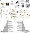
ZIKV alters infant macaque CNS gene expression. To investigate the impact of early-life ZIKV infection on brain development, we performed single-cell transcriptomic profiling of CNS tissues from infant rhesus macaques (Figure 1A). ZIKV viremia peaked at 2–3 days after infection and was cleared from peripheral circulation within 2 weeks (Figure 1B). Prior work from our group has demonstrated that entry of viral RNA into the CNS occurs after peak viremia, with detection by PCR and RNAscope (Advanced Cell Diagnostics) at 14–15 days of infection (28). Based on this finding, 3 infant rhesus macaques were infected with ZIKV at 1 month of life and euthanized 14 days after infection alongside 3 age-matched uninfected controls. Tissues from the amygdala, hippocampus, and striatum were harvested within 10 minutes of euthanasia and immediately processed to minimize postmortem interval confounders, followed by single-cell RNA sequencing (scRNA-seq) using the 10x Genomics platform. One ZIKV-infected infant rhesus macaque died prematurely at 11 days after infection and was excluded from the scRNA-seq analysis; tissues were preserved for histologic examination.
Study design, viral kinetics, and CNS-specific transcriptome changes followFigure 1

Study design, viral kinetics, and CNS-specific transcriptome changes following ZIKV infection in infant rhesus macaques. (A) Study overview. Three 1-month-old infant rhesus macaques were infected subcutaneously with 1 × 10⁵ plaque-forming units (PFU) ZIKV strain PRVABC59. Sex- and age-matched uninfected animals were used as controls. Fourteen days after infection, macaques were euthanized, the amygdala, hippocampus, and striatum of one hemisphere were used for single-cell transcriptomic analyses, and the same regions from the other hemisphere were used for histologic examination. (B) Plasma viral loads were measured by quantitative reverse transcription PCR from time of infection until necropsy. (C) Dimensionality reduction/UMAP analysis of combined Seurat object after quality control. (D) UMAP analysis of combined Seurat object segregated by treatment group and by brain region. (E and F) Gene Ontology (GO) analysis of differentially expressed genes among ZIKV-infected versus control animals showing downregulated and upregulated genes, respectively.

After quality control, 105,421 cells from controls and 94,975 cells from ZIKV-infected animals were retained for downstream analysis. Uniform manifold approximation and projection (UMAP) clustering revealed all age-relevant CNS cell classes along with their subtypes, which we subsequently reclustered for detailed, cell type–specific analyses. Cell type identities were assigned based on cluster-specific marker genes, with representative markers shown in the dot plot (Supplemental Figure 1A; supplemental material available online with this article; https://doi.org/10.1172/jci.insight.203088DS1). UMAP plots grouped by treatment (ZIKV vs. control) and by brain region (amygdala, hippocampus, striatum) are presented in Figure 1, C and D, respectively. The distribution of cell type proportions by treatment group, brain region, and individual animal is summarized in Supplemental Figure 1, B–D, with corresponding cell numbers shown in Supplemental Tables 1–3. Of all major cell types, the proportion of mature astrocytes was consistently lower in ZIKV-infected infants compared with controls, while oligodendrocyte precursor cells, mature oligodendrocytes, excitatory neurons, and ependymal cell numbers were consistently higher in ZIKV-infected animals (Supplemental Figure 1E and Supplemental Table 3). The large increase in choroid plexus cells in the hippocampus was only seen in one ZIKV-infected animal and thus was not further explored.

We first assessed global transcriptional changes associated with ZIKV infection across all cell types. Therefore, we performed differential expression analysis across all cells comparing ZIKV-infected with control animals, identifying significantly differentially expressed genes using an adjusted P value threshold of less than 0.05. Gene Ontology (GO) enrichment analysis of downregulated genes revealed significant suppression of general pathways related to CNS development, including neuron and glial cell development and differentiation, axon development/axonogenesis, cell adhesion, and sterol biosynthesis (Figure 1E). These findings suggest impaired maturation and regulation of key neurodevelopmental programs following ZIKV infection. Conversely, upregulated pathways in ZIKV-infected animals were predominantly associated with immune responses and apoptosis, including cytokine production, activation of immune response, innate immune responses, regulation of cytoskeleton organization, apoptotic signaling pathway, and programmed cell death (Figure 1F). This robust neuroinflammatory response was also accompanied by upregulation of some nervous system development pathways. Together, these data provide evidence that the immune response to postnatal ZIKV infection in the CNS is accompanied by both cell death and dysregulated neurodevelopmental trajectories.

Activation of microglia and interferon signaling. Microglia have a complex, dual role in neurotropic viral infections. They serve as protective agents by defending against the virus but can also contribute to disease progression by promoting neuroinflammation (35). Given the robust upregulation of immune-related pathways observed in the global transcriptomic analysis, we next focused specifically on microglia to evaluate their response to postnatal ZIKV infection. First, we reclustered all myeloid-lineage cells and identified 10 transcriptionally distinct subclusters (MG1–10) (Figure 2A). These subclusters were present in all 3 brain regions and in both control and ZIKV-infected animals (Supplemental Figure 2A). In total, we analyzed 57,477 microglial cells, with 27,212 cells originating from control animals and 30,265 from ZIKV-infected animals. The distribution of cell type proportions by treatment group, brain region, and individual animal is summarized in Supplemental Figure 2, B–D, with corresponding cell numbers in Supplemental Tables 4–6. Only one subcluster (MG7) exhibited consistent cell number differences between ZIKV-infected infants and controls (Supplemental Table 6).
ZIKV infection leads to microglial immune activation and further inductionFigure 2

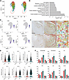
ZIKV infection leads to microglial immune activation and further induction of interferon signaling in infant rhesus macaques. (A) UMAP analysis of microglial subclusters segregated by treatment group. (B) Upregulated biological pathways from GO analysis of differentially expressed genes between ZIKV-infected and control among microglial subcluster MG6. (C) Feature plots of activated microglial markers and MHC class II molecules. (D) Expression of interferon-stimulated genes among ZIKV-infected and control microglial subcluster MG6. (E) Iba-1 staining of microglia from the hippocampus. Regional morphometric characterization of microglial cells was performed by StainAI. Microglia were color-coded as ramified (green), hypertrophic (gold), bushy (orange), ameboid (red), rod (blue), hyper-rod (purple), or unfocused (gray). (F) Quantification of surveillant (ramified [R]) versus activated (hypertrophic [H], bushy [B], and ameboid [A]) microglia in hippocampal gray matter of ZIKV-infected versus control infant rhesus macaques. Rod (RD) and hyper-rod (HR) activated microglia in the hippocampal white matter are also shown. Mean and SD are shown. Teal bars, ZIKV; salmon bars, controls.

To identify specific microglial populations associated with immune activation, we examined gene expression patterns across all subclusters. This analysis revealed that subcluster MG6 had elevated expression of immune response genes. Using GO pathway analysis to compare groups, MG6 cells from ZIKV-infected macaques exhibited enrichment for pathways associated with immunity, including positive regulation of immune system process, defense response, lymphocyte activation, chemotaxis, cytokine-mediated signaling pathway, and immune effector process (Figure 2B). Cluster MG6 cells in ZIKV-infected infants showed elevated expression of the canonical myeloid cell activation markers CD69, CD44, and MHC class II molecules MAMUDRA and MAMUDRB1 (Figure 2C). To comprehensively assess interferon pathway activation, we interrogated the differential expression of a panel of interferon-stimulated genes within both the entire microglial population and the MG6 subcluster in the combined dataset and across individual brain regions (Supplemental Figure 3, A and B, respectively). These analyses revealed widespread and robust microglial induction of interferon-stimulated genes in response to ZIKV infection. Elevated expression of the representative interferon-stimulated genes IFI6, IFI27, MX1, IFIT3, STAT1, and IL18, specifically within the MG6 subcluster, is shown in Figure 2D.

We next used design-based stereology and histologic examination of hippocampi to identify microglia by Iba-1 staining. Microglia were subcategorized into ramified, hypertrophic, bushy, ameboid, rod, and hyper-rod based on their morphologic features in the following regions of the hippocampal formation: dentate gyrus granular and molecular layers, cornu ammonis (CA) subfields 1–3, and fornix. Iba-l staining was quantified for each cell type using machine learning techniques (Figure 2E). The frequency of Iba-1+ activated microglia (categorized as hypertrophic, bushy, and ameboid cells) in the gray matter was increased in ZIKV-infected infants compared with controls across all 6 regions of the hippocampus (Figure 2F). Although the small number of animals precluded formal statistical analysis, this difference was most pronounced in the granular region of the dentate gyrus, where microglia regulate neurogenesis through phagocytosis of dying granule cells (36, 37) as part of a homeostatic process involved in memory formation, pattern separation, and spatial behavior (38–40). These collective findings suggest that postnatal ZIKV infection induces a shift in the microglial population toward an activated, interferon-responsive state, potentially influencing neurogenesis.

ZIKV upregulates neuronal apoptosis pathways. Design-based stereology was used to quantify hippocampal neuronal populations of the pyramidal layer in CA subfields 1–3 in cresyl violet–stained sections and nestin-positive immature neurons in the dentate gyrus. Counts were mostly similar between ZIKV-infected and control infants, with a trend toward reduced CA1 neurons (Supplemental Figure 4, A and B). Notably, mature neurons in the vulnerable CA1 region of ZIKV-infected macaques showed frequent cytoplasmic inclusions, typically a precursor to cell death, that were not seen in controls (Supplemental Figure 4C). Double immunolabeling for activated caspase-3 and the mature neuronal marker NeuN demonstrated caspase-3+ mature neurons in the hippocampus of ZIKV-infected animals, whereas little or no colocalization was observed in controls (Supplemental Figure 4D). Quantification of cleaved caspase-3 by immunohistochemistry further revealed a trend toward increased apoptosis in all regions examined (caudate nucleus, amygdala, hippocampus, and dentate gyrus) (Supplemental Figure 4E). These findings are consistent with the upregulation of apoptosis pathways seen in the initial global transcriptomic analysis (Figure 1F).

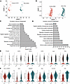
To evaluate how postnatal ZIKV infection influences neuronal subtypes critical for cortical circuit development, we next examined gene expression in both excitatory and inhibitory neuronal populations (a total of 10,077 neurons were studied: 3,137 excitatory and 854 inhibitory neurons from controls and 5,354 excitatory and 732 inhibitory neurons from ZIKV-infected macaques) (Figure 3, A and B). Region-specific distributions of these neuronal populations across the amygdala, hippocampus, and striatum as well as cell proportions by treatment, region, and individual animal are shown in Supplemental Figure 5, with corresponding cell numbers in Supplemental Tables 7–12. GO pathway analysis revealed upregulation of multiple cell death pathways in both neuron subtypes in ZIKV-infected macaques (Figure 3, C and D). Both excitatory and inhibitory neurons of ZIKV-infected infants compared with controls showed increased expression of genes involved in regulating apoptosis (PYCARD, DAPK3, BNIP3L, CASP6, SIAH1, PAK2, BCL2L2-PABPN1, PPIA), autophagy (GABARAP), and mitophagy (SLC25A6) (Figure 3, E and F). These results indicate that induction of cell death signaling pathways is a common feature of postnatal and congenital ZIKV infection, with anticipated consequences for neurodevelopment.
ZIKV infection leads to induction of apoptotic pathways in the limbic regioFigure 3

ZIKV infection leads to induction of apoptotic pathways in the limbic region of infant rhesus macaques. (A) UMAP analysis of excitatory neurons segregated by treatment group. (B) UMAP analysis of inhibitory neurons segregated by treatment group. (C) Upregulated biological pathways from GO analysis of differentially expressed genes between ZIKV-infected and control animals among excitatory neurons. (D) Upregulated biological pathways from GO analysis of differentially expressed genes between ZIKV-infected and control animals among inhibitory neurons. (E) Expression of selected cell death genes in excitatory neurons from ZIKV-infected versus control macaques. (F) Expression of selected cell death genes in inhibitory neurons from ZIKV-infected versus control macaques.

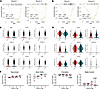
Neurodevelopmental transcriptional program in neurons and behavior changes. We next assessed whether ZIKV-induced gene expression changes in neuronal populations resembled shifts observed in other neurodevelopmental disorders. To do so, we interrogated the transcriptomic signature of neurons using a curated panel of 147 genes (41–45) implicated in the common neurodevelopmental disorder autism spectrum disorder (ASD), and categorized them based on their expression timing in the Epoch-1 (prenatal) and Epoch-2 (late prenatal and early postnatal) phases of neurodevelopment (41, 46). In excitatory neurons, gene set enrichment analysis (GSEA) revealed downregulation of both Epoch-1 and Epoch-2 gene sets in ZIKV-infected macaques (enrichment scores = –0.372 and –0.500, respectively), with a significant normalized enrichment score (NES) for Epoch-2 (NES = –1.49, P = 0.0451) (Figure 4A). In inhibitory neurons, both Epoch-1 and Epoch-2 gene sets were similarly downregulated (enrichment scores = –0.461 and –0.429), with the Epoch-1 gene set demonstrating statistical significance (NES = –1.50, P = 0.0089) (Figure 4B). To test whether these gene expression changes were nonspecific, we performed GSEA using 2 independent gene sets associated with cerebral palsy. These analyses did not reveal significant associations with ZIKV infection (Supplemental Figure 6, A–D). Furthermore, we tested the enrichment of these ASD-associated gene sets in other neural cell populations, including mature oligodendrocytes (Supplemental Figure 6, E and F), oligodendrocyte progenitors, and microglia, none of which showed significant enrichment. These control analyses support the specificity of ASD gene dysregulation in excitatory and inhibitory neurons in the context of ZIKV infection. High-confidence ASD risk genes, including ANK2, CHD8, CUL3, DYRK1A, GRIN2B, KATNAL2, POGZ, SCN2A, and TBR1, were consistently downregulated in ZIKV-infected infants in both excitatory (Figure 4C) and inhibitory neurons (Figure 4D), suggesting that postnatal ZIKV infection broadly affects core neurodevelopmental pathways that control social interaction and communication.
ZIKV infection leads to dysregulation of autism spectrum disorder genes amoFigure 4

ZIKV infection leads to dysregulation of autism spectrum disorder genes among neuronal populations and neurobehavioral changes in infant rhesus macaques. (A) GSEA plots for a panel of 147 autism spectrum disorder (ASD) genes spanning the Epoch-1 (prenatal) and Epoch-2 (late prenatal and early postnatal) phases of neurodevelopment in excitatory neurons of ZIKV-infected infant macaques. NES, normalized enrichment score; ES, enrichment score. (B) GSEA plots for 147 ASD genes spanning the Epoch-1 (prenatal) and Epoch-2 (late prenatal and early postnatal) phases of neurodevelopment in inhibitory neurons of ZIKV-infected infant macaques. (C) Expression of high-confidence ASD genes in excitatory neurons of ZIKV-infected and control infants. (D) Expression of high-confidence ASD genes in inhibitory neurons of ZIKV-infected and control infants. (E) Battery of neurobehavioral tests assessing neuromotor skills, motor ability, orientation, and state control in ZIKV-infected versus control infant macaques from 3 to 6 weeks of life. Differences were assessed using a linear mixed model with group and age as fixed factors and individual animals as a random factor. Teal symbols and lines, ZIKV; salmon symbols and lines, controls.

To relate these molecular findings to functional outcomes, we examined early neurobehavioral development in the same infant animals. We have previously shown that postnatal ZIKV infection at 1 month of life results in reduced prosocial behaviors and increased emotional reactivity at 1 year (27). Here, the Infant Neurobehavioral Assessment Scale, based on the Brazelton Newborn Behavioral Assessment Scale, was used to quantify behaviors related to orientation, motor activity, neuromotor responsiveness, and state control (i.e., level of arousal). We found that, at a very early age, postnatal ZIKV infection was associated with a significant decrease in orientation scores (F3,21 = 3.60, P = 0.049, ηp2 = 0.34) and an even more striking increase in state control scores (F3,21 = 6.84, P = 0.007, ηp2 = 0.49) (Figure 4E). Increased state control scores reflect poorer emotional regulation, higher reactivity, and less consolability. Both groups had similar neuromotor scores and typical age-related increases in motor ability from 3 to 6 weeks. Early alterations in attention and behavioral regulation, as seen here, are phenotypes frequently reported in children with neurodevelopmental disorders (47–50).

Disruption of differentiation and metabolic programs in oligodendrocytes. Among all the differences observed between ZIKV-infected and control infant macaques, oligodendrocyte differentiation was one of the most prominent changes. Oligodendrocytes are glial cells in the CNS responsible for forming myelin sheaths around axons, which insulate the fibers and enhance the speed of electrical signal transmission (51). During brain development, oligodendrocyte precursor cells differentiate into mature oligodendrocytes to create these sheaths, a process essential for building functional neural networks and facilitating sensory information processing (51). To assess how postnatal ZIKV infection affects oligodendrocyte-lineage cells, we performed focused subclustering of oligodendrocytes from single-cell transcriptomic data. From a total of 51,899 cells (22,168 from controls and 29,731 from ZIKV-infected macaques), UMAP analysis revealed 3 major populations: oligodendrocyte progenitor cells (OPCs), newly formed oligodendrocytes, and mature oligodendrocytes (Figure 5A). Subpopulation identity was determined through examination of canonical lineage marker expression, with enrichment of OLIG2 in OPCs, BCAS1 in newly formed oligodendrocytes, and MBP in mature oligodendrocytes (Figure 5B). The distribution of these subclusters in the amygdala, hippocampus, and striatum, with proportions by treatment group, brain region, and individual animal, is depicted in Supplemental Figure 7. Corresponding cell numbers are shown in Supplemental Tables 13–15. The ZIKV-infected macaques had increased proportions of both OPCs and mature oligodendrocytes compared with controls (Supplemental Table 13).
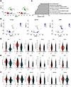
ZIKV infection dysregulates key biological pathways in mature oligodendrocyFigure 5

ZIKV infection dysregulates key biological pathways in mature oligodendrocytes of infant rhesus macaques. (A) UMAP analysis of oligodendrocyte subclusters segregated by treatment group. (B) Feature plots of lineage markers OLIG2, BCAS1, and MBP used to distinguish oligodendrocyte progenitor cells, newly formed oligodendrocytes, and mature oligodendrocytes, respectively. (C) Downregulated biological pathways from GO analysis of differentially expressed genes between ZIKV-infected and control mature oligodendrocytes. (D) Expression of representative genes regulating metabolic processes in mature oligodendrocytes from ZIKV-infected and control infant macaques. (E) Expression of selected genes involved in sterol biosynthesis in mature oligodendrocytes from ZIKV-infected and control infant macaques. (F) Expression of genes controlling oligodendrocyte differentiation in mature oligodendrocytes from ZIKV-infected and control infant macaques.

To determine how ZIKV infection alters mature oligodendrocyte function, we performed differential gene expression and GO analysis between ZIKV-infected and control oligodendrocyte populations. Downregulated pathways (lower in ZIKV) were significantly enriched for terms related to metabolic processes, sterol biosynthesis, and oligodendrocyte and glial cell differentiation (Figure 5C). Volcano plots of representative genes from these pathways further highlight their suppression in ZIKV-infected animals (Supplemental Figure 8). Expression of individual genes involved in cellular metabolism (ATP8, FIS1, PARP6, PGAM1, EIF6, PSEN1) (Figure 5D), sterol biosynthesis (DHCR24, HMGCS1, SQLE, LSS, SREBF2, MVD) (Figure 5E), and oligodendrocyte differentiation and maturation (QKI, MYRF, KCNJ10, SLC45A3, TMEM98, DAAM2) (Figure 5F) was significantly reduced following ZIKV infection. These data indicate that postnatal ZIKV infection perturbs oligodendrocyte development by disrupting core metabolic pathways, suppressing sterol biosynthesis, and impairing terminal differentiation — processes that are critical for CNS maturation and myelination during early development (52–56). Concurrent histologic staining of the corpus callosum and limbic system white matter tracts (cingulum, fornix) showed similar myelin levels in infants with or without ZIKV infection (Supplemental Figure 9), indicating that the transcriptomic changes we observed in oligodendrocytes after 2 weeks of ZIKV infection did not simultaneously impact myelin density in these regions.

Abnormal white matter micro-organization at 3 months of age. To evaluate white matter microstructure and longer-term impact of the CNS transcriptomic changes induced by postnatal ZIKV infection, we followed an additional 24 macaque infants (12 control and 12 ZIKV-infected) until 3 months of age. These infants underwent MRI with diffusion tensor imaging (DTI) of the brain to investigate white matter micro-organization. DTI characterizes the properties of water diffusion in brain tissue; water diffusion is anisotropic in white matter regions with axons that are aligned and densely packed. Fifteen white matter fiber tracts were reconstructed in a high-resolution study-specific DTI atlas, and fractional anisotropy (FA), axial diffusivity (AD), mean diffusivity (MD), and radial diffusivity (RD) were sampled along each tract’s arc length. Figure 6A shows representative examples of corpus callosum dorsal segment 1 (cc1d), cingulum (cg), and fornix in a gray matter surface of a macaque brain in DTI atlas space. We next compared the mean FA, AD, MD, and RD for the ZIKV-infected versus control infants, focusing on the largest white matter tract (corpus callosum) and tracts projecting between limbic system structures. FA, a measure of directional variation in diffusivities that is sensitive to changes in white matter micro-organization, was increased in the cc1d, right cingulum (cg_r; projecting from the cingulate gyrus to entorhinal cortex), and right fornix (major output tract of the hippocampus) of ZIKV-infected macaques (Figure 6, B–D). This increase in FA was paralleled by increased AD, which describes diffusion parallel to the fibers and is sensitive to axon loss or injury. Mean MD and RD were similar compared with controls. This pattern can be a sign of accelerated white matter maturation, consistent with inflammation-induced axonal damage accompanied by repair-triggered enhanced myelination (57, 58). Comparisons of FA for all 15 white matter tracts between ZIKV-infected and control infants as well as within each group by sex are shown in Supplemental Table 16. In conclusion, these data reveal mechanisms of disrupted neurodevelopment that involve CNS immune activation, programmed cell death, cortical circuit regulation, and oligodendrocyte maturation resulting in microstructural changes to white matter and increased emotional reactivity.
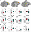
Postnatal ZIKV infection alters corpus callosum and cingulate gyrus micro-oFigure 6

Postnatal ZIKV infection alters corpus callosum and cingulate gyrus micro-organization. (A) Diffusion tensor images of white matter tracts of corpus callosum dorsal segment 1 (cc1d), cingulum (cg), and fornix visualized by projection onto a gray matter surface in DTI atlas space. (B) Fractional anisotropy (FA), axial diffusivity (AD), mean diffusivity (MD), and radial diffusivity (RD) of ZIKV-infected versus control infants for cc1d. (C) FA, AD, MD, and RD of ZIKV-infected versus control infants for the right cingulum. (D) FA, AD, MD, and RD of ZIKV-infected versus control infants for the right fornix. Two-factor ANOVA was used to investigate effects of group (ZIKV and control) and interaction term on FA, AD, RD, and MD. Boxes represent the interquartile range (25th-75th percentiles), with the central line indicating the median and whiskers extending to the minimum and maximum values.
Discussion

The specific set of ZIKV-induced changes in the CNS transcriptome that we report is highly suggestive of impaired neurodevelopment following early-life ZIKV infection. Results of neuroimaging and behavioral testing performed in this study and prior research show longer-term brain structural and functional abnormalities resulting from postnatal exposure to ZIKV (27, 28). Human infants and children infected with ZIKV do not display the more severe anatomic deformities that encompass congenital ZIKV syndrome (59, 60), though developmental disabilities have been noted (17), similar to reports of normocephalic newborns with congenital exposure (61, 62). Here, using ZIKV infection of 4-week-old rhesus macaque infants, we demonstrate massive divergence from uninfected control macaques, with aberrant microglial activation, transcriptomic evidence of neuronal apoptosis, and reduced expression of genes governing neurodevelopment, as well as altered oligodendrocyte maturation and disorganized myelin tracts.

The ZIKV outbreak of 2015–2016 in Central and South America is believed to have resulted in over 700,000 infections (63). With ZIKV now endemic in many countries, ongoing infection is assured, although detection outside of pregnancy is more variable. With this backdrop and considering the low to middle income status of countries with ZIKV in circulation, an epidemiologic assessment of the onset of developmental disability following early-life ZIKV infection is next to impossible. Prospective cohort and observational studies have shown delayed neurodevelopment in some countries but not others (17–21). Additionally, the multiple descriptions of congenital ZIKV infection resulting in postnatal onset of microcephaly (61, 62, 64) further reinforce the dynamic and critical nature of early-life brain development. Therefore, the nonhuman primate model employed here provides an important system to better understand the consequences of ZIKV infection in infancy.

We have previously shown virus dissemination into the central and peripheral nervous system by 2 weeks after infection using PCR and RNAscope (28). It is likely that direct infection plays a role in the negative outcomes of ZIKV infection, but the consequences of the antiviral immune response and immune activation may also be substantial. To that end, we observed that postnatal ZIKV infection induced a subset of activated microglia characterized by upregulation of transcription factors involved in interferon signaling, leading to increased expression of interferon-stimulated genes including IFI6, IFI27, and MX1. A similar immune activation signature is seen in infection with other neurotropic viruses, such as West Nile, dengue, herpes simplex, and HIV (65, 66), where neuroinflammation causes substantial pathology. The presence of microglia has also been associated with a neuroprotective effect in human brain organoids, possibly through interferon-mediated reduction in ZIKV replication as well as phagocytosis of apoptotic infected cells (67). A role for type I interferon, specifically IFN-β, in limiting apoptosis in ZIKV-infected cells in human fetal brain explants has also recently been proposed (68).

In congenital ZIKV infection, virus-triggered apoptosis of fetal intermediate progenitor cells has been associated with ventricular enlargement and microcephaly (5–9). We did not identify these neural progenitor cells in our single-cell preparations, likely because of the age of the macaques at the time of necropsy. However, we uncovered an apoptosis signature in both excitatory and inhibitory neurons, with activation of caspase-3 observed in hippocampal neurons. Collectively, these findings indicate that loss of neurons in the developing brain is a hallmark of both congenital ZIKV syndrome and postnatal ZIKV infection and that innate immune activation may both limit infection and promote immunopathology. Others have identified a transcriptional shift from neurogenesis to gliogenesis in ZIKV-infected neurons in the fetal explant system (68), and we have noted gliosis and glial nodules as common findings in postnatal ZIKV infection (28).

With regard to linking gene expression data to potential outcomes, we noted downregulation of genes expressed by neurons that are known to be associated with ASD risk (41). The specificity of this result is highlighted by the fact that CP gene sets were not differentially expressed in ZIKV-infected macaques and that only neurons exhibited the ASD transcriptional program. Together with the observation of increased emotional reactivity on behavioral testing and our prior finding of abnormal social interactions (27), these data support a link between altered socioemotional behavior and the specific genes linked to increased ASD risk. Thus, we postulate that there is potential for an ASD-like neurodevelopmental disorder to arise in some individuals exposed to ZIKV in early life. Poor personal-social performance on a standardized developmental screening tool has in fact been documented in children with ZIKV exposure (17). Our data cannot distinguish between ASD-specific mechanisms and overlap with ASD gene programs; we postulate that the ZIKV-induced gene expression changes in neurons reflect an ASD-like delay in neuronal maturation. Our analysis was limited by the nature of RNA sequencing that permits an understanding of the relative levels of gene expression but not specific variants or epigenetic factors, and it should be recognized that many genes linked to ASD risk are not merely downregulated but instead have mutations or are loss-of-function variants (69, 70).

Investigation of white matter tracts by magnetic resonance DTI in 3-month-old macaques demonstrated the ongoing impact of prior ZIKV infection at 1 month of age. Somewhat surprisingly, given the downregulation of oligodendrocyte metabolism and differentiation genes at 2 weeks after infection, at 3 months ZIKV-infected infants had evidence of accelerated white matter maturation in the cingulum, fornix, and corpus callosum. Increased FA, as we found with ZIKV, can be also seen in white matter tracts following preterm birth (71–73) and in adolescents with severe perinatal brain injury (74) as well as in the settings of mild cognitive impairment (75, 76) and mild traumatic brain injury (77). One interpretation of these findings is that a repair-driven compensatory response to early-life insult (viral or otherwise) results in disorganized myelination of white matter and/or enhanced remyelination following oligodendrocyte loss (78). A caveat is that, given the need to sacrifice macaques for single-cell analysis of the brain, the magnetic resonance DTI results were obtained using a different cohort of macaques; thus, links between the transcriptional changes and imaging findings are indirect. Intriguingly, in a pig-tailed macaque model of congenital infection, ZIKV was associated with downregulation of genes regulating myelination and myelin decompaction at late fetal gestation in the deep white matter (79). Ex vivo cultures from Ifnar1-knockout mice showed diminished myelin and axons, and in vivo perinatal infection of these immunodeficient mice confirmed direct infection and loss of myelinating oligodendrocytes (80, 81). Thus, age at infection, immune status, and duration of follow-up after infection are important factors in interpreting the structural implications of differential gene expression, particularly in light of the remarkable plasticity of the brain.

There were limitations to this work. First, a limited number of animals were subjected to transcriptomic analysis. This limitation was partially mitigated by the large number of single cells studied and analysis of 3 separate brain regions. ZIKV infection led to relatively consistent changes across amygdala, hippocampus, and striatum, which may be related to global viral impact and/or technical limitations with single-cell preparations that may cause data homogenization. Future studies using spatial transcriptomics approaches would allow for in situ gene expression profiling with a deeper examination of regional cell organization and interaction. In addition, we note that studying gene expression does not capture the impact of epigenetic and posttranslational modifications that influence protein activity. Multiomic technologies coupled with longer-term behavioral and cognitive assessments will likely provide a more complete understanding of postnatal ZIKV infection.

Using a clinically relevant infant nonhuman primate model, this research provides mechanistic insight into the cells and gene signatures that are modulated by ZIKV infection with indications as to how this response disrupts the typical neurodevelopmental trajectory. We show that postnatal ZIKV infection of 4-week-old rhesus macaques has a distinct, nuanced, and mutable impact on the brain, underscoring the potential vulnerability of newborns and infants with ZIKV exposure.

Methods

Sex as a biological variable

Thirteen male and 17 female infant Indian rhesus macaques (Macaca mulatta) were included in this study. Sex was considered as a biological variable for MRI data.

Animals

Fifteen infants were infected with ZIKV at 4 weeks of age, and 15 infants served as age-, sex-, and rearing-matched uninfected controls. The infants were delivered naturally by their dams while housed in indoor/outdoor social groups at the Emory National Primate Research Center (ENPRC). All dams were from the specific pathogen–free colony (negative for herpes B, SIV, simian retrovirus, and simian T-lymphotropic virus type 1), had not been previously used for infectious disease or vaccine studies, and did not have any clinical signs of infection during pregnancy. Infants were then removed from their dams at 12–16 days of age and transported to the ENPRC nursery facility. All infants rotated daily between being singly housed or pair-housed in warming incubators for the first 3–4 weeks. When singly housed, infants had visual and auditory contact with conspecifics. Infants were fed formula by hand every 2–3 hours for the first month, then via self-feeders for the remainder of the study, per standard ENPRC protocol. Soft blankets, plush toys, or fleece were provided and changed daily. Soft chow (Purina Primate Chow, Old World Monkey formulation) and fruits were introduced starting at 1 month of age. Water was provided ad libitum. At 5 weeks of age, infants were transitioned into age-appropriate caging and housed in trios including full tactile, visual, and auditory contact with conspecifics. During the ZIKV-infected period, infants were housed in an animal biosafety level 2 enhanced room until virus was no longer detected in blood or urine. All housing was indoors on a 12-hour light/12-hour dark cycle.

Virus and infection

The ZIKV of Puerto Rican origin (PRVABC59, GenBank accession number KU501215.1) used in this study had been passaged 4 times and titrated on Vero cells. Experimental infections were performed via the subcutaneous route using 1 × 10⁵ plaque-forming units of ZIKV PRVABC59. One ZIKV-infected infant rhesus macaque died at 11 days after infection and had to be excluded from scRNA-seq analysis, although tissues were preserved for histologic examination. The cause of death was determined to be lung congestion due to pneumonia. PCR of lung tissues was negative for SARS-CoV-2 and positive for ZIKV.

ZIKV-RNA detection by quantitative reverse transcription PCR

Total RNA was extracted from 140 μL of plasma using the QIAamp Viral RNA Mini Kit (QIAGEN). RNA from urine was isolated using the QIAamp Circulating Nucleic Acid Kit (QIAGEN). ZIKV-RNA standard was generated by annealing of 2 oligonucleotides spanning the target ZIKV prM-E gene region and performing of in vitro transcription using the MEGAscript T3 Transcription Kit (Ambion). Purified RNA was reverse-transcribed using a High-Capacity cDNA Reverse Transcription Kit (Applied Biosystems) and random hexamers. For quantitation of viral RNA, a standard curve was generated using 10-fold dilutions of ZIKV-RNA standard, and quantitative reverse transcription PCR was performed using TaqMan Gene Expression Master Mix (Applied Biosystems) and ZIKV primers (1 μM) and probe (250 nM). The ZIKV primer-probe set targeting the prM-E gene region included forward primer ZIKV/PR 907 (5′-TTGGTCATGATACTGCTGATTGC-3′), reverse primer ZIKV/PR 983c (5′-CCTTCCACAAAGTCCCTATTGC-3′), and probe ZIKV/PR 932 FAM (5′-CGGCATACAGCATCAGGTGCATAGGAG-3′). The probe was labeled with 6-carboxyfluorescein (FAM) at the 5′ end and 2 quenchers, an internal ZEN quencher and 3′ end Iowa Black FQ (Integrated DNA Technologies). The standard curve had R2 value greater than 0.99. ZIKV RNA copies were interpolated from the standard curve using the sample Ct value and are represented as copies per milliliter of plasma.

Neurobehavioral assessment

Neurobehavior was evaluated with a validated assessment developed for infant macaques, the Infant Neurobehavioral Assessment Scale (INAS), adapted from Schneider and Suomi (82–84), which is based on the human Brazelton Newborn Behavioral Assessment Scale (85). Twenty-six test items in the INAS aligned with the neurodevelopmental areas of interest and make up the orientation, motor maturity and activity (ability), neuromotor (sensory) responsiveness, and state control constructs (Supplemental Table 17). This neurobehavioral test has been used to define neonatal development of prenatally ZIKV-exposed infants (86, 87). Ratings were based on a 5-point Likert scale ranging from 0 to 2. The INAS was administered weekly between 3 and 6 weeks of age (Supplemental Table 18). INAS testing was performed at 6 days after infection to avoid potential confounders from handling or sedation and to minimize the potential influence of peak viremia on behavior. Before the study began, 3 examiners trained in standardized INAS administration and scoring procedures, achieving inter-rater reliability greater than 90% and intra-rater reliability greater than 96%. Two examiners were blinded. The third examiner could not be blinded and thus assisted only with task administration. Items were administered in a consistent sequence to optimize performance and decrease handling time. Assessments were hand-scored on a printout of the scoring form. Higher orientation, motor, and neuromotor scores reflect the optimal condition, whereas higher state control scores reflect poor emotional regulation, higher reactivity, and less consolability.

Brain dissection for transcriptomics and histology

Fourteen days after infection, 5 animals received an overdose of pentobarbital and were transcardially perfused with phosphate buffer solution (0.2 M, pH 7.4). After removal of the brain from the skull, the right and left hemispheres were immediately dissected (within 5 minutes). The left hemisphere was placed in 4% paraformaldehyde until further processing, then blocked into 2 cm slabs, cryoprotected in graded buffered sucrose solutions (10%–30%), and frozen in isopentane. Blocks of tissue were then systematically sectioned (50 μm) in 10 parallel series with the first series for cresyl violet staining and the remaining 9 series placed into antigen preserve (1% polyvinyl pyrrolidone, 50% ethylene glycol in 0.1 M PBS, pH 7.4) (88, 89). The right hemisphere was systematically dissected to remove the entire amygdala, caudate nucleus, and hippocampus for transcriptomic analysis.

Histologic analysis

Immunohistochemistry. Sections were washed 3 times in PBS and pretreated with 20% methanol/3% hydrogen peroxide in 0.1 M PBS for 30 minutes at room temperature. Sections were washed in 0.1 M PBS, blocked with 3% normal horse serum in PBS for 1 hour, and washed. For single labeling, sections were incubated overnight with rabbit anti–Iba-1 (1:1,000; Wako 019-19741), rabbit anti-nestin (1:1,000; Millipore ABD69), or rabbit anti–cleaved caspase-3 (1:500 in PBS/0.3% Triton X-100; Cell Signaling Technology 9579). For double labeling, sections were incubated overnight in blocking solution with rabbit anti–cleaved caspase-3 (1:500 in PBS/0.3% Triton X-100; Cell Signaling Technology 9579) and mouse anti-NeuN (1:1,000; MilliporeSigma MAB377). The following day, sections were washed in PBS. Single-labeled sections were incubated with biotinylated horse anti-rabbit IgG (1:200; Vector Laboratories BA-1100), washed, incubated with ABC (Vector Laboratories PK6100), and visualized with diaminobenzidine (Sigma-Aldrich). Double-labeled sections were incubated with goat anti-rabbit Alexa Fluor 488 (1:200; Sigma-Aldrich SAB4600036) or goat anti-mouse Alexa Fluor 555 (1:200; Sigma-Aldrich SAB4600060) and washed in PBS. Single-labeled sections were mounted on gelatin-coated slides, dehydrated through graded alcohols, cleared in xylenes, and coverslipped with Permount mounting medium (Fisher SP15-500). Double-labeled sections were mounted on gelatin-coated slides, dehydrated through graded alcohols, and coverslipped with DPX mounting medium (Electron Microscopy Sciences 13512). Negative-control sections processed without primary antibody showed no staining.

Quantification of caspase-3+ cells. Caspase-3+ cells were quantified using the Olympus cellSens Software program. Areas of interest were outlined and caspase-3+ cells were quantified under a ×40 objective.

StainAI. The StainAI model has been trained to detect 6 activation classes corresponding to ramified, hypertrophic, bushy, ameboid, rod-shaped, and hypertrophic rod-shaped microglial cells (90). Iba-1–immunostained sections were scanned at ×20 (0.37 μm/pixel) and analyzed using previously published methods (90). Briefly, a YOLO+UNet pipeline detected and segmented microglia. YOLOv5 detected Iba-1–stained microglial somata and processes, and 119 × 119 μm2 bounding boxes were superimposed on each cell for UNet segmentation of cell bodies and processes. UNet-generated cell masks were classified using a C5.0 decision tree based on 28 morphometric parameters: 6 area, 5 perimeter, 6 span-length, 4 span-length ratio, 2 circularity, 3 fractal, and 2 intercellular-distance parameters. Single-cell masks were color-coded as ramified (green), hypertrophic (yellow), bushy (orange), ameboid (red), rod-shaped (cyan), or hypertrophic rod-shaped (blue). Out-of-focus cells or cells smaller than 30 μm2 were grayed out and excluded. MATLAB (MathWorks, Inc) was used to define regions of interest (ROIs) for quantification of microglial phenotypes from the YOLO+UNet pipeline (90).

Design-based stereology. Pyramidal neuronal populations of the CA1–CA3 fields of the hippocampus were quantified as previously described (91). Every 30th cresyl violet section was sampled. Subfields were identified using a ×4 objective, and 1,600 μm3 counting frames were generated in MicroBrightField Stereo Investigator. Standard x-y grids (CA1, 500 × 500 μm; CA2, 250 × 250 μm; CA3, 350 × 350 μm) were used, and neurons were counted at ×100 (1.3 NA). For nestin-positive neurons, the dentate gyrus granular/subgranular region was delineated at ×10, 1,600 μm3 counting frames were generated in Stereo Investigator, a 100 × 100 μm x-y grid was applied, and neurons were counted at ×100 (1.3 NA). Myelin was stained with 0.2% gold chloride (92). Sections were removed from antigen preserve, washed in PBS, mounted on gelatin-coated slides, dried overnight, incubated in 0.2% gold chloride for 2 hours at room temperature, washed in 3 changes of water, fixed in 2.5% sodium thiosulfate for 5 minutes, and rinsed under running tap water for 30 minutes. Sections were dehydrated through graded alcohols (50%–95%), cleared in xylenes, and coverslipped with Permount. Images were acquired at ×200 with an Olympus microscope, DP23 camera, and cellSens software and analyzed using ImageJ/Fiji (NIH). For fine fibers, images were converted to 8-bit and thresholded to isolate particles, and ROI was selected. The Fit Rectangle tool standardized counting-area size, followed by Analyze Particles to quantify myelin density; particle outlines and counts were generated for each sample. For larger fiber bundles (corpus callosum, fornix, and cingulum), ROI was selected and mean density measured. Embedded scale bars were used to calibrate pixels to microns.

Tissue dissociation and single-cell sorting

All dissociation steps were performed in the presence of Protector RNase inhibitor (Sigma-Aldrich), the transcription inhibitor actinomycin D (Sigma-Aldrich, catalog A1410), and the translation inhibitor anisomycin (Sigma-Aldrich, catalog A9789) (93). Tissue was dissociated into a single-cell suspension using previously published methods (94). Briefly, tissue was chopped using a scalpel and incubated in papain (20 U/mL) at 34°C for 45 minutes before rinsing with a protease inhibitor solution (ovomucoid) and mechanical trituration. Dissociated cells were incubated with 50 nM calcein-AM and 4 μM ethidium homodimer-1 from the LIVE/DEAD Viability/Cytotoxicity Kit (Thermo Fisher Scientific) before cell sorting. Live (calcein-positive, ethidium-negative) cells were sorted on a BD FACSAria II into 4% BSA with RNase inhibitor.

Single-cell RNA sequencing

Cell suspensions were loaded onto the 10x Genomics Chromium Controller using the Chromium NextGEM Single Cell 5′ Library & Gel Bead kit to capture individual cells and barcoded gel beads within droplets for reverse transcription (95). Libraries were prepared according to manufacturer instructions and sequenced on an Illumina NovaSeq 6000 with a paired-end 26 × 91 configuration targeting a depth of 50,000 reads per cell. Cell Ranger software (10x Genomics) was used to perform demultiplexing of cellular transcript data, and mapping and annotation of unique molecular identifiers (UMIs) and transcripts for downstream data analysis.

Bioinformatics pipeline

Alignment, filtering, barcode counting, and UMI counting were performed with Cell Ranger v6.0.2 using a reference built from a composite of the rhesus Mmul10 genome with Ensembl annotation v100. scRNA-seq data were analyzed using the Seurat package (v4.0.0) in R (v2024.09.0+375). For quality control, cells with less than 500 detected features or with more than 20% of reads mapping to mitochondrial genes or less than 200 detected features per cell were excluded. After filtering, datasets were normalized, and variable features were identified using variance-stabilizing transformation method. Metadata annotations for each Seurat object included animal ID, brain region, and treatment group, with additional metadata added post hoc to ensure consistent labeling across samples. Following preprocessing, all individual datasets were integrated using Seurat’s integration workflow and scaled, and principal component analysis was performed. Dimensionality reduction was conducted using uniform manifold approximation and projection (UMAP). Cell clustering was performed at multiple resolution parameters (0.1–0.5) to explore clustering granularity. UMAP projections were visualized, and cell type identities were assigned based on canonical marker gene expression. A total of 105,421 control and 94,975 ZIKV-infected cells were successfully captured and profiled.

Diffusion MRI data acquisition and processing

Diffusion-weighted imaging (DWI) scans were acquired on a 3T Siemens Trio scanner at the ENPRC Imaging Center using an 8-channel phase array coil following published protocols (96). Subjects were scanned supine under 0.8%–1% isoflurane anesthesia using a head holder with ear bars and mouthpiece to avoid motion artifacts. A vitamin E capsule was taped on the right temple to identify the right hemisphere. Animals were intubated, given dextrose/NaCl (i.v.) for hydration, and placed on an MRI-compatible heating pad to maintain body temperature. Physiologic measures were monitored and maintained throughout scans following ENPRC and IACUC protocols. After scanning and complete recovery from anesthesia, subjects were returned to home cages. DWI scans were acquired with a single-shot double-spin-echo diffusion-weighted echo-planar imaging sequence (voxel size 1.0 × 1.0 × 1.0 mm3, b = 0 and 1,000 s/mm2Y, 128 directions).

Preprocessing of DWI images included automatic removal of artifact-rich images using DTIPrep (97), susceptibility-related distortion correction, eddy current, and motion correction using topup (98) and eddy_openmp tools (99). Weighted least-squares estimation was used to produce a diffusion tensor image for each subject. Multi-atlas templates were used to create brain masks for subjects using deformable registration (100) and majority voting. DTI images were skull stripped, and a study-specific atlas was built using DTI Atlas Builder (101). Fifteen fiber tracts — corpus callosum dorsal segment 1 (cc1d), corpus callosum dorsal segment 2 (cc2d), corpus callosum dorsal segment 3 (cc3d), genu of corpus callosum (ccg), splenium of corpus callosum (ccs), cingulum left and right (cg_l, cg_r), cortico-spinal left and right (cst_l, cst_r), inferior longitudinal left and right (ilf_l, ilf_r), uncinate left and right (uf_l, uf_r), and fornix left and right (fornix_l, fornix_r) — were propagated into a study-specific atlas from the high-resolution DTI rhesus macaque atlas. Propagated tracts were voxelized and used as seed maps for fiber tractography. Tractography was performed automatically using Autotract (102), followed by manual inspection to further clean tract definitions. Processed tracts from the study-specific atlas were then mapped back into each subject’s DTI image space using deformation fields calculated during the atlas building stage. Fractional anisotropy (FA), axial diffusivity (AD), mean diffusivity (MD), and radial diffusivity (RD) were sampled along the length of each tract’s arc using DTI Atlas Fiber Analyzer (101). These process steps yielded diffusion profiles for each tract, and mean FA, AD, MD, and RD per tract were computed for the final analyses.

Head coil change adjustment

During data acquisition, the MRI head coil became inoperative and was replaced with the same model. The new coil led to significantly increased FA values. Given the small sample size, we adjusted for coil effect using mean shift method instead of using coil as an additional covariate in the analysis. We randomly selected 2 male and 2 female subjects from both ZIKV and control groups scanned before and after coil change (8 scans before coil change and 8 after). This approach was chosen to avoid overparameterization, particularly because coil assignment was not fully independent of sex distribution in the sample. For each white matter fiber tract, we calculated mean FA, AD, RD, and MD values obtained using both coils. We computed differences in these values and subtracted them from data obtained using the newer coil.

Statistics

To determine whether infants randomly assigned to each group differed at baseline, physical measurements were analyzed using 2-tailed independent-samples t test and Cohen’s d effect size. Potential neurobehavioral differences were assessed by analysis of the 4 INAS scores using linear mixed models with group and age as fixed factors and individual animal as a random factor. Results were graphed using Prism 10.01 (GraphPad Software). Behavioral data were analyzed using SPSS 29 for Windows (IBM). Significance was set at a P value of less than 0.05, and effect sizes were calculated as ηp2.

Differential gene expression between ZIKV-infected and control animals was analyzed at the single-cell level using Seurat (FindMarkers) with MAST framework and treatment as grouping variable. P values were Bonferroni adjusted, with an adjusted P value of less than 0.05 defining significantly differentially expressed genes. Gene Ontology (GO) enrichment analysis of filtered gene sets was performed using the g:Profiler tool (https://biit.cs.ut.ee/gprofiler/gost).

Gene set enrichment analysis (GSEA) was performed using the fgsea R package (v1.30.0) to assess enrichment of curated custom gene sets among transcriptional changes between ZIKV-infected and control animals. Seurat differential expression results were ranked by average log2 fold change and compared with custom gene sets using fgsea with 1,000 permutations and fixed seed for reproducibility. Normalized enrichment score (NES), enrichment score, and nominal P values were reported.

Data were analyzed using 2-way analysis of variance (ANOVA) with group (Zika vs. control) and sex (male vs. female) as between-subjects factors. For each tract/region, ANOVA models tested main effects of group and sex. Where appropriate, Tukey’s post hoc multiple comparisons examined group differences while controlling the family-wise error rate. Model assumptions were assessed by residual inspection and Levene’s test for homogeneity of variances. Sums of squares, degrees of freedom, F statistics, and P values were reported for each effect.

Study approval

This study was conducted in strict accordance with US Department of Agriculture regulations and recommendations in the NIH Guide for the Care and Use of Laboratory Animals (National Academies Press, 2011), approved by the Emory University Institutional Animal Care and Use Committee, and conducted in an Association for Assessment and Accreditation of Laboratory Animal Care International–accredited facility.

Data availability

All data generated during this study are included in this published article and the Supporting Data Values file. Single-cell RNA-seq data are available through the NCBI’s Gene Expression Omnibus database under accession number GSE342497. Code for scRNA-seq analysis will be shared upon request.

All unique reagents, including available nonhuman primate samples, generated from this study are available from the corresponding author with a completed material transfer agreement. Requests for further information and for resources and reagents should be directed to, and will be fulfilled by, the corresponding author.

Manuscript information

Author contributions. Conceptualization: SAS, JR, and AC. Investigation: animal care, infection, neurobehavioral assessment, and brain dissection: SF, RR, and JR. ZIKV-RNA detection by PCR: NS, KM, DB, and MSS. Histologic analysis: AH, YMA, SA, EN, AM, CHH, TWT, ST, and MB. Tissue dissociation and single-cell sorting: M Sampson and SAS. Single-cell RNA sequencing and bioinformatics pipeline: MJL, GT, and SB. Diffusion MRI data acquisition and processing: JR, M Styner, and RV. Statistical analysis: VVE, RV, JR, and AC. Writing: original draft: VVE, SAS, RV, MB, MJL, GT, MMS, JR, and AC. Writing: review and editing: VVE, SAS, RV, MB, M Sampson, KM, MMS, JR, and AC.

AI disclosure. The authors used ChatGPT (OpenAI; model versions GPT-4 and GPT-5) between October 2024 and April 2026 to assist with R coding, script optimization, and troubleshooting for single-cell RNA sequencing data analysis, including Seurat, edgeR and pseudobulk workflows. AI assistance was limited to computational coding support. All analytical outputs were reviewed and validated by the authors, who retained responsibility for the accuracy and interpretation of the results.

Conflict of interest. The authors have declared that no conflict of interest exists.

Funding. This study was supported by the following:

  • NIH grant R01NS120182 (to AC and JR).
  • Additional funding from institutional NIH grants P51OD011132 and U42OD011023 supporting the Emory National Primate Research Center (ENPRC).
  • Next-generation sequencing services were provided by the ENPRC Genomics Core (RRID:SCR_026418), which is supported in part by NIH P51OD011132.
  • Sequencing data were acquired on an Illumina NovaSeq 6000 funded by NIH S10OD026799.

This work is the result of NIH funding, in whole or in part, and is subject to the NIH Public Access Policy. Through acceptance of this federal funding, the NIH has been given a right to make the work publicly available in PubMed Central.

Supplemental material

View Supplemental data

View Supporting data values

Acknowledgments

We thank the Emory National Primate Research Center veterinarians and Division of Research Resources staff for their expert assistance.

Address for correspondence. Ann Chahroudi, Health Science Research Building, Suite N447, 1750 Haygood Drive NE, Atlanta, Georgia 30322, USA. Email: ann.m.chahroudi@emory.edu.

Footnotes

Copyright: © 2026, Edara et al. This is an open access article published under the terms of the Creative Commons Attribution 4.0 International License.

Reference information: JCI Insight. 2026;11(21):e203088.https://doi.org/10.1172/jci.insight.203088.

References
  1. Tang H, et al. Zika virus infects human cortical neural progenitors and attenuates their growth. Cell Stem Cell. 2016;18(5):587–590.
    View this article via: CrossRef PubMed Google Scholar
  2. Cugola FR, et al. The Brazilian Zika virus strain causes birth defects in experimental models. Nature. 2016;534(7606):267–271.
    View this article via: CrossRef PubMed Google Scholar
  3. Adams Waldorf KM, et al. Congenital Zika virus infection as a silent pathology with loss of neurogenic output in the fetal brain. Nat Med. 2018;24(3):368–374.
    View this article via: CrossRef PubMed Google Scholar
  4. Lin MY, et al. Zika virus infects intermediate progenitor cells and post-mitotic committed neurons in human fetal brain tissues. Sci Rep. 2017;7(1):14883.
    View this article via: CrossRef PubMed Google Scholar
  5. Dang J, et al. Zika virus depletes neural progenitors in human cerebral organoids through activation of the innate immune receptor TLR3. Cell Stem Cell. 2016;19(2):258–265.
    View this article via: CrossRef PubMed Google Scholar
  6. Souza BSF, et al. Zika virus infection induces mitosis abnormalities and apoptotic cell death of human neural progenitor cells. Sci Rep. 2016;6(1):39775.
    View this article via: CrossRef PubMed Google Scholar
  7. Hanners NW, et al. Western Zika virus in human fetal neural progenitors persists long term with partial cytopathic and limited immunogenic effects. Cell Rep. 2016;15(11):2315–2322.
    View this article via: CrossRef PubMed Google Scholar
  8. Li C, et al. Zika virus disrupts neural progenitor development and leads to microcephaly in mice. Cell Stem Cell. 2016;19(1):120–126.
    View this article via: CrossRef PubMed Google Scholar
  9. Shao Q, et al. Zika virus infection disrupts neurovascular development and results in postnatal microcephaly with brain damage. Development. 2016;143(22):4127–4136.
    View this article via: CrossRef PubMed Google Scholar
  10. Walker CL, et al. Zika virus and the nonmicrocephalic fetus: why we should still worry. Am J Obstet Gynecol. 2019;220(1):45–56.
    View this article via: CrossRef PubMed Google Scholar
  11. Rasmussen SA, et al. Zika virus and birth defects—reviewing the evidence for causality. N Engl J Med. 2016;374(20):1981–1987.
    View this article via: CrossRef PubMed Google Scholar
  12. Moore CA, et al. Characterizing the pattern of anomalies in congenital Zika syndrome for pediatric clinicians. JAMA Pediatr. 2017;171(3):288–295.
    View this article via: CrossRef PubMed Google Scholar
  13. Oehler E, et al. Zika virus infection complicated by Guillain-Barré syndrome—case report, French Polynesia, December 2013. Euro Surveill. 2014;19(9):20720.
    View this article via: CrossRef PubMed Google Scholar
  14. Pan American Health Organization. Epidemiological Update, Neurological syndrome, congenital anomalies, and Zika virus infection (17 January 2016). https://iris.paho.org/handle/10665.2/50659 Accessed September 24, 2026.
  15. Salgado DM, et al. Clinical, laboratory and immune aspects of Zika virus-associated encephalitis in children. Int J Infect Dis. 2020;90:104–110.
    View this article via: CrossRef PubMed Google Scholar
  16. Raper J, Chahroudi A. Clinical and preclinical evidence for adverse neurodevelopment after postnatal Zika virus infection. Trop Med Infect Dis. 2021;6(1):10.
    View this article via: CrossRef PubMed Google Scholar
  17. Pacheco O, et al. Neurodevelopmental findings in children 20-30 months of age with postnatal Zika infection at 1-12 months of age, Colombia, September-November 2017. Paediatr Perinat Epidemiol. 2021;35(1):92–97.
    View this article via: CrossRef PubMed Google Scholar
  18. Lannuzel A, et al. Long-term outcome in neuroZika: when biological diagnosis matters. Neurology. 2019;92(21):e2406–e2420.
    View this article via: CrossRef PubMed Google Scholar
  19. Asturias EJ, et al. Postnatal Zika and dengue infection and their effects on neurodevelopment among children living in rural Guatemala. Pediatr Infect Dis J. 2025;44(4):290–298.
    View this article via: CrossRef PubMed Google Scholar
  20. Lebov JF, et al. Cohort study: neurological and cognitive-behavioral sequelae of acquired Zika virus infection among Nicaraguan children. Pediatr Res. 2025;97(2):687–694.
    View this article via: CrossRef PubMed Google Scholar
  21. Lebov JF, et al. Neurological and neuropsychological sequelae of Zika virus infection in children in León, Nicaragua. Rev Panam Salud Publica. 2022;46:e90.
    View this article via: CrossRef PubMed Google Scholar
  22. Gilmore JH, et al. Imaging structural and functional brain development in early childhood. Nat Rev Neurosci. 2018;19(3):123–137.
    View this article via: CrossRef PubMed Google Scholar
  23. Knickmeyer RC, et al. A structural MRI study of human brain development from birth to 2 years. J Neurosci. 2008;28(47):12176–12182.
    View this article via: CrossRef PubMed Google Scholar
  24. Ha S, et al. Characteristics of brains in autism spectrum disorder: structure, function and connectivity across the lifespan. Exp Neurobiol. 2015;24(4):273–284.
    View this article via: CrossRef PubMed Google Scholar
  25. Bast T. The hippocampal learning-behavior translation and the functional significance of hippocampal dysfunction in schizophrenia. Curr Opin Neurobiol. 2011;21(3):492–501.
    View this article via: CrossRef PubMed Google Scholar
  26. Medina A, et al. Treatment with sofosbuvir attenuates the adverse neurodevelopmental consequences of Zika virus infection in infant rhesus macaques. J Neuroimmunol. 2023;381:578148.
    View this article via: CrossRef PubMed Google Scholar
  27. Raper J, et al. Long-term alterations in brain and behavior after postnatal Zika virus infection in infant macaques. Nat Commun. 2020;11(1):2534.
    View this article via: CrossRef PubMed Google Scholar
  28. Mavigner M, et al. Postnatal Zika virus infection is associated with persistent abnormalities in brain structure, function, and behavior in infant macaques. Sci Transl Med. 2018;10(435):eaao6975.
    View this article via: CrossRef PubMed Google Scholar
  29. Workman AD, et al. Modeling transformations of neurodevelopmental sequences across mammalian species. J Neurosci. 2013;33(17):7368–7383.
    View this article via: CrossRef PubMed Google Scholar
  30. Zhu Y, et al. Spatiotemporal transcriptomic divergence across human and macaque brain development. Science. 2018;362(6420):eaat8077.
    View this article via: CrossRef PubMed Google Scholar
  31. Carryl H, et al. Of mice and monkeys: can animal models be utilized to study neurological consequences of pediatric HIV-1 infection? ACS Chem Neurosci. 2015;6(8):1276–1289.
    View this article via: CrossRef PubMed Google Scholar
  32. Davis EP, et al. Early life exposure to unpredictable parental sensory signals shapes cognitive development across three species. Front Behav Neurosci. 2022;16:960262.
    View this article via: CrossRef PubMed Google Scholar
  33. Fonseca JA, et al. More than the infinite monkey theorem: NHP models in the development of a pediatric HIV cure. Curr HIV/AIDS Rep. 2024;21(1):11–29.
    View this article via: CrossRef PubMed Google Scholar
  34. Li A, et al. Role of non-human primate models in accelerating research and developing countermeasures against Zika virus infection. Lancet Microbe. 2025;6(6):101030.
    View this article via: CrossRef PubMed Google Scholar
  35. Klein RS, et al. Neuroinflammation during RNA viral infections. Annu Rev Immunol. 2019;37:73–95.
    View this article via: CrossRef PubMed Google Scholar
  36. Sierra A, et al. Microglia shape adult hippocampal neurogenesis through apoptosis-coupled phagocytosis. Cell Stem Cell. 2010;7(4):483–495.
    View this article via: CrossRef PubMed Google Scholar
  37. Kettenmann H. Neuroscience: the brain’s garbage men. Nature. 2007;446(7139):987–989.
    View this article via: CrossRef PubMed Google Scholar
  38. Yassa MA, Stark CE. Pattern separation in the hippocampus. Trends Neurosci. 2011;34(10):515–525.
    View this article via: CrossRef PubMed Google Scholar
  39. Gilbert PE, et al. Memory for spatial location: role of the hippocampus in mediating spatial pattern separation. J Neurosci. 1998;18(2):804–810.
    View this article via: CrossRef PubMed Google Scholar
  40. Gemma C, Bachstetter AD. The role of microglia in adult hippocampal neurogenesis. Front Cell Neurosci. 2013;7:229.
    View this article via: CrossRef PubMed Google Scholar
  41. Courchesne E, et al. Prenatal origins of ASD: the when, what, and how of ASD development. Trends Neurosci. 2020;43(5):326–342.
    View this article via: CrossRef PubMed Google Scholar
  42. Dawson G, et al. Early social attention impairments in autism: social orienting, joint attention, and attention to distress. Dev Psychol. 2004;40(2):271–283.
    View this article via: CrossRef PubMed Google Scholar
  43. Fodstad JC, et al. Social and communication behaviours in infants and toddlers with autism and pervasive developmental disorder–not otherwise specified. Dev Neurorehabil. 2009;12(3):152–157.
    View this article via: CrossRef PubMed Google Scholar
  44. Matson JL, et al. The effects of inattention/impulsivity and ASD symptom severity on social skills in toddlers. Dev Neurorehabil. 2010;13(6):408–412.
    View this article via: CrossRef PubMed Google Scholar
  45. Elsabbagh M, et al. Social and attention factors during infancy and the later emergence of autism characteristics. Prog Brain Res. 2011;189:195–207.
    View this article via: PubMed CrossRef Google Scholar
  46. Courchesne E, et al. The ASD living biology: from cell proliferation to clinical phenotype. Mol Psychiatry. 2019;24(1):88–107.
    View this article via: CrossRef PubMed Google Scholar
  47. Canals J, et al. Neonatal behavioral assessment scale as a predictor of cognitive development and IQ in full-term infants: a 6-year longitudinal study. Acta Paediatr. 2011;100(10):1331–1337.
    View this article via: CrossRef PubMed Google Scholar
  48. Maxwell JR, et al. Abnormal neurobehavior profiles observed in the newborn period following low-to-moderate prenatal alcohol exposure. Alcohol Clin Exp Res (Hoboken). 2025;49(4):818–828.
    View this article via: CrossRef PubMed Google Scholar
  49. Shoaff JR, et al. Early infant behavioural correlates of social skills in adolescents. Paediatr Perinat Epidemiol. 2021;35(2):247–256.
    View this article via: CrossRef PubMed Google Scholar
  50. Sucharew H, et al. NICU network neurobehavioral scale profiles predict developmental outcomes in a low-risk sample. Paediatr Perinat Epidemiol. 2012;26(4):344–352.
    View this article via: CrossRef PubMed Google Scholar
  51. Bradl M, Lassmann H. Oligodendrocytes: biology and pathology. Acta Neuropathol. 2010;119(1):37–53.
    View this article via: CrossRef PubMed Google Scholar
  52. Dimas P, et al. CNS myelination and remyelination depend on fatty acid synthesis by oligodendrocytes. Elife. 2019;8:e44702.
    View this article via: CrossRef PubMed Google Scholar
  53. Mathews ES, Appel B. Cholesterol biosynthesis supports myelin gene expression and axon ensheathment through modulation of P13K/Akt/mTor signaling. J Neurosci. 2016;36(29):7628–7639.
    View this article via: CrossRef PubMed Google Scholar
  54. Bujalka H, et al. MYRF is a membrane-associated transcription factor that autoproteolytically cleaves to directly activate myelin genes. PLoS Biol. 2013;11(8):e1001625.
    View this article via: CrossRef PubMed Google Scholar
  55. Huang H, et al. Interactive repression of MYRF self-cleavage and activity in oligodendrocyte differentiation by TMEM98 protein. J Neurosci. 2018;38(46):9829–9839.
    View this article via: CrossRef PubMed Google Scholar
  56. Liu JY, et al. Inward rectifying Kir4.1 channels regulate oligodendrocyte precursor cell differentiation and CNS myelination in vivo. Neurosci Lett. 2023;807:137278.
    View this article via: CrossRef PubMed Google Scholar
  57. Beaulieu C. The basis of anisotropic water diffusion in the nervous system — a technical review. NMR Biomed. 2002;15(7-8):435–455.
    View this article via: CrossRef PubMed Google Scholar
  58. Liang H, et al. Abnormal brain diffusivity in participants with persistent neuropsychiatric symptoms after COVID-19. NeuroImmune Pharm Ther. 2023;2(1):37–48.
    View this article via: PubMed CrossRef Google Scholar
  59. Tolosa N, et al. Zika virus disease in children in Colombia, August 2015 to May 2016. Paediatr Perinat Epidemiol. 2017;31(6):537–545.
    View this article via: CrossRef PubMed Google Scholar
  60. Burger-Calderon R, et al. Age-dependent manifestations and case definitions of paediatric Zika: a prospective cohort study. Lancet Infect Dis. 2020;20(3):371–380.
    View this article via: CrossRef PubMed Google Scholar
  61. van der Linden V, et al. Description of 13 infants born during October 2015–January 2016 with congenital Zika virus infection without microcephaly at birth — Brazil. MMWR Morb Mortal Wkly Rep. 2016;65(47):1343–1348.
    View this article via: CrossRef PubMed Google Scholar
  62. Rice ME, et al. Vital signs: Zika-associated birth defects and neurodevelopmental abnormalities possibly associated with congenital Zika virus infection — U.S. territories and freely associated states, 2018. MMWR Morb Mortal Wkly Rep. 2018;67(31):858–867.
    View this article via: CrossRef PubMed Google Scholar
  63. Ikejezie J, et al. Zika virus transmission — region of the Americas, May 15, 2015–December 15, 2016. MMWR Morb Mortal Wkly Rep. 2017;66(12):329–334.
    View this article via: CrossRef PubMed Google Scholar
  64. Aragao MFVV, et al. Nonmicrocephalic infants with congenital Zika syndrome suspected only after neuroimaging evaluation compared with those with microcephaly at birth and postnatally: how large is the Zika virus “iceberg”? AJNR Am J Neuroradiol. 2017;38(7):1427–1434.
    View this article via: CrossRef PubMed Google Scholar
  65. Carty M, et al. Innate antiviral signalling in the central nervous system. Trends Immunol. 2014;35(2):79–87.
    View this article via: CrossRef PubMed Google Scholar
  66. Tang Y, et al. Persistent type I interferon signaling within the brain of people with HIV on ART with cognitive impairment. PLoS Pathog. 2025;21(8):e1013411.
    View this article via: CrossRef PubMed Google Scholar
  67. Abdelbasset M, et al. Differential contributions of fetal mononuclear phagocytes to Zika virus neuroinvasion versus neuroprotection during congenital infection. Cell. 2024;187(26):7511–7532.
    View this article via: CrossRef PubMed Google Scholar
  68. Stokes C, et al. The human neural cell atlas of Zika virus infection in developing brain tissue. Cell Rep Med. 2025;6(6):102189.
    View this article via: CrossRef PubMed Google Scholar
  69. Zhou X, et al. Integrating de novo and inherited variants in 42,607 autism cases identifies mutations in new moderate-risk genes. Nat Genet. 2022;54(9):1305–1319.
    View this article via: CrossRef PubMed Google Scholar
  70. Choi L, An JY. Genetic architecture of autism spectrum disorder: lessons from large-scale genomic studies. Neurosci Biobehav Rev. 2021;128:244–257.
    View this article via: CrossRef PubMed Google Scholar
  71. Groeschel S, et al. Identification and interpretation of microstructural abnormalities in motor pathways in adolescents born preterm. Neuroimage. 2014;87:209–219.
    View this article via: CrossRef PubMed Google Scholar
  72. Brossard-Racine M, et al. Cerebellar microstructural organization is altered by complications of premature birth: a case-control study. J Pediatr. 2017;182:28–33.
    View this article via: CrossRef PubMed Google Scholar
  73. Hollund IMH, et al. White matter alterations and their associations with motor function in young adults born preterm with very low birth weight. Neuroimage Clin. 2018;17:241–250.
    View this article via: CrossRef PubMed Google Scholar
  74. Allin MPG, et al. White matter and cognition in adults who were born preterm. PLoS One. 2011;6(10):e24525.
    View this article via: CrossRef PubMed Google Scholar
  75. Liu X, et al. Abnormal white matter integrity of anterior-inferior hypothalamus in mild cognitive impairment patients with depression symptoms. J Affect Disord. 2024;362:225–229.
    View this article via: CrossRef PubMed Google Scholar
  76. Huang C, et al. White matter connectivity in incident mild cognitive impairment: a diffusion spectrum imaging study of world trade center responders at midlife. J Alzheimers Dis. 2021;80(3):1209–1219.
    View this article via: CrossRef PubMed Google Scholar
  77. Li X, et al. Tract-specific white matter hyperintensities disrupt brain networks and associated with cognitive impairment in mild traumatic brain injury. Hum Brain Mapp. 2024;45(17):e70050.
    View this article via: CrossRef PubMed Google Scholar
  78. Traka M, et al. A genetic mouse model of adult-onset, pervasive central nervous system demyelination with robust remyelination. Brain. 2010;133(10):3017–3029.
    View this article via: CrossRef PubMed Google Scholar
  79. Tisoncik-Go J, et al. Disruption of myelin structure and oligodendrocyte maturation in a macaque model of congenital Zika infection. Nat Commun. 2024;15(1):5173.
    View this article via: CrossRef PubMed Google Scholar
  80. Cumberworth SL, et al. Zika virus tropism and interactions in myelinating neural cell cultures: CNS cells and myelin are preferentially affected. Acta Neuropathol Commun. 2017;5(1):50.
    View this article via: CrossRef PubMed Google Scholar
  81. Schultz V, et al. Oligodendrocytes are susceptible to Zika virus infection in a mouse model of perinatal exposure: implications for CNS complications. Glia. 2021;69(8):2023–2036.
    View this article via: CrossRef PubMed Google Scholar
  82. Schneider ML, Suomi SJ. Neurobehavioral assessment in rhesus monkey neonates (Macaca mulatta): developmental changes, behavioral stability, and early experience. Infant Behav Dev. 1992;15(2):155–177.
    View this article via: CrossRef Google Scholar
  83. Schneider ML, et al. The impact of prenatal stress, fetal alcohol exposure, or both on development: perspectives from a primate model. Psychoneuroendocrinology. 2002;27(1-2):285–298.
    View this article via: CrossRef PubMed Google Scholar
  84. Raper J, et al. Multiple anesthetic exposure in infant monkeys alters emotional reactivity to an acute stressor. Anesthesiology. 2015;123(5):1084–1092.
    View this article via: CrossRef PubMed Google Scholar
  85. Brazelton TB. Assessment of the infant at risk. Clin Obstet Gynecol. 1973;16(1):361–375.
    View this article via: CrossRef PubMed Google Scholar
  86. Ausderau K, et al. Neonatal development in prenatally Zika virus-exposed infant macaques with dengue immunity. Viruses. 2021;13(9):1878.
    View this article via: CrossRef PubMed Google Scholar
  87. Moadab G, et al. Prenatal Zika virus infection has sex-specific effects on infant physical development and mother-infant social interactions. Sci Transl Med. 2023;15(719):eadh0043.
    View this article via: CrossRef PubMed Google Scholar
  88. Burke MW, et al. Dissecting the non-human primate brain in stereotaxic space. J Vis Exp. 2009;29:1–5.
    View this article via: PubMed CrossRef Google Scholar
  89. Burke MW, et al. Brain banking: making the most of your research specimens. J Vis Exp. 2009;29:1260.
    View this article via: PubMed CrossRef Google Scholar
  90. Hsu CH, et al. StainAI: quantitative mapping of stained microglia and insights into brain-wide neuroinflammation and therapeutic effects in cardiac arrest. Commun Biol. 2025;8(1):462.
    View this article via: CrossRef PubMed Google Scholar
  91. Carryl H, et al. Hippocampal neuronal loss in infant macaques orally infected with virulent simian immunodeficiency virus (SIV). Brain Sci. 2017;7(4):40.
    View this article via: CrossRef PubMed Google Scholar
  92. Schmued LC. A rapid, sensitive histochemical stain for myelin in frozen brain sections. J Histochem Cytochem. 1990;38(5):717–720.
    View this article via: CrossRef PubMed Google Scholar
  93. Marsh SE, et al. Dissection of artifactual and confounding glial signatures by single-cell sequencing of mouse and human brain. Nat Neurosci. 2022;25(3):306–316.
    View this article via: CrossRef PubMed Google Scholar
  94. Zhang Y, et al. Purification and characterization of progenitor and mature human astrocytes reveals transcriptional and functional differences with mouse. Neuron. 2016;89(1):37–53.
    View this article via: CrossRef PubMed Google Scholar
  95. Zheng GX, et al. Massively parallel digital transcriptional profiling of single cells. Nat Commun. 2017;8:14049.
    View this article via: CrossRef PubMed Google Scholar
  96. Howell BR, et al. Disentangling the effects of early caregiving experience and heritable factors on brain white matter development in rhesus monkeys. Neuroimage. 2019;197:625–642.
    View this article via: CrossRef PubMed Google Scholar
  97. Oguz I, et al. DTIPrep: quality control of diffusion-weighted images. Front Neuroinform. 2014;8:4.
    View this article via: CrossRef PubMed Google Scholar
  98. Andersson JLR, et al. How to correct susceptibility distortions in spin-echo echo-planar images: application to diffusion tensor imaging. Neuroimage. 2003;20(2):870–888.
    View this article via: CrossRef PubMed Google Scholar
  99. Andersson JLR, Sotiropoulos SN. An integrated approach to correction for off-resonance effects and subject movement in diffusion MR imaging. Neuroimage. 2016;125:1063–1078.
    View this article via: CrossRef PubMed Google Scholar
  100. Avants BB, et al. The Insight ToolKit image registration framework. Front Neuroinform. 2014;8:44.
    View this article via: CrossRef PubMed Google Scholar
  101. Verde AR, et al. UNC-Utah NA-MIC framework for DTI fiber tract analysis. Front Neuroinform. 2014;7:51.
    View this article via: CrossRef PubMed Google Scholar
  102. Prieto JC, et al. Autotract: automatic cleaning and tracking of fibers. Proc SPIE Int Soc Opt Eng. 2016;9784:978408.
    View this article via: PubMed CrossRef Google Scholar
Version history
  • Version 1 (September 24, 2026): In-Press Preview
  • Version 2 (November 9, 2026): Electronic publication

Article tools

  • View PDF
  • Download citation information
  • Send a comment
  • Terms of use
  • Standard abbreviations
  • Need help? Email the journal

Metrics

  • Article usage
  • Citations to this article

Go to

  • Top
  • Abstract
  • Introduction
  • Results
  • Discussion
  • Methods
  • Manuscript information
  • Supplemental material
  • Acknowledgments
  • Footnotes
  • References
  • Version history
Advertisement
Advertisement

Copyright © 2026 American Society for Clinical Investigation
ISSN 2379-3708

Sign up for email alerts