
Research ArticleCell biologyDevelopmentNephrology
Open Access |  10.1172/jci.insight.203950
Loss of ADAMTS9 disrupts ciliogenesis and collagen homeostasis, resulting in nephronophthisis-like polycystic kidneys
Sydney Fischer,1 Karyn L. Robert,1 Manu Ahmed,1 Griffin I. Kane,2,3 Matthew A. Kavanaugh,4 Wei Wang,4 Pamela V. Tran,4 Prabhani U. Atukorale,2,3 and Sumeda Nandadasa1
1Department of Pediatrics, University of Massachusetts Chan Medical School, Worcester, Massachusetts, USA.
2Department of Biomedical Engineering, University of Massachusetts Amherst, Amherst, Massachusetts, USA.
3Division of Innate Immunity, Department of Medicine, University of Massachusetts Chan Medical School, Worcester, Massachusetts, USA.
4Department of Cell Biology and Physiology and the Jared Grantham Kidney Institute, University of Kansas Medical Center, Kansas City, Kansas, USA.
Address for correspondence. Sumeda Nandadasa, Department of Pediatrics, UMass Chan Medical School, 55 Lake Avenue North, S6-210 Medical School Building, Worcester, Massachusetts 01655, USA. Phone: 508.856.3962; Email: Sumeda.Nandadasa@Umassmed.edu.
Find articles by Fischer, S. in: PubMed | Google Scholar
1Department of Pediatrics, University of Massachusetts Chan Medical School, Worcester, Massachusetts, USA.
2Department of Biomedical Engineering, University of Massachusetts Amherst, Amherst, Massachusetts, USA.
3Division of Innate Immunity, Department of Medicine, University of Massachusetts Chan Medical School, Worcester, Massachusetts, USA.
4Department of Cell Biology and Physiology and the Jared Grantham Kidney Institute, University of Kansas Medical Center, Kansas City, Kansas, USA.
Address for correspondence. Sumeda Nandadasa, Department of Pediatrics, UMass Chan Medical School, 55 Lake Avenue North, S6-210 Medical School Building, Worcester, Massachusetts 01655, USA. Phone: 508.856.3962; Email: Sumeda.Nandadasa@Umassmed.edu.
Find articles by Robert, K. in: PubMed | Google Scholar
1Department of Pediatrics, University of Massachusetts Chan Medical School, Worcester, Massachusetts, USA.
2Department of Biomedical Engineering, University of Massachusetts Amherst, Amherst, Massachusetts, USA.
3Division of Innate Immunity, Department of Medicine, University of Massachusetts Chan Medical School, Worcester, Massachusetts, USA.
4Department of Cell Biology and Physiology and the Jared Grantham Kidney Institute, University of Kansas Medical Center, Kansas City, Kansas, USA.
Address for correspondence. Sumeda Nandadasa, Department of Pediatrics, UMass Chan Medical School, 55 Lake Avenue North, S6-210 Medical School Building, Worcester, Massachusetts 01655, USA. Phone: 508.856.3962; Email: Sumeda.Nandadasa@Umassmed.edu.
Find articles by Ahmed, M. in: PubMed | Google Scholar
1Department of Pediatrics, University of Massachusetts Chan Medical School, Worcester, Massachusetts, USA.
2Department of Biomedical Engineering, University of Massachusetts Amherst, Amherst, Massachusetts, USA.
3Division of Innate Immunity, Department of Medicine, University of Massachusetts Chan Medical School, Worcester, Massachusetts, USA.
4Department of Cell Biology and Physiology and the Jared Grantham Kidney Institute, University of Kansas Medical Center, Kansas City, Kansas, USA.
Address for correspondence. Sumeda Nandadasa, Department of Pediatrics, UMass Chan Medical School, 55 Lake Avenue North, S6-210 Medical School Building, Worcester, Massachusetts 01655, USA. Phone: 508.856.3962; Email: Sumeda.Nandadasa@Umassmed.edu.
Find articles by Kane, G. in: PubMed | Google Scholar
1Department of Pediatrics, University of Massachusetts Chan Medical School, Worcester, Massachusetts, USA.
2Department of Biomedical Engineering, University of Massachusetts Amherst, Amherst, Massachusetts, USA.
3Division of Innate Immunity, Department of Medicine, University of Massachusetts Chan Medical School, Worcester, Massachusetts, USA.
4Department of Cell Biology and Physiology and the Jared Grantham Kidney Institute, University of Kansas Medical Center, Kansas City, Kansas, USA.
Address for correspondence. Sumeda Nandadasa, Department of Pediatrics, UMass Chan Medical School, 55 Lake Avenue North, S6-210 Medical School Building, Worcester, Massachusetts 01655, USA. Phone: 508.856.3962; Email: Sumeda.Nandadasa@Umassmed.edu.
Find articles by Kavanaugh, M. in: PubMed | Google Scholar
1Department of Pediatrics, University of Massachusetts Chan Medical School, Worcester, Massachusetts, USA.
2Department of Biomedical Engineering, University of Massachusetts Amherst, Amherst, Massachusetts, USA.
3Division of Innate Immunity, Department of Medicine, University of Massachusetts Chan Medical School, Worcester, Massachusetts, USA.
4Department of Cell Biology and Physiology and the Jared Grantham Kidney Institute, University of Kansas Medical Center, Kansas City, Kansas, USA.
Address for correspondence. Sumeda Nandadasa, Department of Pediatrics, UMass Chan Medical School, 55 Lake Avenue North, S6-210 Medical School Building, Worcester, Massachusetts 01655, USA. Phone: 508.856.3962; Email: Sumeda.Nandadasa@Umassmed.edu.
Find articles by Wang, W. in: PubMed | Google Scholar
1Department of Pediatrics, University of Massachusetts Chan Medical School, Worcester, Massachusetts, USA.
2Department of Biomedical Engineering, University of Massachusetts Amherst, Amherst, Massachusetts, USA.
3Division of Innate Immunity, Department of Medicine, University of Massachusetts Chan Medical School, Worcester, Massachusetts, USA.
4Department of Cell Biology and Physiology and the Jared Grantham Kidney Institute, University of Kansas Medical Center, Kansas City, Kansas, USA.
Address for correspondence. Sumeda Nandadasa, Department of Pediatrics, UMass Chan Medical School, 55 Lake Avenue North, S6-210 Medical School Building, Worcester, Massachusetts 01655, USA. Phone: 508.856.3962; Email: Sumeda.Nandadasa@Umassmed.edu.
Find articles by
Tran, P.
in:
PubMed
|
Google Scholar
|

1Department of Pediatrics, University of Massachusetts Chan Medical School, Worcester, Massachusetts, USA.
2Department of Biomedical Engineering, University of Massachusetts Amherst, Amherst, Massachusetts, USA.
3Division of Innate Immunity, Department of Medicine, University of Massachusetts Chan Medical School, Worcester, Massachusetts, USA.
4Department of Cell Biology and Physiology and the Jared Grantham Kidney Institute, University of Kansas Medical Center, Kansas City, Kansas, USA.
Address for correspondence. Sumeda Nandadasa, Department of Pediatrics, UMass Chan Medical School, 55 Lake Avenue North, S6-210 Medical School Building, Worcester, Massachusetts 01655, USA. Phone: 508.856.3962; Email: Sumeda.Nandadasa@Umassmed.edu.
Find articles by Atukorale, P. in: PubMed | Google Scholar
1Department of Pediatrics, University of Massachusetts Chan Medical School, Worcester, Massachusetts, USA.
2Department of Biomedical Engineering, University of Massachusetts Amherst, Amherst, Massachusetts, USA.
3Division of Innate Immunity, Department of Medicine, University of Massachusetts Chan Medical School, Worcester, Massachusetts, USA.
4Department of Cell Biology and Physiology and the Jared Grantham Kidney Institute, University of Kansas Medical Center, Kansas City, Kansas, USA.
Address for correspondence. Sumeda Nandadasa, Department of Pediatrics, UMass Chan Medical School, 55 Lake Avenue North, S6-210 Medical School Building, Worcester, Massachusetts 01655, USA. Phone: 508.856.3962; Email: Sumeda.Nandadasa@Umassmed.edu.
Find articles by Nandadasa, S. in: PubMed | Google Scholar
Published September 24, 2026 - More info
JCI Insight. 2026;11(21):e203950. https://doi.org/10.1172/jci.insight.203950.
© 2026 Fischer et al. This work is licensed under the Creative Commons Attribution 4.0 International License. To view a copy of this license, visit http://creativecommons.org/licenses/by/4.0/.
Received: December 22, 2025; Accepted: September 21, 2026


-
Introduction
Nephronophthisis (NPHP) is the most common genetic cause of end-stage renal disease in children and young adults, and results from autosomal recessive mutations affecting the formation or function of renal primary cilia (1). Key characteristics of NPHP include cyst formation in the corticomedullary region, high interstitial fibrosis, and disruption of the tubular basement membrane (2). NPHP patients can also co-manifest other ciliopathies, including Joubert syndrome (JBTS), which affects cerebellum formation; retinal abnormalities such as retinitis pigmentosa and coloboma; and heart, liver, and skeletal abnormalities (3, 4). Unlike polycystic kidney disease (PKD), which arises from autosomal dominant (ADPKD) or recessive (ARPKD) mutations of the polycystin multiprotein complex (5) and is characterized by highly enlarged polycystic kidneys, NPHP kidneys form smaller or normal-sized cystic kidneys. To date, NPHP is associated with over 20 genes, which are involved in the formation or the function of primary cilia (6). While defective cilia function remains the only common denominator among different forms of cystic kidney disease, the underlying molecular mechanisms that lead to cyst formation downstream of cilia function remain highly debated and elusive (7). The specific factors that determine cystic kidney size and subtype across different ciliopathy variants are also unknown.
Until recently, the extracellular matrix (ECM) metalloproteinase ADAMTS9 (a disintegrin and metalloproteinase with thrombospondin motifs, family member 9) was thought to exclusively act on ECM clearance by proteolytically cleaving two chondroitin sulfate proteoglycans, versican and aggrecan (8–13). Unexpectedly, we discovered that homozygous recessive mutations of ADAMTS9 result in the ciliopathies NPHP and JBTS, and its loss of function disrupts ciliogenesis in humans and mice (14, 15). Six additional ADAMTS9 variants have since been identified, and 8 variants are now known to cause NPHP and JBTS (14, 16–18) (Figure 1A). To uncover its ciliary substrates, we conducted an advanced proteomics screen using the iTRAQ-TAILS (isobaric tagging for relative and absolute quantitation combined with terminal amine isotopic labeling of substrates) N-terminomics technique (19). This identified TMEM67, a key component of the ciliary transition zone (TZ), as a novel substrate of ADAMTS9 (20, 21). Characterizing ADAMTS9-mediated TMEM67 cleavage revealed that this cleavage event is required for the formation of the MKS/B9 module of the ciliary TZ, as it generates a TMEM67 C-terminal functional form (TMEM67 Δ342) that translocates to the cilium (21). The MKS/B9 module, in part, forms the extracellular and transmembrane compartment of the TZ, also known as the TZ necklace (22). Generation of a novel TMEM67 cleavage–deficient mouse model and characterization of TMEM67 ciliopathy patient variants of the cleavage site revealed that TMEM67 cleavage is essential for normal mammalian kidney development (21). Adamts9-null mice die during gastrulation, and ADAMTS9’s function during kidney development or NPHP was unknown (23). Although ADAMTS9 loss in human kidney organoids further demonstrated the importance of ADAMTS9 for normal kidney organoid development (18), the role of ADAMTS9 in mouse kidney development has remained uninvestigated.
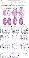 Figure 1
Deletion of Adamts9 in the proximal nephron causes small highly cystic kidneys. (A) Ciliopathy patient variants of ADAMTS9 mapped to its domain structures. S, signal peptide; Pro, pro-domain; Cat, catalytic domain; Dis, disintegrin domain; T1–15, thrombospondin type 1 repeats 1–15; CRD, cysteine-rich domain; L1, linker 1; L2, linker 2; Gon-1, Gon-1 domain. (B) Male kidney sections stained with hematoxylin and eosin (H&E) (n = at least 3 per group). Adamts9 conditional deletion in the proximal nephron results in highly cystic but relatively small polycystic kidneys in comparison with Ift88-cKO and Tmem67-KO kidneys. Mutation of the ADAMTS cleavage site of the versican GAG-β domain (VcanAA/AA mice), conditional deletion of versican in the mouse kidney (Vcan cKO), or conditional deletion of Adamts9 in the ureteric lineage (collecting ducts) does not result in polycystic kidneys. Scale bar: 1 mm. (C–E) Body weight, kidney weight, and combined kidney weight/body weight percentage of male and female Adamts9-cKO, Ift88-cKO, and Tmem67-KO mice and their control littermates. Data are shown as mean ± SEM; *P < 0.05, **P < 0.01, ***P < 0.001, ****P < 0.0001 calculated by 2-way ANOVA plus Tukey’s multiple-comparison test. (F) Kidney area (mm2) quantification at the indicated ages (n = at least 3 per sex per genotype). Data are shown as mean ± SEM; **P < 0.01, ****P < 0.0001 calculated by 2-way ANOVA plus Tukey’s multiple-comparison test.
Here, we conditionally deleted Adamts9 in the murine kidney nephric mesenchyme using the proximal tubule– and podocyte-specific Six2-Cre driver, and in the ureteric bud lineage using the distal nephron–specific Ksp-Cre driver (24, 25). Loss of ADAMTS9 in the proximal nephron mimicked the human disease NPHP and resulted in small but highly cystic kidneys with high interstitial collagen. Conditional deletion in males resulted in a more severe phenotype in comparison with females, and ADAMTS9 was required for TMEM67 cleavage and TZ assembly in renal cilia. ADAMTS9 deletion also led to a complete loss of the MKS/B9 module of the TZ. Characterization of TMEM67 cleavage using the TMEM67 cleavage–deficient mouse model revealed key differences in canonical Wnt signaling in comparison with Tmem67-null and Adamts9-deficient cystic kidneys, excluding elevated canonical Wnt signaling as the common driving force for renal cyst formation. Functional analysis of ADAMTS9 ciliopathy variants highlighted the requirement of ADAMTS9-mediated TMEM67 cleavage in disease etiology, revealing loss of TMEM67 C-terminus localization to the ciliary TZ as a crucial regulatory step. Loss of ciliogenesis coupled with loss of collagen homeostasis, unique to Adamts9 deletion by its proteolytic cleavage of MT1-MMP (MMP-14), led to characteristically small but highly cystic kidneys, which modeled NPHP within mice.
-
Results
ADAMTS9 deletion in the proximal nephron causes cystic kidney development. Homozygous loss-of-function mutations in ADAMTS9 cause NPHP. To date, 8 disease-causing alleles have been reported, whose mutations are spread throughout the different domains of the metalloproteinase (Figure 1A). However, neither the underlying mechanism of cyst formation nor its function in mammalian kidney development is known. To assess the impact of Adamts9 deletion, we determined its expression pattern in the mouse nephron using the Kidney Cell Explorer, developed by comparison of single-cell RNA sequencing (scRNA-seq) data from male and female mouse kidneys (26). Adamts9 expression was limited to the proximal end (podocytes, parietal epithelium, and proximal tubules) in both male and female nephrons, but male mice showed a markedly higher level of expression in the third segment of the proximal tubules (Supplemental Figure 1A; supplemental material available online with this article; https://doi.org/10.1172/jci.insight.203950DS1). Strikingly, the novel ADAMTS9 substrate TMEM67 also showed a similar expression pattern (Supplemental Figure 1, A and B). Other ADAMTS family versicanases or Vcan did not show this expression bias (Supplemental Figure 1C). We therefore carried out Adamts9 conditional deletion using the Six2-Cre driver, which expresses Cre recombinase in the nephrogenic lineage (24), targeting the proximal nephron (hereafter referred to as Adamts9-cKO mice). Male Adamts9-cKO mice developed highly cystic, small kidneys by 21 days of age (P21) (Figure 1B). Strikingly, female Adamts9-cKO mice only showed highly cystic kidneys by 3 months of age (Supplemental Figure 1D). At the terminal stage, Adamts9-cKO male mice took on a hunched posture and were distinctly small with an abnormal gait, whereas cKO females were indistinguishable from healthy mice (Supplemental Videos 1 and 2 and Supplemental Figure 2A). We used conditional deletion of Ift88 using the same Cre driver as a model lacking renal ciliogenesis, and Tmem67-KO mice as a model lacking the ADAMTS9 substrate of the TZ, in comparison with Adamts9-cKO kidneys. Deletion of Ift88 results in a complete loss of cilia formation (27), while deletion of TMEM67 results in abnormally long or short cilia in the murine kidney (21). Both models developed highly cystic kidneys that were greatly enlarged compared with Adamts9-cKO kidneys (Figure 1, B–F).
We previously developed a versican cleavage–deficient mouse model (VcanAA/AA) in which the ADAMTS9 cleavage site of the versican GAG-β domain is mutated uncleavable (28). Neither the VcanAA/AA mice nor conditional deletion of versican in the kidney (Six2-Cre Vcanfl/fl [Vcan cKO] or Ksp-Cre Vcanfl/fl [data not shown]) resulted in cystic kidneys (Figure 1B). This indicated that cystic kidney development was unrelated to ADAMTS9’s versican GAG-β domain cleavage function in the murine kidney. This supported our previous findings in human RPE-1 (retinal pigment epithelial) cells (15), and also suggested that versican and its cleavage are dispensable for normal kidney development. Adamts9 deletion using Ksp-Cre did not develop cystic kidneys, validating the Kidney Cell Explorer expression data, and potentially indicating that ADAMTS9 function may be specific to the proximal nephron (Figure 1B). Adamts9-cKO kidneys had similar or significantly lower kidney weights compared with littermate control mice (Figure 1C). In stark contrast, Ift88-cKO mice and Tmem67-KO mice had significantly higher kidney weights and kidney weight/body weight ratios (Figure 1, D and E). Quantification of kidney area also showed a similar result, where Adamts9-cKO kidneys remained small, and Ift88-cKO and Tmem67-KO kidneys were enlarged (Figure 1F). Periodic acid–Schiff (PAS) staining, a valuable tool to assess kidney injury, showed proteinaceous, polysaccharide-rich nephric casts throughout the Adamts9-cKO cystic kidneys (Supplemental Figure 2, B and C). These data revealed that loss of ADAMTS9 in the proximal nephron of the murine kidney produces small but highly cystic kidneys, modeling the human disease NPHP, and thereby providing a valuable platform to study the disease mechanism by which loss of ADAMTS9 causes NPHP.
Adamts9-cKO kidneys undergo normal nephrogenesis and form cysts postnatally. To investigate when cyst formation occurred, we harvested kidneys on the last day of embryonic development (E18.5) and at 7 days of age (P7), and analyzed their histopathology in midline sections. Neither male nor female Adamts9-cKO kidneys had distinct cystic dilations by H&E staining at E18.5 (Figure 2A). Ift88-cKO and Tmem67-KO kidneys showed many nephric tubules that were abnormally dilated at E18.5. High-magnification analysis of Adamts9-cKO kidney medullary regions showed large areas filled with mesenchymal cells in comparison with WT kidneys (Figure 2B). At P7, all 3 models showed cystic dilations, and Ift88-cKO kidneys were considerably larger than the others (Figure 2A). To investigate whether Adamts9-cKO kidneys, despite being highly cystic, remained smaller as a result of reduced nephrogenesis during embryonic development, we first quantified the number of glomeruli observed in midline sections of E18.5 and P7 kidneys (Figure 2C). This analysis revealed no significant differences in the number of glomeruli present in the cystic kidney models. To evaluate the total number of mature glomeruli in whole kidneys, we used the acid maceration technique (29–31) (Figure 2D). This also revealed a comparable number of mature glomeruli present in both sexes of Adamts9-cKO kidneys at birth to that of control littermates. To examine whether there are differences in cell proliferation and apoptosis of nephrons, we immunostained for the cell proliferation marker phospho–histone H3 and the apoptosis marker cleaved caspase-3, and compared Adamts9-cKO mice with Ift88-cKO mice. These analyses revealed comparably increased cell proliferation in both cystic kidney models and no distinct changes in the number of apoptotic cells (Supplemental Figure 4, A and B, and Supplemental Figure 5, A and B). Staining of WT and Adamts9-cKO kidneys at E18.5 with the nephron segmental markers LTL (proximal tubules) and DBA (collecting duct) revealed comparable staining for both lineages (Figure 2E). However, high-magnification images showed that the LTL staining of the Adamts9-cKO kidney cortices was morphologically distinct from the WT kidneys, with tubular dilations, presumably marking the beginning of cystic expansion. Ift88-cKO kidneys showed a similar result with distinctly dilated proximal tubules (Supplemental Figure 3A). Combined, these results suggested that ADAMTS9 enzymatic activity is required for normal kidney development in mice and its absence leads to postnatal cystic expansion.
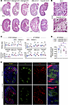 Figure 2
Adamts9-cKO kidneys develop cysts postnatally. (A and B) H&E staining of E18.5 and P7 kidneys (n = 3–4 kidneys per sex per genotype). Boxed areas are shown at higher magnification in B. *Large areas with mesenchyme-like cells present in the Adamts9-cKO kidney sections. (C) Quantification of kidney area (mm2) and number of glomeruli in midline kidney sections of E18.5 and P7 kidneys (n = 3–4 per sex per genotype). Data are shown as mean ± SEM; statistical significance was calculated by 2-way ANOVA plus Tukey’s multiple-comparison test. (D) Quantification of mature glomeruli in whole kidneys of P0 Adamts9-cKO and littermate control kidneys by the acid maceration method (n = 3–6 per sex per genotype). Data are shown as mean ± SEM; statistical significance was calculated by 2-way ANOVA plus Tukey’s multiple-comparison test. (E) E18.5 male WT and Adamts9-cKO kidneys stained for proximal tubules (LTL, green) and collecting ducts (DBA, red) show comparable levels. The white boxes indicate magnified kidney medullary regions, showing abnormally enlarged morphologies of Adamts9-cKO proximal tubules (green) in comparison with WT tubules. The zoomed-in images are approximately ×10 in relative magnification. Scale bars: 500 μm (top) and 1 mm (bottom) in A, 50 μm in B, and 500 μm in E.
ADAMTS9 is required for ciliary necklace and renal primary cilia formation. To investigate ciliogenesis in Adamts9-cKO kidneys, we first used freeze-fracture scanning electron microscopy (SEM), which revealed distinct primary cilia in the renal tubules of WT kidneys, but their nearly complete absence in the Adamts9-cKO kidneys (Figure 3A). Very few cystic epithelial cells in Adamts9-cKO kidneys possessed primary cilia, which were extremely short (red arrowheads, Figure 3A). Analysis of cilia ultrastructure by transmission electron microscopy (TEM) showed a complete loss of the ciliary “necklace,” the distinct transmembrane and extracellular compartment of the TZ (22), in the short cilia observed in Adamts9-cKO kidneys (Figure 3B). Numerous partially formed cilia were also observed, which had failed to extend a long axoneme beyond the TZ. To assess ciliogenesis in Adamts9-cKO kidneys by immunostaining, we stained kidney cryosections with the ciliary membrane marker ARL13B and the basal body marker FGFR1 oncogene partner (FOP). This revealed a nearly complete loss of ciliogenesis except for a few extremely short primary cilia present in the Adamts9-cKO kidneys (Figure 3C). To investigate whether loss of ciliogenesis was due to loss of TZ assembly in the Adamts9-cKO kidneys, we cultured 21-day-old kidneys and analyzed TZ formation in ice-cold methanol-fixed primary renal cultures. We previously identified 6 molecules belonging to the MKS/B9 module of the TZ that were lost by ADAMTS9 or TMEM67 deletion in RPE-1 cells (21). We investigated TZ localization of these molecules in renal primary cilia (Figure 3D). TCTN1, TCTN2, TCTN3, TMEM237, and B9D2 showed significantly lower TZ localization in Adamts9-cKO kidneys, while CC2D2A levels were not significantly affected (Figure 3D). These results supported our TEM observations, in which the TZ necklace, composed of the tectonic complex (TCTN1–3) and other transmembrane components (22), was completely lost in Adamts9-cKO cilia.
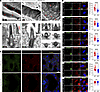 Figure 3
Loss of ciliogenesis in Adamts9-cKO kidneys. (A) Freeze-fracture SEM imaging of male P21 WT and Adamts9-cKO kidneys (n = 3) showing a complete loss of ciliogenesis or extremely short cilia (red arrowheads) present in Adamts9-cKO cystic renal tubular epithelium compared with the long cilia seen in WT. (B) TEM imaging showing that the TZ necklace (red arrowheads) seen in WT cilia is absent in the short Adamts9-cKO cilia, while most mutant cilia were not extended beyond the cell surface. (C) Immunostaining for ARL13B (green) and FOP (red) in P21 male WT and Adamts9-cKO kidneys showing loss of ciliation in the mutant kidneys. (D) Adamts9-cKO kidney primary cell cultures immunostained for basal body (γ-tubulin, green) and the TZ markers (red), and the mean pixel intensity quantifications of the TZ markers (n = 3 experiments). Data are shown as mean ± SEM; ****P < 0.0001 calculated by 2-tailed unpaired t test. Scale bars: 5 μm in A, 200 nm (left and middle) and 400 nm (right) in B, 10 μm in C, and 10 μm in D.
Adamts9 is required for ECM homeostasis in the murine kidney. Since ADAMTS9 is also a bona fide ECM metalloproteinase, we investigated the overall impact of ADAMTS9 loss on renal ECM homeostasis. Masson’s trichrome analysis showed large accumulations of collagen (blue) surrounding the glomeruli and in the interstitial matrix surrounding the cystic tubules in Adamts9-cKO kidneys (Figure 4A). Collagen IV, limited to the glomeruli in WT kidneys, showed high levels of staining throughout the Adamts9-cKO cystic kidneys and in the glomerular basement membrane (GBM) (Figure 4B). The loss of only a few other ECM-associated proteins is known to cause PKD. The lamin-511 α subunit, laminin α5, is the best characterized among these, and its deletion has previously been shown to cause polycystic kidneys in mice (32, 33). Laminin α5 immunostaining showed a decreased, diffused, and non-fibrillar staining pattern in the GBM of Adamts9-cKO kidneys compared with WT kidneys (Figure 4C). Ultrastructure analysis of the GBM by TEM showed thicker GBMs present in Adamts9-cKO cystic kidneys (Figure 4D). To our surprise, laminin α5 and collagen IV immunostaining in Ift88-cKO cystic kidneys showed the opposite staining pattern (Supplemental Figure 3, B–D). Laminin α5 staining was highly upregulated and collagen IV staining was decreased in Ift88-cKO glomeruli. These results indicated that while the GBM composition and morphology and overall ECM homeostasis are also severely impaired by loss of ADAMTS9 in the murine kidney, the underlying molecular mechanism driving these ECM dynamics may differ from those caused by loss of renal primary cilia alone (i.e., Ift88 cKO).
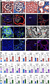 Figure 4
Adamts9-cKO cystic kidneys have impaired ECM homeostasis. (A) Masson’s trichrome staining of P21 male WT and Adamts9-cKO kidneys shows collagen accumulation (blue) surrounding glomeruli (left) and cystic tubules (right) (n = 3 kidneys per group). (B) Collagen IV (red) immunostaining, which is limited to glomeruli in WT kidneys, shows intense staining surrounding the Adamts9-cKO glomeruli (left) and throughout the kidney. Calbindin D28K immunostaining (green), which marks the distal convoluting tubules and collecting duct tubules, shows smaller tubules among larger cystic tubules in Adamts9-cKO kidneys (n = 3 kidneys per group). (C) Laminin α5 (green) staining seen in the WT glomerular basement membrane (GBM) is decreased in Adamts9-cKO glomeruli (n = 3 kidneys per group). (D) TEM images showing thickening of the GBM of Adamts9-cKO kidneys in comparison with WT (n = 3, each group). PC, podocytes; En, endothelium; RBC, red blood cell. (E and F) Kidney sections stained with the renal fibroblast marker MEIS1/2/3 (green) (E) and the fibrosis marker αSMA (red) (F) show that Adamts9-cKO kidneys are undergoing renal fibrosis (n = 3 kidneys per group). (G–I) Quantitative real-time PCR analysis of the ECM-related genes Tgfb1, Fn1, Col1a1, Col4a1, Has2, and Lama5, the kidney injury marker Havcr1, and the fibrosis marker Acta2 in male P21 Adamts9-cKO and Tmem67-KO and P14 Ift88-cKO kidneys in comparison with their respective WT littermates (n = 4 kidneys per group). Data are shown as mean ± SEM; *P < 0.05, **P < 0.01, ***P < 0.001, ****P < 0.0001 calculated by 2-tailed unpaired t test. Scale bars: 10 μm (left) and 20 μm (right) in A, 10 μm (left) and 50 μm (right) in B, 10 μm in C, 500 nm in D, and 50 μm in E and F.
The homeobox protein MEIS1, which marks renal myofibroblasts (34, 35), showed accumulation of renal myofibroblasts throughout the interstitial ECM surrounding the cystic kidney tubules in the Adamts9-cKO kidneys (Figure 4E). Similarly, α-smooth muscle actin (αSMA), which is limited to the vasculature in WT kidneys, also showed staining throughout the Adamts9-cKO and Ift88-cKO kidneys surrounding the cystic renal tubular epithelial cells and glomeruli (Figure 4F and Supplemental Figure 3E).
Together, these results indicated that Adamts9-cKO cystic kidneys undergo fibrosis and have increased ECM deposition. Since Adamts9-cKO and Ift88-cKO kidneys showed different staining patterns, we inquired whether the ECM dynamics result from dysregulated cell signaling and transcriptional changes to ECM-related genes. Quantitative real-time PCR analysis for TGF-β (Tgfb1), a master regulator of ECM-related gene transcription and fibrosis (36), showed significantly upregulated levels in the Adamts9-cKO kidneys. Similarly, transcript levels of the ECM molecules fibronectin (Fn1), collagens type I and type IV (Col1a, Col4a), and hyaluronan synthase 2 (Has2) were highly upregulated in the Adamts9-cKO cystic kidneys (Figure 4G). Laminin α5, which had decreased staining in the Adamts9-cKO kidneys, showed decreased transcript levels (Lama5), but not to a statistically significant degree. The renal fibrosis indicator smooth muscle α-2 actin (Acta2) and the kidney injury marker Kim1 (Havcr1) also showed significantly high transcript levels in the Adamts9-cKO kidneys (Figure 4G). Ift88-cKO and Tmem67-KO mice also showed similar dysregulations of these ECM transcripts, indicating that loss of cilia function can indeed regulate ECM dynamics via dysregulated gene transcription (Figure 4, H and I). Intriguingly, Ift88-cKO kidneys, which showed highly increased laminin α5 in the GBM and a moderate decrease in collagen IV staining, showed correlating Lama5 and Col4a1 transcript levels. In Tmem67-KO kidneys, all ECM-related genes analyzed were upregulated (Figure 4I). Combined, these data revealed the presence of highly complex etiologies in different cystic kidney models leading to abnormal ECM homeostasis, which may be governed in part by transcriptional changes. Since neither loss of versican nor mutation of its known ADAMTS9 cleavage site in the GAG-β domain led to cystic kidney development, we did not further investigate versican or its proteolysis in this study.
ADAMTS9-mediated TMEM67 cleavage in the murine kidney is required for normal kidney development. Using N-terminomics (19), we recently identified and validated the TZ molecule TMEM67 as a novel substrate of ADAMTS9 (21). ADAMTS9-mediated TMEM67 extracellular domain cleavage resulted in shedding of the TMEM67 cysteine-rich domain and translocation of the cleaved TMEM67 C-terminal half to the TZ (Figure 5A). To investigate whether ADAMTS9 is required for TMEM67 cleavage in the murine kidney, we performed Western blot analysis of WT and Adamts9-cKO kidney primary cultures. This revealed significantly reduced TMEM67 N-terminal fragment in the Adamts9-cKO culture medium, indicating that Adamts9-cKO kidneys are deficient for TMEM67 cleavage (Figure 5B). To study TMEM67 cleavage directly, we mutated the ADAMTS9 cleavage sites of TMEM67 and generated a TMEM67 cleavage–deficient mouse model (Tmem67ΔCLE/ΔCLE mice) (21). Similarly to the Tmem67-null, Tmem67ΔCLE/ΔCLE mice also developed large cystic kidneys by P14 (Figure 5C), and had significantly reduced body weights, increased kidney weights, and increased kidney/body weight ratios and increased kidney areas (Figure 5D). Analysis of TMEM67 cleavage comparing WT and Tmem67ΔCLE/ΔCLE renal primary cultures revealed a lack of TMEM67 cleavage in Tmem67ΔCLE/ΔCLE kidneys (Supplemental Figure 6A). PAS staining in Tmem67ΔCLE/ΔCLE and Tmem67-null cystic kidneys showed proteinaceous, polysaccharide-rich nephric casts throughout the cystic kidneys (Supplemental Figure 6B). Similarly to Tmem67-null, Tmem67ΔCLE/ΔCLE mice also showed cystic dilations at E18.5 (Supplemental Figure 6, C and D). E18.5 Tmem67ΔCLE/ΔCLE kidneys stained with DBA and LTL lectins showed overall comparable staining levels to that of WT kidneys, but with distinctly larger LTL-labeled tubules in the cortex (Supplemental Figure 6E). We previously showed that Tmem67ΔCLE/ΔCLE kidneys had reduced ciliogenesis and formed both significantly long and short renal cilia by EM analysis, and both these cilia types lacked formation of the TZ necklace (21). Immunostaining with ARL13B and FOP confirmed these observations and showed abnormal ciliogenesis in both Tmem67-null and Tmem67ΔCLE/ΔCLE kidneys (Figure 5E). Intriguingly, the abnormally long primary cilia in the Tmem67-mutant kidneys showed very low ARL13B intensities while the extremely short cilia showed intense ARL13B levels (Figure 5F), indicating that ARL13B and membrane trafficking dynamics may be severely impaired in these abnormal cilia. We also observed significant nuclear staining of ARL13B in the Tmem67-mutant cystic kidneys (Figure 5, E and G). Interestingly, the ARL13B antibody used in these assays targets the unique C-terminus of ARL13B, and C-terminally GFP-tagged ARL13B has been previously shown to localize to the nucleus (37). This ARL13B nuclear localization was not observed in the Adamts9-cKO cystic kidney model, which suggested that the dynamic regulation of the ARL13B C-terminus may be unique to the TMEM67 loss-of-function cystic epithelium. We hypothesize that this is due to aberrant ciliary signaling by the aberrantly long or short cilia formed in the Tmem67-deficient kidneys, which are absent in the Adamts9-cKO kidneys. Combined, these data revealed that ADAMTS9 mediates TMEM67 cleavage in the murine kidney, and is required to prevent polycystic kidney development in mice.
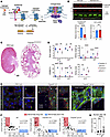 Figure 5
ADAMTS9 is essential for TMEM67 cleavage and ciliogenesis in the kidney. (A) Cartoon summarizing ADAMTS9 cleavage of the TMEM67 extracellular domain and identification of 2 functional forms of TMEM67. The full-length form present at the cell surface is essential for non-canonical Wnt signaling, and the cleaved TMEM67 C-terminus is required for ciliogenesis. (B) Western blot analysis of conditioned medium and cell lysates from kidney primary cultures (n = 3 cultures per group). Red, TMEM67; green, GAPDH. Data are shown as mean ± SEM; ****P < 0.0001 calculated by 2-tailed unpaired t test. (C) H&E staining of male P14 WT or Tmem67ΔCLE/ΔCLE kidney sections (n = 3–4 per group). (D) Body weight, kidney weight, combined kidney weight (KW)/body weight (BW), and kidney area (mm2) quantifications of P14 and P21 WT and Tmem67ΔCLE/ΔCLE mice (n = at least 3 per sex per genotype). Data are shown as mean ± SEM; *P < 0.05, **P < 0.01, ***P < 0.001, ****P <0.0001 calculated by 2-way ANOVA plus Tukey’s multiple-comparison test. (E) WT, Tmem67-KO, and Tmem67ΔCLE/ΔCLE (ΔCLE) kidneys stained with ARL13B (green) and FOP (red) show extremely short (white arrowheads) and abnormally long (open arrowheads) cilia in both Tmem67-KO and ΔCLE kidneys (n = 3 kidneys per group). (F) Quantification of cilia length and ARL13B pixel intensity correlation show abnormally long cilia with low ARL13B intensities (red boxes) and abnormally short cilia with intense ARL13B staining (blue boxes) in Tmem67-KO and ΔCLE kidneys (n = 3 kidneys, 30 cilia per genotype). Statistical significance was determined by 2-tailed non-parametric Spearman’s correlation (WT, –rs = –0.2184, P = 0.1275; Tmem67 KO, –rs = –0.7908, P < 0.0001; Tmem67 ΔCLE, –rs = –0.7272, P < 0.0001). (G) Nuclear ARL13B quantification shows increased staining in Tmem67-KO and ΔCLE kidneys (n = 3 kidneys, 30 nuclei per genotype). ***P < 0.001, ****P < 0.0001 by 2-way ANOVA plus Tukey’s multiple-comparison test for statistical significance. Scale bars: 1 mm in C and 10 μm in E.
Defective TZ assembly in TMEM67 cleavage–deficient kidneys. We previously showed that ADAMTS9-mediated TMEM67 cleavage is required for normal TZ assembly in RPE-1 cells and that Tmem67ΔCLE/ΔCLE renal primary cilia lack the TZ necklace (21). To investigate renal primary cilia TZ assembly further, we established primary cultures from Tmem67ΔCLE/ΔCLE and Tmem67-KO kidneys and compared them with WT kidney cultures from their corresponding littermates (Supplemental Figure 7, A–F). Intriguingly, similarly to Adamts9-cKO renal primary cultures, TCTN1–3, TMEM237, and B9D2 showed significantly decreased TZ localization, while CC2D2A TZ localization was unaffected (Supplemental Figure 7F). This also suggested that CC2D2A stability at the TZ may be differentially regulated or differentially dependent on TMEM67 levels when renal primary cilia are compared with RPE-1 cells. Combined, these results showed that loss of ADAMTS9, TMEM67, or its cleavage severely impaired normal renal primary cilia formation and disrupted the formation of the MKS/B9 module that forms the TZ necklace. Compared with Adamts9-cKO kidneys, Tmem67 mutants with both extremely long and short cilia suggested that ADAMTS9 may have more ciliary substrates that have not been identified yet, leading to a more severe loss of ciliation in the cystic renal tubular epithelium.
Defective ECM homeostasis in TMEM67 cleavage–deficient cystic kidneys. To investigate whether loss of TMEM67 cleavage can result in ECM homeostasis defects, we analyzed the ECM signature of Tmem67ΔCLE/ΔCLE kidneys. TEM analysis of glomeruli showed thicker GBM present in the Tmem67ΔCLE/ΔCLE kidneys (Supplemental Figure 8A). Masson’s trichrome staining confirmed these observations and showed increased levels of collagen deposited surrounding the glomeruli and cysts (Supplemental Figure 8B). As in Adamts9-cKO cystic kidneys, but not Ift88-cKO kidneys, collagen IV showed increased staining in the glomeruli and surrounding the cysts of both Tmem67 models (Supplemental Figure 8C). Strikingly, laminin α5 staining showed intense staining in the Tmem67-mutant glomeruli similar to that in Ift88-cKO but dissimilar to that in Adamts9-cKO kidneys (Supplemental Figure 8D). αSMA showed robust staining throughout Tmem67-mutant kidneys (Supplemental Figure 8E). Quantitative real-time PCR analysis of ECM markers showed a similar upregulation to that seen in Tmem67-null kidneys, which indicated that these molecular changes were from dysregulated gene transcription and cell signaling events downstream of defective cilia formation (Supplemental Figure 8F). Notably, consistent with the immunostaining data, Lama5 transcript levels were upregulated in both Tmem67-KO and Tmem67ΔCLE/ΔCLE kidneys and in Ift88-cKO kidneys in contrast to Adamts9-cKO kidneys, which showed decreased Lama5 transcription. Intriguingly, in ADPKD, laminin α5 expression is highly upregulated and has been shown to promote proliferation of cystic renal epithelial cells (38, 39). Thus the increased laminin α5 levels in Tmem67-mutant and Ift88-cKO cystic kidneys indicated greater similarity to the molecular signature of ADPKD cystic kidneys than to that of Adamts9-cKO kidneys.
Differential regulation of canonical Wnt signaling in Adamts9-cKO and Tmem67ΔCLE/ΔCLE kidneys. To investigate what signaling pathways may be differentially regulating these differential gene expression patterns observed among the interconnected cystic kidney models, we investigated the Wnt signaling pathway. Balanced Wnt singling is crucial for normal kidney development, and disruption of both canonical and non-canonical Wnt signaling pathways has been associated with cystic kidney disease (40, 41). More importantly to this study, in addition to its role in ciliary TZ assembly, TMEM67 is also involved in Wnt signal transduction through its N-terminal cysteine-rich domain, which forms a frizzled-like receptor for transducing non-canonical Wnt signaling mediated through Wnt5a, by forming a complex with ROR2 (42–46). We recently showed that TMEM67 cleavage by ADAMTS9 mediates the balance between the full-length form present at the cell surface, which functions as a Wnt signaling receptor, and the cleaved C-terminal half, which translocates to the TZ for ciliogenesis (21) (summarized in Figure 5A). In Tmem67-KO mouse brains and mouse embryonic fibroblasts (MEFs), non-canonical Wnt singling is lost and canonical Wnt signaling is highly upregulated. In contrast, Tmem67ΔCLE/ΔCLE mouse brains and MEFs, which form a stable uncleavable full-length protein at the cell surface, transduce completely normal Wnt signaling (21).
To investigate the Wnt signaling signatures of the cystic kidneys, we first analyzed active β-catenin antibody staining, which specifically recognizes non-phosphorylated β-catenin, as a readout for canonical Wnt signaling. The Adamts9- and Ift88-cKO kidneys showed highly elevated active β-catenin in the renal epithelial cells lining the cystic dilations, while a very low level of staining was observed in kidney tubules from WT littermates (Figure 6, A, B, and E). Strikingly, a similar increase in active β-catenin staining was only seen in the Tmem67-null kidneys and not in the Tmem67ΔCLE/ΔCLE kidneys (Figure 6, C–E). These observations phenocopied our previous observations in the brains of these mice and Wnt signaling assays conducted in MEFs, identifying canonical Wnt signaling as a major difference between the Tmem67-null and the Tmem67ΔCLE/ΔCLE models (21). To investigate non-canonical Wnt signaling signatures in the cystic kidneys, we investigated the expression of Plod2, Lcor, and Hadh, known downstream targets of the non-canonical Wnt signaling pathway, which were also differentially regulated in Tmem67-KO and Tmem67ΔCLE/ΔCLE MEFs (21). Adamts9- and Ift88-cKO kidneys showed no distinct regulatory pattern for these markers, while Tmem67-KO kidneys showed significantly lower levels of Plod2 and Hadh transcription (Figure 6F). The Tmem67ΔCLE/ΔCLE kidneys, on the other hand, which had constitutively cell surface–localized full-length TMEM67, showed highly elevated Plod2, Lcor, and Hadh transcript levels (Figure 6F). These results suggested that while both canonical and non-canonical Wnt signaling pathways were disrupted, the increased canonical Wnt signaling alone may not be sufficient to drive cystic kidney formation or essential to drive cystogenesis in the Tmem67-null kidneys, since Tmem67ΔCLE/ΔCLE kidneys are highly cystic without having elevated canonical Wnt signaling (Figure 6D).
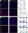 Figure 6
Differential canonical and non-canonical Wnt signaling in polycystic kidney models. (A–D) Immunostaining for active β-catenin (red) at the terminal stage of male cystic kidneys and their respective WT littermate kidneys (n = 3 kidneys per genotype). (E) Quantification of active β-catenin pixel intensities (n = 3 kidneys per 30 tubules per genotype). Data are shown as mean ± SEM; ****P < 0.0001 calculated by 2-tailed unpaired t test. (F) Quantitative real-time PCR analysis of the non-canonical Wnt signaling target genes Plod2, Lcor, and Hadh in male Adamts9-cKO, Ift88-cKO, Tmem67-KO, and Tmem67 ΔCLE kidneys in comparison with their respective WT kidneys (n = 4 kidneys per genotype). Data are shown as mean ± SEM; **P < 0.01, ***P < 0.001, ****P < 0.0001 calculated by 2-tailed unpaired t test. Scale bars: 20 μm in A and 50 μm in B–D.
Differential recruitment of macrophages and upregulation of inflammatory chemokines in Adamts9-cKO and other cystic kidneys. Inflammation and macrophage accumulation within the cystic kidney microenvironment contribute to disease burden (47–49), and inhibition of inflammation has been shown to slow down cystic kidney progression (50, 51). We therefore analyzed the immune microenvironment in ADAMTS9- and TMEM67-deficient cystic kidney models in comparison with WT littermates and Ift88-cKO cystic kidneys. This analysis was driven by our observations during freeze-fracture SEM and TEM, in which many macrophages colonizing the cystic lumens of Adamts9-cKO kidneys were observed (Supplemental Figure 9, A and B), and by the fact that we did not observe similar large colonies of macrophages in the lumen of any other cystic kidney models investigated in this study (21) (Supplemental Figure 9C). To probe this further, we investigated the immune signatures of all 4 cystic kidney models by flow cytometry analysis, in comparison with their WT littermates of both sexes (Supplemental Figure 9, D–F). Remarkably, despite having the smallest cystic kidney size, the Adamts9-cKO kidneys showed the highest level of immune activity compared with Ift88-cKO or Tmem67-deficient cystic kidneys. Overall, there were high percentages of CD45+ immune cells, F4/80+ macrophages, and CD11C+ dendritic cells or macrophages in all 4 cystic kidney models compared with their littermates. Both male and female cystic kidneys showed increased levels comparable to one another and did not show any sex-specific bias in immune activation (Supplemental Figure 9, D–F). To investigate chemokine signaling known to stimulate macrophage recruitment, we investigated expression of monocyte chemoattractant protein-1 (MCP-1; CCL2) (52). Adamts9-cKO kidneys had an approximately 20-fold increase in MCP-1 (Ccl2) expression compared with WT littermates. While Ift88-cKO and Tmem67-mutant cystic kidney models also showed significantly increased MCP-1, the upregulations were not as prominent as in Adamts9-cKO kidneys (Supplemental Figure 9G). CCR2, which acts as the primary receptor for MCP-1 (52), was transcriptionally upregulated in Adamts9- and Ift88-cKO kidneys, but downregulated in Tmem67-deficient kidneys (Supplemental Figure 9G). CCR4, a chemokine receptor primarily found on T cells (53), showed a similar trend and showed upregulation in Adamts9- and Ift88-cKO kidneys but was either downregulated or not affected in the Tmem67-deficient cystic kidneys. Tumor necrosis factor-α, which is a master regulator of inflammation and immune cell activity (54), was highly upregulated in all 4 cystic kidney models, with Ift88-cKO kidneys showing the highest level of upregulation. These results showed, in general, a highly immunogenic and inflamed kidney microenvironment present in all 4 cystic kidney models, but with key differences in the types of molecules involved in the immune responses. The proinflammatory cytokines are produced by interstitial fibroblasts, inflammatory cells, and tubular epithelial cells in cystic kidneys (55); thus we hypothesize that the high levels of ECM present in the Adamts9-cKO kidneys may be sequestering these cytokines, contributing to the increased immunogenic activity in them.
Adamts9-cKO kidneys are uniquely defective in collagen homeostasis. To further investigate the unique properties of Adamts9-cKO kidneys, we compared the collagen levels in the 4 cystic kidney models. Strikingly, both male and female Adamts9-cKO kidneys had a very high level of intestinal collagen, dramatically distinguishing them among the 4 cystic kidney models investigated in this study (Supplemental Figure 10, A–E). While the other models did show elevated ECM-related gene transcription and collagen deposition compared with their WT littermates, the amount of collagen was distinctly lower in comparison with Adamts9-cKO kidneys. It is unknown why loss-of-function mutations relating to ciliogenesis and cilia function are able to cause a spectrum of dramatically different cystic kidneys such as the highly enlarged kidneys of ADPKD and the small cystic kidneys in NPHP. Our findings suggested that renal fibrosis and interstitial ECM levels in the cystic kidney model may be an important determining factor of ADPKD-like versus NPHP-like phenotypic outcomes. To test this hypothesis directly, we analyzed the collagen level in mouse kidneys with conditional deletion of Pkd1 and Pkd2. Strikingly, both Pkd1- and Pkd2-cKO kidneys, despite being highly cystic and enlarged, showed only sparse amounts of collagen (Supplemental Figure 11, A and B). Collagen IV staining showed a similar result, and moderately low interstitial staining was seen in Pkd1- and Pkd2-cKO kidneys (Supplemental Figure 11, C and D). Col1a1 and Col4a1 transcript levels showed either no change in Pkd1-cKO kidneys or downregulation of Col4a1 levels in Pkd2-cKO kidneys (Supplemental Figure 11, E and F).
We theorized that interstitial collagen level may be the deciding factor governing kidney enlargement and type (NPHP-like or ADPKD-like) (see below). To investigate when collagen deposition is first observed in Adamts9-cKO kidneys, we conducted Masson’s trichrome staining in E18.5 and P7 kidneys compared with Ift88-cKO kidneys. By the end of gestation there was no discernible collagen staining in WT or either cystic kidney model, even though Ift88-cKO kidneys contained many cystic expansions (Supplemental Figure 12, A and B). However, at P7, despite being less cystic than Ift88-cKO kidneys, both male and female Adamts9-cKO kidneys showed large interstitial collagen depositions surrounding the cysts (Supplemental Figure 13, A and B). The Ift88-cKO kidneys did not show these deposits but showed collagen staining associated with the basement membrane, similar to that seen in WT kidneys.
Since ADAMTS9 is not a direct collagenase, we further investigated whether the increased collagen seen in Adamts9-cKO kidneys is due to loss of MT1-MMP cleavage, which we previously identified by our N-terminomics screen identifying novel ADAMTS9 substrates (20). MT1-MMP is a membrane-anchored collagenase responsible for directly degrading type I, II, and III interstitial fibrillar collagens (56). We previously showed that ADAMTS9-mediated cleavage releases the MT1-MMP catalytic domain from the cell surface, generating a soluble form of MT1-MMP (20). Furthermore, MT1-MMP cell surface proteolytic activity triggers a cascade of events that can lead to deregulated collagen deposition. Cell surface MT1-MMP together with TIMP-2 activates MMP-2 by pro-domain cleavage, and activated MMP-2, in tandem, activates MMP-9 (57, 58). Both MMP-2 and MMP-9 are well-characterized collagenases (59, 60). We previously showed that ADAMTS9-null RPE-1 cells and their conditioned medium have increased collagenase activity due to increased cell surface activation of MMP-2 and MMP-9, caused by loss of MT1-MMP cleavage (20). We tested whether Adamts9-cKO kidney primary cultures had impaired collagen homeostasis using these two assays. First, we cultured primary Adamts9-cKO or WT kidney cells on chamber slides coated with dye-quenched (DQ) collagen type I. This showed increased DQ-collagen signal surrounding the Adamts9-cKO cells, indicative of more cell surface collagenase activity (Supplemental Figure 14, A and B). Second, we collected the serum-free conditioned medium of the primary cultured WT and Adamts9-cKO kidneys and conducted collagen zymography. This revealed increased levels of active MMP-2 released by Adamts9-cKO kidneys (Supplemental Figure 14, C and D), confirming our previous findings in ADAMTS9-null cells. These results suggested that in addition to ADAMTS9-mediated regulation of ciliogenesis via TMEM67 cleavage, the ability to regulate collagen homeostasis via MT1-MMP cleavage is also severely impaired in Adamts9-cKO kidneys.
YAP/TAZ signaling in Adamts9-cKO cystic renal tubular epithelium and the other cystic kidney models. The Hippo signaling pathway transducers YAP and TAZ are known to be modulated by interstitial ECM stiffness, and are also known to be dysregulated in polycystic kidneys (61–63). We therefore investigated whether this pathway is differentially affected in the Adamts9-cKO kidneys at their terminal stage, when large accumulations of collagen are seen. Immunostaining for YAP, which marks the active form of the pathway and regulates cell proliferation, showed comparable staining patterns with slightly higher intensities in the Adamts9-cKO cystic tubular epithelium (Supplemental Figure 15A). Phospho-YAP (p-YAP) also showed positive signal in WT and Adamts9-cKO kidneys, but, interestingly, the intense signal in Adamts9-cKO kidneys marked the non-cystic tubules while the cystic epithelium had low intensities (Supplemental Figure 15A). Since phosphorylation of YAP marks it for targeted degradation, this indicated that YAP-mediated cell proliferation may be occurring in the cystic renal tubular epithelium and actively degraded in the non-cystic tubules. TAZ staining did not reveal any distinct signal pattern or difference in signal intensities, while p-TAZ staining (targeted for degradation) was significantly more intense in the cystic renal tubules of Adamts9-cKO kidneys (Supplemental Figure 15B). To evaluate relative YAP/TAZ signaling level in all 4 kidney models, we analyzed the expression of key YAP/TAZ target genes. We assayed the expression of CYR61 (Ccn1), a key regulator of cell proliferation (64); connective tissue growth factor (CTGF; Ccn2), a matricellular protein with key roles in ECM remodeling and wound healing (65); and the receptor tyrosine kinase AXL (Axl), which regulates cell survival, migration, and immune regulation (66). All 3 markers showed varying degrees of upregulation in the 4 cystic kidney models with the exception of Ccn2 in Adamts9-cKO kidneys, which was not significantly changed (Supplemental Figure 15C). Hence these findings suggested that while the complex YAP/TAZ signaling pathway is active in Adamts9-cKO kidneys, it was not distinctly different from that in the other cystic kidney models investigated in this study, despite the increased levels of collagen seen in them.
Loss of TMEM67 C-terminus from the ciliary TZ is a common disease etiology in ADAMTS9 ciliopathy patients. Polycystic kidney diagnosis is common to nearly all known ADAMTS9 ciliopathy patients. Therefore, we investigated whether these patient variants affected ciliogenesis and whether loss of TMEM67 cleavage is part of their disease etiology. To investigate the patient variant functionality in ciliogenesis, we generated all 8 variants using site-directed mutagenesis and conducted rescue experiments in comparison with WT, full-length human ADAMTS9 expression in ADAMTS9-null RPE-1 cells. Loss of ADAMTS9 resulted in a loss of ciliogenesis and cilium length in RPE-1 cells, which was fully rescued by the introduction of WT ADAMTS9 but not the patient variants (Figure 7A). Only the S1826N variant, which occurs in the C-terminus (in the unique Gon-1 domain), partially increased the percentage of ciliated cells and resulted in cilia lengths that were comparable to those seen with WT ADAMTS9 expression (Figure 7, C and D).
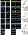 Figure 7
ADAMTS9 ciliopathy patient variants are defective in ciliogenesis and TMEM67 C-terminus TZ localization. (A, C, and D) Immunostaining for primary cilia (yellow arrowheads) by acetylated α-tubulin (Ac-α-tubulin) (green) and γ-tubulin (red) staining in serum-starved WT or ADAMTS9-KO RPE-1 cells transfected with empty vector (EV), WT, and T65R, S636F, R647Q, C1024*, R1203W, Q1525H-f/s, P1665S, and S1826N ADAMTS9 variants (A) shows significantly lower ciliogenesis (C) and cilia lengths (D) in comparison with WT ADAMTS9 transfection (n = 3 experiments, 100 cells or 30 cilia per group). Data are shown as mean ± SEM; **P < 0.01, ****P < 0.0001 by 2-way ANOVA plus Tukey’s multiple-comparison test. (B and E) Immunostaining for TMEM67 C-terminus (red) and γ-tubulin (green) shows significantly reduced TZ-localized TMEM67 C-terminus in ADAMTS9-null RPE-1 cells and cells transfected with ciliopathy variants (n = 3 experiments, 30 TZs per group). Data are shown as mean ± SEM; ****P < 0.0001 by 2-way ANOVA plus Tukey’s multiple-comparison test. Scale bars: 25 μm in A and 5 μm in B.
We previously demonstrated that ADAMTS9-null RPE-1 cells lose the TMEM67 C-terminal half generated by its cleavage and subsequent localization to the TZ, and expression of the TMEM67 C-terminal fragment (TMEM67 Δ342) alone can partially restore both cilium and TZ assembly (21). Using an antibody generated against the TMEM67 C-terminus (TMEM67-C) (43) and ice-cold methanol fixation, we investigated TMEM67 C-terminal localization of the ADAMTS9 patient variants (Figure 7B). All variants showed reduced localization of the TMEM67 C-terminus to the TZ in comparison with WT ADAMTS9 (Figure 7E). These results indicated that TMEM67 cleavage may be at the center of a common disease mechanism affecting ciliogenesis in ADAMTS9 ciliopathy patients, leading to loss of ciliary function and NPHP kidney pathogenesis.
-
Discussion
Here we demonstrated that loss of ADAMTS9 in the mouse kidney mimics the human cystic kidney disease NPHP, and ADAMTS9-mediated TMEM67 cleavage is essential for normal kidney development. We used 4 different cystic kidney models related to TMEM67 cleavage or loss of ciliogenesis (Figure 8A) to investigate the similarities and differences in their molecular signatures comparing their phenotypic manifestations (Figure 8B). Our findings highlight that Adamts9 conditional deletion in the nephrogenic lineage specifically causes small polycystic kidneys with high interstitial collagen, resembling the hallmarks of the human disease NPHP. In contrast, Tmem67 loss-of-function mutants or Ift88 nephrogenic-lineage conditional deletion led to highly enlarged cystic kidneys, which are more reminiscent of an ADPKD-like) phenotype (Figure 8B). While the 4 cystic kidney models in this study shared some common phenotypic manifestations, such as cystogenesis, renal fibrosis, defective gene transcription, and immune response, there were marked differences in their Wnt signaling signatures, their cilia phenotypes, and their ECM levels (Figure 8B). While both Adamts9 and Ift88 conditional deletion resulted in a complete loss of cilia, both Tmem67 mutant models had a substantial number of ciliated cells, albeit they were either severely short or long. Since at the time of the study, a floxed mouse allele for Tmem67 was not available, one limitation of our study is that the Tmem67 cystic kidney models used here affect the entire kidney. Although we have not directly compared data from Adamts9- and Ift88-cKO kidneys to Tmem67-deficient models for statistical analysis, the differential molecular signatures seen in these models in cross-comparisons may be partly due to differences arising from global versus conditional gene deletion status in these cystic models.
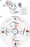 Figure 8
ADAMTS9 loss in the murine kidney leads to small NPHP-like kidneys, while loss of IFT88 or TMEM67 function leads to large ADPKD-like kidneys. (A) Graphical summary depicting the functionality of ADAMTS9 in regulating ciliogenesis, cell signaling, and collagen homeostasis in the mouse kidney. (B) Graphical summary depicting the key differences of the 4 cystic kidney models investigated and their phenotypic outcome causing either large ADPKD-like kidneys (Ift88-cKO, Tmem67-KO, and ΔCLE kidneys) or small NPHP-like kidneys in the Adamts9 cKO.
We propose here that this increased interstitial collagen seen in Adamts9-cKO kidneys may act as a very stiff “ECM scaffold,” mechanically preventing the Adamts9-cKO cystic kidneys from enlarging, resulting in the distinctly small cystic kidney phenotype compared with the others (Figure 8B). We believe the cascade of events leading to deregulated MMP activity triggered by loss of MT1-MMP cleavage, unique to Adamts9-cKO kidneys but not the 3 other models, may be the distinguishing factor for the very high levels of collagen seen in them. Since Adamts9-cKO kidneys have more active MMP-2 in kidney primary culture medium and increased cell surface collagen cleavage, our data potentially suggest that the soluble MT1-MMP form, which is lost in Adamts9-cKO kidneys, may play an important role in regulating interstitial collagen level in cystic kidneys. It is also possible that increased collagen seen in the Adamts9-cKO kidneys may have resulted from the overall increased immune response in them and is formed by dysregulated transcriptional networks, as opposed to decreased MMP-2– and MMP-9–mediated proteolytic cleavage of collagen. However, it is also possible that increased MMP-2– and MMP-9–mediated collagen cleavage may have a positive role in overall collagen deposition in the interstitial ECM of cystic kidneys. Supporting this theory, MMP-9 (activated by MT1-MMP) has been shown to be a positive mediator of collagen levels in the kidney, and mice lacking MMP-9 have reduced interstitial collagen in obstructive nephropathy (67). The broad-spectrum MMP inhibitor marimastat (which can inhibit MT1-MMP, MMP-2, and MMP-9) was recently used to inhibit MMP activity in PKD mouse models with promising, beneficial outcomes (68). Interestingly, marimastat treatment decreased collagen levels and fibrosis in the PKD mouse kidneys. Thus far, this strategy has not been utilized in an NPHP-like cystic kidney model with high collagen levels, and it is unclear whether decreasing collagen levels in an NPHP kidney will decrease the disease burden or increase the pathological outcome, making it more similar to an enlarged PKD-like kidney. We plan to investigate this further in future studies, using the Adamts9-cKO cystic kidney model.
The increased ECM may also act as an “extracellular reservoir” for signaling molecules and proinflammatory cytokines, explaining the comparatively high immunogenic response in the Adamts9-cKO kidneys. Our results also suggest that cyst formation, regardless of the initiating genetic cause, may converge on common pathways, but utilizing different receptors for immune activation, which may be exacerbated by loss of ECM remodeling in the kidney. Defining the specific roles and activation states of the infiltrating immune populations, such as distinguishing proinflammatory versus reparative macrophage subsets, or assessing antigen-presenting functions of dendritic cells, will be crucial for designing cystic kidney subtype–specific immunotherapeutics. Testing whether modulation of inflammatory pathways can also modify cyst progression in Adamts9-cKO kidneys in future studies may provide valuable insight into the relationship of interstitial fibrosis level and therapeutic outcome in the treatment of cystic kidney disease.
Because the mice with conditional deletion of versican and the versican cleavage–deficient mice have normal renal histopathology and lifespans, we conclude here that ADAMTS9-mediated versican proteolysis is unrelated to cystic kidney formation. However, it should be noted that TMEM67 and MT1-MMP may not be the only targets of this crucial metalloproteinase in the kidney. Since loss of ADAMTS9 leads to a more severe loss of cilia phenotype while loss of TMEM67, or its cleavage, leads to abnormal cilia formation, it is most likely that ADAMTS9 may proteolytically cleave other proteins important for cilia assembly. A future proteomics study aimed at investigating the N-terminome of Adamts9-cKO kidneys may shed valuable light on uncovering these substrates.
Differential regulation of laminin α5 in the 4 different cystic kidney models is also an interesting observation made in this study, and supports our theory on increased MT1-MMP activity in Adamts9-cKO kidneys. Decreased laminin α5 immunostaining in the GBM was unique to Adamts9-cKO kidneys, while the other 3 models significantly upregulated its expression and showed intense staining in the GBM. The signaling pathway responsible for this unique transcriptional upregulation of Lama5 is unknown. Currently, there is no evidence to suggest that laminin α5 might be a direct substrate of ADAMTS9. However, MT1-MMP has been previously shown to cleave the α5 chain of purified laminin-10 (69). We therefore suggest that the increased cell surface MT1-MMP present in Adamts9-cKO kidney podocytes may have led to increased degradation of laminin α5 in the GBM, resulting in its decreased staining in Adamts9-cKO kidneys, while transcriptional upregulation of Lama5 led to increased laminin α5 levels in Ift88-cKO and Tmem67-deficient kidneys.
While we used Six2-Cre–mediated conditional deletion of Ift88 as a control model lacking primary cilia in this study, it is also important to note that this is, to our knowledge, the first study to report the outcome of deleting Ift88 specifically in the nephrogenic lineage. Intriguingly, loss of Ift88 in the ureteric lineage or proximal tubules or postnatally by inducible Cre expression throughout the kidney leads to relatively slow-onset cystic kidney formation (27, 70, 71). Our results suggest that nephrogenic-lineage deletion is comparably more severe and results in rapid-onset cystic kidney development and lethality of Ift88-cKO mice by P14. Furthermore, E18.5 Ift88-cKO kidneys showed onset of tubular enlargements but otherwise had normal kidney histopathology. On the other hand, ablation of primary cilia in the nephron progenitors by the conditional deletion of Kif3a using Six2-Cre has been shown to reduce the number of nephrons by affecting the survival of metanephric mesenchymal cells, highlighting the importance of normal cilia formation for embryonic kidney development (72). Although the formation of small, highly fibrotic, NPHP-like kidneys is commonly associated with many TZ gene variants, mutations of the centrosome- and basal body–associated gene CEP120 can also cause similar kidney pathologies (73, 74). Intriguingly, Six2-Cre–mediated deletion of Cep120 also causes increased ECM deposition and downregulation of non-canonical Wnt signaling during embryonic kidney development (75).
The factors regulating differential Adamts9 and Tmem67 expression in male versus female proximal tubules are unknown. It is unknown whether simply higher levels of Adamts9 expression in male proximal tubules caused male Adamts9-cKO kidneys to have a more severe pathology when Adamts9 was deleted. The male sex-specific phenotypic burden is unique to Adamts9 loss of function, as both Tmem67-null and Tmem67ΔCLE/ΔCLE mice did not show a sex bias. Expression of Six2-Cre is most likely not sex specific, since Six2-Cre–mediated deletion of Ift88 did not show a sex bias. It is possible that Adamts9 expression may be differentially regulated in male and female cKO kidneys, downstream of the kidney injury response, which, importantly, has sex-specific gene expression differences in both humans and mice (76). It will be interesting to investigate whether ADAMTS9 ciliopathy patients show any sex-specific bias in survival and renal function in future clinical studies focused on answering this question. Based on the publicly available information of the small cohort of ADAMTS9 patients identified currently, a definitive interpretation cannot be made regarding a sex bias in them. Of the first two ADAMTS9 patients identified (14), F179-21 (Gln1525Hisfs*60) was diagnosed with NPHP plus JBTS and was female, while A5408-21 (Thr65Arg), diagnosed with NPHP, was male. Interestingly, the male patient was diagnosed with end-stage renal disease (ESRD) in infancy, while the female patient was not diagnosed with ESRD until age 5. A patient with biallelic compound heterozygosity for Ser636Phe and Cys1024* is a female with NPHP and JBTS. She received a donor kidney transplant at the age of 9, and a heart transplant at the age of 7 for hypoplastic left heart syndrome (18). The patient with biallelic compound heterozygosity for Pro1665Ser and Ser1826Asn was diagnosed with NPHP and also manifested heart and liver abnormalities. However, neither the sex nor the age at ESRD diagnosis was reported (17). The patient with biallelic compound heterozygosity for Arg647Gln and Arg1203Trp is a male diagnosed with JBTS without any renal manifestations reported (16).
Finally, when the very first patients carrying ADAMTS9 mutations were reported (14), the fact that they were ciliopathy patients, diagnosed with NPHP and JBTS, was a puzzling new development. This was because ADAMTS9 was a bona fide ECM metalloproteinase that had been extensively studied in relation to ECM homeostasis and had no apparent connection to the cilium or ciliary function. The concurrent development of Adamts9-floxed mice and hypomorphic mice (Adamts9Gt), which allowed us to study the function of ADAMTS9 beyond gastrulation and identification of novel substrates using N-terminomics, has now solidified ADAMTS9 as a crucial extracellular protease for ciliogenesis.


