
Research ArticleCardiologyMuscle biology
Open Access |  10.1172/jci.insight.204852
Combination S100A1 and ARC gene therapy as a treatment for Duchenne muscular dystrophy cardiomyopathy
David W. Hammers,1,2 Cora C. Hart,1,2 Eli Zerpa,1,2 Karen I. Laurent,1,2 Young il Lee,1,2 Meg M. Sleeper,2,3 and H. Lee Sweeney1,2
1Department of Pharmacology & Therapeutics and
2Myology Institute, University of Florida College of Medicine, Gainesville, Florida, USA.
3Small Animal Clinical Sciences, University of Florida College of Veterinary Medicine, Gainesville, Florida, USA.
Address correspondence to: David W. Hammers or H. Lee Sweeney, Department of Pharmacology & Therapeutics, University of Florida, 1200 Newell Dr., Gainesville, Florida, 32610, USA. Email: dhammers@ufl.edu (DWH); lsweeney@ufl.edu (HLS).
Find articles by
Hammers, D.
in:
PubMed
|
Google Scholar
|

1Department of Pharmacology & Therapeutics and
2Myology Institute, University of Florida College of Medicine, Gainesville, Florida, USA.
3Small Animal Clinical Sciences, University of Florida College of Veterinary Medicine, Gainesville, Florida, USA.
Address correspondence to: David W. Hammers or H. Lee Sweeney, Department of Pharmacology & Therapeutics, University of Florida, 1200 Newell Dr., Gainesville, Florida, 32610, USA. Email: dhammers@ufl.edu (DWH); lsweeney@ufl.edu (HLS).
Find articles by Hart, C. in: PubMed | Google Scholar
1Department of Pharmacology & Therapeutics and
2Myology Institute, University of Florida College of Medicine, Gainesville, Florida, USA.
3Small Animal Clinical Sciences, University of Florida College of Veterinary Medicine, Gainesville, Florida, USA.
Address correspondence to: David W. Hammers or H. Lee Sweeney, Department of Pharmacology & Therapeutics, University of Florida, 1200 Newell Dr., Gainesville, Florida, 32610, USA. Email: dhammers@ufl.edu (DWH); lsweeney@ufl.edu (HLS).
Find articles by Zerpa, E. in: PubMed | Google Scholar
1Department of Pharmacology & Therapeutics and
2Myology Institute, University of Florida College of Medicine, Gainesville, Florida, USA.
3Small Animal Clinical Sciences, University of Florida College of Veterinary Medicine, Gainesville, Florida, USA.
Address correspondence to: David W. Hammers or H. Lee Sweeney, Department of Pharmacology & Therapeutics, University of Florida, 1200 Newell Dr., Gainesville, Florida, 32610, USA. Email: dhammers@ufl.edu (DWH); lsweeney@ufl.edu (HLS).
Find articles by Laurent, K. in: PubMed | Google Scholar
1Department of Pharmacology & Therapeutics and
2Myology Institute, University of Florida College of Medicine, Gainesville, Florida, USA.
3Small Animal Clinical Sciences, University of Florida College of Veterinary Medicine, Gainesville, Florida, USA.
Address correspondence to: David W. Hammers or H. Lee Sweeney, Department of Pharmacology & Therapeutics, University of Florida, 1200 Newell Dr., Gainesville, Florida, 32610, USA. Email: dhammers@ufl.edu (DWH); lsweeney@ufl.edu (HLS).
Find articles by
Lee, Y.
in:
PubMed
|
Google Scholar
|
1Department of Pharmacology & Therapeutics and
2Myology Institute, University of Florida College of Medicine, Gainesville, Florida, USA.
3Small Animal Clinical Sciences, University of Florida College of Veterinary Medicine, Gainesville, Florida, USA.
Address correspondence to: David W. Hammers or H. Lee Sweeney, Department of Pharmacology & Therapeutics, University of Florida, 1200 Newell Dr., Gainesville, Florida, 32610, USA. Email: dhammers@ufl.edu (DWH); lsweeney@ufl.edu (HLS).
Find articles by Sleeper, M. in: PubMed | Google Scholar
1Department of Pharmacology & Therapeutics and
2Myology Institute, University of Florida College of Medicine, Gainesville, Florida, USA.
3Small Animal Clinical Sciences, University of Florida College of Veterinary Medicine, Gainesville, Florida, USA.
Address correspondence to: David W. Hammers or H. Lee Sweeney, Department of Pharmacology & Therapeutics, University of Florida, 1200 Newell Dr., Gainesville, Florida, 32610, USA. Email: dhammers@ufl.edu (DWH); lsweeney@ufl.edu (HLS).
Find articles by Sweeney, H. in: PubMed | Google Scholar
Published June 9, 2026 - More info
JCI Insight. 2026;11(15):e204852. https://doi.org/10.1172/jci.insight.204852.
© 2026 Hammers et al. This work is licensed under the Creative Commons Attribution 4.0 International License. To view a copy of this license, visit http://creativecommons.org/licenses/by/4.0/.
Received: January 20, 2026; Accepted: June 5, 2026


-
Results
Progressive diastolic dysfunction and LV restriction in D2.mdx mice. The mild progressing course of disease in traditional mdx mouse models of DMD (on C57-based genetic backgrounds) requires advanced aging or use of stress-inducing stimuli to elicit functional deficits (39, 40), which has limited the potential for mechanistic insights and therapeutic development for DMD cardiomyopathy. The backcrossing of the dystrophin nonsense mdx mutation onto the DBA/2J genetic background (known as the D2.mdx mouse) results in more severe muscle pathology than that exhibited by C57-based mdx mouse lines (41, 42). We have previously described and utilized this emerging D2.mdx model to evaluate translational therapeutics for the severe pathological features of DMD skeletal muscle (16, 43–45). The increased disease severity on the DBA/2J background is attributed to a deletion in the latent TGF-β binding protein 4 (LTBP4) polyproline-rich hinge (46, 47), which makes the DBA/2J variant of the LTBP4 hinge closer in size to that of other mammalian species, including rats, pigs, dogs, and humans (Supplemental Figure 1; supplemental material available online with this article; https://doi.org/10.1172/jci.insight.204852DS1), and more susceptible and more susceptible to activating TGF-β (47). To gain a better insight into this murine model’s utility for understanding and testing therapeutic interventions for DMD cardiomyopathy, we performed a comprehensive evaluation of cardiomyopathy development in D2.mdx mice using ECG, echocardiography, and histology.
Electrical abnormalities detected using ECG are typically the first clinical symptoms of DMD cardiomyopathy (6–8); therefore, we analyzed ECGs from DBA/2J WT (D2.WT) and D2.mdx mice from 2 to 12 months of age. Similar to observations in younger boys with DMD, ECG abnormalities are evident in D2.mdx hearts as early as 2 months of age (Supplemental Table 1). This includes significant genotype effects for prolonged QT, JT, and Tpeak to Tend intervals, as well as increased T wave amplitude, which are indicative of impaired LV repolarization. These data confirm that, like boys with DMD, D2.mdx mice exhibit electrical abnormalities early in life.
Diastolic dysfunction and restrictive LV filling are becoming more frequently reported aspects of DMD cardiomyopathy prior to the onset of systolic deficits and DCM development (10–13, 15). To analyze the emergence of these features in D2.mdx mice, we performed echocardiography on D2.WT and D2.mdx mice at 6, 10, 12, and 18 months. M-mode short axis (Figure 1A) analyses revealed no age- or genotype-associated differences in EF (Figure 1B); however, D2.mdx mice exhibited progressive declines in end diastolic volume (EDV; Figure 1C), end systolic volume (ESV; Figure 1D), and SV (Figure 1E) beginning at 10 months, with significant age and genotype effects. Four-chamber pulsed-wave Doppler measurements revealed significant D2.mdx-associated reductions in mitral valve (MV) E/A ratio (Figure 1F), a key indicator of diastolic dysfunction. Significant age- and genotype-associated increases in other measures of impaired LV relaxation and diastolic filling, including LV isovolumic relaxation time (Figure 1G), MV deceleration time (Figure 1H), and LV myocardial performance index (Figure 1I), were also found. Additionally, LV isovolumic contraction time was significantly increased in D2.mdx mice (Figure 1J), suggesting that defects in LV contractility, pressure development, and/or aortic valve opening may be aspects of cardiomyopathy in the D2.mdx mouse. These measures of impaired D2.mdx diastolic function were not due to faster heart rates than WT counterparts (Supplemental Figure 2A). Although evidence of LV wall thinning at diastole were not observed to confirm progression to DCM in those mice (Supplemental Figure 2, B and C), individual instances of markedly reduced EF may indicate that systolic failure begins around 18 months in this model. Although some features of LV diastolic dysfunction and restriction may be interpreted as development of cardiac hypertrophy, we identified no evidence of D2.mdx-associated cardiac enlargement at the organ or cardiomyocyte level in 12-month-old mice (Supplemental Table 2). Furthermore, we identified many of these LV restriction phenotypes in historical data from GRMD dogs, an established large animal model to study DMD cardiomyopathy (17, 48, 49). This analysis revealed affected GRMD dogs exhibit reduced LV end diastolic parameters, SVs, and MV E/A ratios compared with normal and carrier canines (Supplemental Table 3). These findings indicate that diastolic dysfunction with LV restriction is a translational feature of DMD cardiomyopathy that is prominently exhibited in the D2.mdx mouse model of DMD.
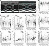 Figure 1
Progressive diastolic dysfunction and LV restriction in D2.mdx mice. Echocardiography was performed on D2.WT (WT; n = 9–17) and D2.mdx (mdx; n = 12–18) mice at the ages of 6, 10, 12, and 18 months. (A) Short-axis M-mode was used to measure the left ventricular (LV) parameters of (B) ejection fraction, (C) end diastolic volume, (D) end systolic volume, and (E) stroke volume. Pulsed wave Doppler was used to determine (F) mitral valve E/A ratios, (G) LV isovolumic relaxation time, (H) mitral valve deceleration time, (I) LV myocardial performance index, and (J) LV isovolumic contraction time. Data are displayed as violin plots (individual values indicated by circles; means indicated by black diamonds) and were analyzed using 2-factor ANOVA (age and genotype effects) followed by multiple t tests with Bonferroni’s correction (α = 0.05; significant P values are indicated).
Similar to the severe histopathology exhibited by D2.mdx skeletal muscle (44), the hearts of these mice also showed increased fibrosis and tissue pathology, as demonstrated by Picrosirius red staining (Figure 2). This is consistent with features found in boys with DMD and most preclinical models of DMD (17, 50–53). Because cardiac fibrosis is a potential cause of LV restriction via impairment of proper cardiomyocyte elongation during diastole, we employed the cardiac myosin inhibitor mavacamten (54), also known as MYK-461, to answer whether the reduced diastolic function and LV filling of D2.mdx mice is due to cardiomyocyte-intrinsic (i.e., impaired relaxation) or cardiomyocyte-extrinsic (i.e., fibrosis) factors. First, we confirmed that D2.WT mice were responsive to oral dosing of mavacamten (2.5 mg/kg), as evidenced by a significant decline in fractional shortening 3 hours after administration (Supplemental Figure 3A). This treatment resulted in a modest but nonsignificant increase in D2.WT EDV (Supplemental Figure 3B). We next repeated this experiment in 10-month-old D2.mdx mice, with the addition of a 3-day posttreatment washout period (diagramed in Supplemental Figure 3C). Similar to the effect seen in D2.WT mice, D2.mdx hearts exhibited a significant reduction in fractional shortening 3 hours after mavacamten dosing, which was completely reversed after washout (Supplemental Figure 3D). In contrast to D2.WT hearts, however, mavacamten caused D2.mdx hearts to show a significant approximately 68% increase in EDV, which was also reversed by washout (Supplemental Figure 3E). These findings support the hypothesis that the restricted LV filling of D2.mdx cardiomyopathy involves cardiomyocyte-intrinsic intracellular mechanisms, such as elevated resting calcium levels (16, 55), which potentially represent a targetable feature to treat DMD cardiomyopathy.
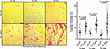 Figure 2
Progressive cardiac fibrosis in D2.mdx mice. Cardiac fibrosis was visualized and quantified using Picrosirius red staining of heart sections from 6-, 12-, and 18-month-old mice of each genotype (scale bar: 100 μm). Data are displayed as violin plots (individual values indicated by circles; means indicated by black diamonds) and were analyzed using 2-factor ANOVA (age and genotype effects) followed by multiple t tests with Bonferroni’s correction (α = 0.05; significant P values are indicated).
S100A1 treatment alleviates LV restriction and ARC prolongs lifespan in D2.mdx mice. Multiple gene therapies have been designed to address calcium handling issues in DMD cardiomyopathy by increasing SERCA activity (26–28). Although effective in mitigating elevated cytosolic calcium concentrations, these strategies also increase ATP demands in already energetically challenged dystrophic cardiomyocytes (20). As an alternative approach to address this calcium dysregulation with gene therapy, we sought to individually test the efficacy of overexpressing either S100A1, a pleiotropic calcium-binding protein that exerts several benefits in failing cardiomyocytes (35), or ARC, a protein that represses calcium dysregulation’s ultimate downstream effect of cell death (36, 37, 56).
To confirm that S100A1 and/or ARC are not naturally upregulated during dystrophic cardiomyopathy, immunoblotting for these proteins in 12-month-old D2.mdx hearts revealed trends of downregulation relative to age-matched WT levels (Supplemental Figure 4). To test the hypothesis that overexpression of each of these proteins would benefit dystrophic hearts, we developed individual self-complementary AAV vectors containing codon-optimized murine ARC and S100A1 driven by the cardiac troponin T (cTnT; Tnnt2 gene) promoter (57) and packaged in AAVrh10 serotype vector (58, 59). These vectors were administered to 1-month-old male D2.mdx mice via tail vein injections using a dose of 6.33 × 10¹³ gc/kg (experiment depicted in Figure 3A), with echocardiography performed at 12 months and 18–22 months (endpoint determined when S100A1 treatment mice reached the point of mean survival). Both transgenes extended mean survival time beyond the course of control D2.mdx mice; however, this effect was significant with ARC treatment (Figure 3B). S100A1 and ARC also improved LV histopathology and reduced cardiac fibrosis compared with control hearts (Figure 3, C and D), in agreement with the observed survival benefits. Although echocardiography revealed no changes to EF across ages or treatments (Figure 3E), significant effects were observed in the key diastolic function parameters of EDV, SV, and MV E/A ratio (Figure 3, F, G, and H) at the older time point. Notably, S100A1 treatment prevents the decline of EDV and SV associated with D2.mdx cardiomyopathy, whereas ARC treatment preserves MV E/A ratio. These data indicate that cardiac expression of both ARC and S100A1 are beneficial for the treatment of D2.mdx cardiomyopathy with therapeutically distinct outcomes.
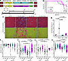 Figure 3
Therapeutic effect of S100A1 and ARC in D2.mdx mouse hearts. (A) Male D2.mdx received systemic treatment with 6.33 × 10¹³ gc/kg scAAVrh10.cTnT vector containing the coding sequence for either S100A1 (n = 6) or ARC (n = 16) at 1 month of age and underwent echocardiography at the 12-month and 18–22-month time points. Control D2.mdx mice (Con; n = 10) were included in this cohort for survival analysis, whereas additional control values are included in echocardiography measurements. (B) Mouse survival is depicted in a Kaplan-Meier plot. (C) Representative images of LV histopathology as visualized by H&E and Picrosirius red (PSR) staining (scale bar: 100 μm) and (D) fibrosis quantification. Echocardiography determined values for (E) ejection fraction, (F) end diastolic volume, (G) stroke volume, and (H) mitral valve (MV) E/A ratio for 12-month and 18–22-month time points. Data are displayed as (B) survival curve and (D–H) violin plots (individual values indicated by circles; means indicated by black diamonds). Data were analyzed using (A) Kaplan-Meier estimate followed by Wilcoxon’s nonparametric comparison test (α = 0.05; *P < 0.0001 vs. Con survival) and (D–H) 1-factor ANOVA (age and treatment effects) followed by multiple t tests with Bonferroni’s correction (α = 0.05; *P < 0.05 vs. age-matched Con values; significant P values are indicated).
Bicistronic S100A1-ARC vector improves D2.mdx cardiac phenotype. Given the improvements in diastolic function incurred by S100A1 overexpression and the survival benefit of ARC overexpression, we sought to combine these therapeutic transgenes into a single bicistronic vector utilizing an internal ribosomal entry site (60). The canine versions of these transgenes were used in this self-complementary therapeutic construct, which was also driven by the cTnT promoter and packaged in AAVrh10 vector (Figure 4A), to support further translational studies in larger mammals. Systemic treatment of this vector at a dose of 2 × 10¹⁴ gc/kg (16, 61) achieved approximately 10-fold elevation of ARC levels and approximately 2-fold elevation of S100A1 levels in D2.mdx mice that were injected at 1 month of age (Figure 4, B and C). Expression of canine S100A1 and ARC in this manner did not result in anti-transgene immune responses, as predicted by the high homology between murine and canine S100A1 (94%) and ARC (78%; primary differences in repetitive proline-glutamic acid–rich C-terminal tail). At the study’s endpoint (20 months), S100A1-ARC–treated hearts exhibited fewer signs of histopathology, including better cardiomyocyte organization and inflammation/infiltration (Figure 4D) and significantly reduced fibrosis (Supplemental Figure 5) than control D2.mdx hearts. Consistent with these findings and in agreement with the individual transgene treatments, this bicistronic vector had no effect on EF (Figure 4E) but did significantly reduce the progressive EDV and SV decline (Figure 4, F and G), LV isovolumic relaxation time elongation (Figure 4H), and worsening of LV myocardial performance index (Figure 4I). Additionally, the MV E/A ratio was significantly higher at both time points in S100A1-ARC–treated hearts (Figure 4J). It is important to note that although the S100A1-ARC treatment group underwent longitudinal measures in this study, no mice in the 12-month control group survived to the 20-month time point. We therefore utilized D2.mdx mice that survived to this time point for age-matched control data. These findings indicate that a bicistronic vector consisting of S100A1 and ARC provides substantial benefits to the dystrophic myocardium, particularly for the improvement of diastolic function.
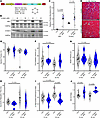 Figure 4
Bicistronic S100A1-ARC provides long-term benefits to D2.mdx cardiac function. (A) Male D2.mdx mice received control (Con; n = 12) or scAAVrh10.cTnT.S100A1-ARC (S100A1-ARC; n = 8) treatments at 1 month of age and underwent echocardiography at 12 and 20 months. (B and C) Cardiac ARC and S100A1 protein levels were assessed at study endpoint. (D) Representative images of left ventricle (LV) histopathology are provided, as analyzed by H&E staining (scale bar: 100 μm). Echocardiography measures of (E) ejection fraction, (F) end diastolic volume, (G) stroke volume, (H) isovolumic relaxation time, (I) LV myocardial performance index, and (J) mitral valve (MV) E/A ratios are shown for both 12-month and 20-month time points. Data are displayed as violin plots (individual values indicated by circles; means indicated by black diamonds) and were analyzed using (C) 2-tailed Welch’s t test (α = 0.05; *P < 0.05 vs. Con values) and (E–J) 2-factor ANOVA (age and treatment effects) followed by multiple t tests with Bonferroni’s correction (α = 0.05; *P < 0.05 vs. age-matched Con values; significant P values are indicated).
S100A1-ARC treatment prevents μDys-associated heart failure. Systemic μDys gene therapies are in the clinic to treat DMD-affected individuals with a miniaturized dystrophin transgene designed to be expressed in all striated muscles (62). In previous work, we identified that specific μDys designs have the ability to promote cardiac dysfunction and premature death in D2.mdx mice, whereas other versions of these human μDys transgenes were either beneficial or had no effect on long-term cardiac outcomes (16). In particular, the lethality caused by the ΔR3-R21 ΔCT μDys (16, 63) at approximately 11 months after treatment has been confirmed to be caused by heart failure, as determined by heart failure gene expression markers of increased Nppb (Supplemental Figure 6A), decreased Myh6 (Supplemental Figure 6B), and increased Myh7 and Myh7/Myh6 ratios (Supplemental Figure 6, C and D). To evaluate whether S100A1-ARC treatment can rescue this phenotype, we treated 1-month-old D2.mdx mice with ΔR3-R21 ΔCT μDys alone or in combination with S100A1-ARC (Figure 5A). Although there was no effect of this combination on μDys content (Figure 5B and Supplemental Figure 6E), ARC and S100A1 transgene levels were robustly expressed in only the dual vector treatment group (Supplemental Figure 6E). Immunofluorescence confirmed both μDys and ARC are uniformly expressed across combination-treated cardiomyocytes (Supplemental Figure 6F). Importantly, S100A1-ARC treatment significantly increased mouse survival from a mean survival time of 336 days to 625 days (Figure 5C). Because age-matched μDys-only mice were not available for comparison of heart function, we compared echocardiography data from 22-month-old μDys+S100A1-ARC–treated mice to those from 20–22-month-old D2.WT and 20-month-old D2.mdx groups. This comparison revealed no significant differences in EF across the groups, whereas SV and EDV from the μDys+S100A1-ARC group more closely resembled D2.WT values than those of untreated D2.mdx mice (Figure 5, D–F). Cardiac histopathology was also markedly improved in the combination treatment group, as shown by both H&E and Picrosirius red staining (Figure 5, G and H). These findings demonstrate that the S100A1-ARC bicistronic vector prevents the heart failure associated with cardiotoxic μDys transgenes and may represent an adjunct therapeutic, if μDys treatment promotes similar forms of heart failure in the clinic.
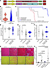 Figure 5
S100A1-ARC gene therapy prevents micro-dystrophin cardiotoxicity. (A) Male D2.mdx mice were treated with the ΔR3-R21ΔCT micro-dystrophin (μDys) gene therapy with (n = 8) and without (n = 15) cotreatment with the bicistronic cardiac S100A1-ARC vector at 1 month of age and were evaluated for survival. (B) Cardiac expression of μDys was measured by immunoblotting at respective humane endpoints of the study. (C) Survival analysis reveals significant life-extension by cotreatment with S100A1-ARC. Echocardiographic measures of (D) ejection fraction, (E) stroke volume, and (F) end diastolic volume are shown for 20–22-month-old D2.WT (n = 6) mice, 20-month-old control D2.mdx (mdx-Con; n = 7) mice, and 22-month-old D2.mdx mice that received both micro-dystrophin and S100A1-ARC treatments at 1 month (mdx-μDys+S100A1-ARC; n = 5). (G) Representative H&E and Picrosirius red (PSR) stained images of left ventricles (LV) from D2.WT, D2.mdx-Con, D2.mdx- μDys+S100A1-ARC, and 12-month-old D2.mdx-μDys mice only (reached humane endpoint; scale bar: 100 μm) demonstrate differential histopathology across groups. (H) Cardiac fibrosis was quantified from PSR-stained samples. Data are displayed as (B and D–F) violin plots with individual values indicated by circles and (C) a survival curve. Data were analyzed using (B) a 2-tailed Welch’s t test (α = 0.05; P value indicated), (C) a Kaplan-Meier estimator analysis (α = 0.05), and (D–F) 1-factor ANOVA followed by multiple t tests with Bonferroni’s correction (α = 0.05; *P < 0.05 vs. WT values). Control D2.mdx survival data in C are also reported in Figure 3B.
S100A1-ARC gene therapy safety in GRMD canine model of DMD. We next sought to test the safety of myocardial S100A1-ARC expression in the GRMD canine model of DMD. Prior to S100A1-ARC development, we investigated AAV serotypes capable of efficient transduction of canine hearts via i.v. administration. Although AAV9 exhibited lower transduction than AAV8 in the initial testing (Supplemental Figure 7A), we identified AAVrh10 as a vector that efficiently transduces canine striated muscle at an i.v. delivered dose of 5 × 10¹² gc/kg compared with AAV8 and AAVrh8 (Supplemental Figure 7, B and C). To further reduce amounts of vector required to specifically transduce canine hearts with therapeutic AAVs, we investigated the transduction efficiency of intracoronary AAV delivery, which revealed high transduction efficiency of the LV, septum, and right ventricle at doses of 2 × 10¹³ gc and 5 × 10¹³ gc of total vector delivery (Figure 6, A and B). Ensuing therapeutic treatments of scAAVrh10.cTnT.S100A1-ARC therefore utilized 5 × 10¹³ gc of vector delivered via intracoronary injections.
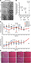 Figure 6
S100A1-ARC gene therapy in GRMD canines. (A) Representative epifluorescence images of EGFP expression in the left ventricle (LV), septum, and right ventricle (RV) of canine hearts 1 week after intracoronary delivery of 1 × 10¹³ or 2 × 10¹³ gc of AAVrh10.EGFP. (B) AAV vector genomes were quantified in the LV, septum, and RV of GRMD hearts after intracoronary treatment with 5 × 10¹³ gc of AAVrh10 vector. Data are displayed as violin plots (individual values indicated by circles; means indicated by black diamonds) and were analyzed using 2-tailed Welch’s t test (α = 0.05; P value and effect size indicated). (C) Fractional shortening and stroke volume measurements, as determined by echocardiography, from S100A1-ARC gene therapy–treated GRMD dogs (GT1 in blue, GT2 in orange, and GT3 in red; color-matched arrows indicate respective age of treatment). Historical GRMD values from the colony for each age range are shown in black (means as large circles with SEM shown; individual values as small circles). Age ranges having 2 or more GT values were analyzed using 2-tailed Welch’s t test (α = 0.05; P value and effect size indicated). (D) Representative H&E staining of LV cross-sections and longitudinal sections from S100A1-ARC–treated GRMD GT2 and GT3 with comparably age-matched control GRMD samples (scale bar: 100 μm).
The bicistronic scAAVrh10.cTnT.S100A1-ARC vector was delivered to 3 GRMD dogs: GT1 at 5 months, GT2 at 10 months, and GT3 at 24 months. Because GRMD cardiomyopathy is progressive with age (17, 48), we chose this spectrum of treatment ages to verify the safety of vector delivery across ages representing different degrees of preexisting cardiac pathology. All treatments were well tolerated, and no overt signs of cardiomyopathy were exhibited during the course of this study, which was followed for years without signs of safety concern from the vector treatment. The longevity and eventual endpoints of these dogs are summarized in Supplemental Table 4.
The low sample size of this safety evaluation limited interpretation of efficacy from this treatment. However, echocardiography data from the treated canines of this group compared with historical age-matched data from the colony revealed large effect sizes for fractional shortening improvements at 18–20, 21–24, and 25–30 months and SV increases at 18–20 months for GT1 and GT2 (Figure 6C). In agreement with possible functional benefits associated with these GRMD treatments, tissue sections of GT2 and GT3 LV samples exhibited markedly reduced signs of histopathology compared with comparably age-matched untreated GRMD samples (Figure 6D). Immunofluorescence confirmed both the loss of dystrophin staining in GRMD LV sections and the persistence of ARC transgene in S100A1-ARC-treated LV (Supplemental Figure 8). Together, these data provide evidence of both safety and improved cardiac phenotype by intracoronary delivery of S100A1-ARC gene therapy in the GRMD heart.
Skeletal muscle phenotypes improved by striated muscle expression of S100A1-ARC. Given the protective effect of S100A1-ARC in the dystrophic heart, we sought to determine whether dystrophic skeletal muscle can also benefit from treatment with this bicistronic transgene, including the reduction in severity of muscle degeneration that occurs early in life for D2.mdx mice (44). We investigated systemic treatment of S100A1-ARC driven by the CK8e striated muscle promoter (64) (Figure 7A) delivered at 1 month, with an endpoint at 2 months to gauge potential protection at the peak of D2.mdx skeletal myopathy. At the endpoint, the quadriceps of treated mice exhibited robust levels of vector transduction (Figure 7B); however, this transduction was lower than that found in the heart, similar to previous reports (16, 65). Importantly, treated extensor digitorum longus muscles exhibited greater maximum force production than controls, whereas the soleus muscles of treated mice demonstrated significantly higher specific tension (tetanic force normalized to muscle cross-sectional area; Figure 7, C and D). In agreement with the improved muscle force production in the extensor digitorum longus and soleus, S100A1-ARC–treated quadriceps and gastrocnemius muscles showed less histopathology than control muscles, as observed with H&E staining (Figure 7E). To quantify aspects of muscle pathology associated with this early-stage D2.mdx disease progression, we stained quadriceps and gastrocnemius sections with fluorescently tagged wheat germ agglutinin (WGA), which delineates muscle fibers from the interstitium, and anti-mouse IgG, which marks serum protein leakage into damaged muscle. As shown in Supplemental Figure 9A, this staining combination revealed different forms of muscle pathology, including inflammatory lesions, regenerative foci, myofiber damage/necrosis, and edema, which can be cumulatively quantified as total muscle pathology using the method illustrated in Supplemental Figure 9B. Pathology analysis using this methodology revealed that significant reductions in muscle pathology of the quadriceps and gastrocnemius were achieved as a result of S100A1-ARC treatment (Figure 7, F and G). These data suggest that S100A1-ARC effectively reduces the extent of muscle fiber death and preserves muscle function during early-stage D2.mdx skeletal muscle pathology. Therefore, the protective effect of this therapeutic dual transgene strategy is not restricted to the myocardium and may represent an impactful therapy for all muscles affected by DMD.
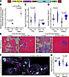 Figure 7
S100A1-ARC benefits D2.mdx skeletal muscle. (A) Male D2.mdx mice received treatment with control (Con; n = 11) or S100A1-ARC driven by the striated muscle–specific CK8e promoter (Tx; n = 4) at 1 month of age and underwent ex vivo functional measures of the soleus and extensor digitorum longus (EDL) muscles at 2 months. (B) Vector genomes were quantified in the quadriceps and hearts from Con and Tx mice. (C) Maximum tetanic force production and (D) specific tension were performed on the EDL and soleus muscles of Con and Tx D2.mdx mice at study endpoint. (E) Representative H&E staining of quadriceps (Quad) and gastrocnemius (Gastroc) muscles from Con and Tx D2.mdx are shown at study endpoint (scale bar: 100 μm). (F) Representative wheat germ agglutinin (WGA) and endogenous mouse IgG staining of Con and Tx Gastroc (scale bar: 500 μm) and (G) accompanying quantification of total muscle pathology are shown for Quad and Gastroc muscle sections from this study. Data are displayed as violin plots with individual values indicated by circles and were analyzed using (B) 2-factor ANOVA (treatment and muscle effects) followed by multiple t tests with Bonferroni’s correction (α = 0.05; *P < 0.05 vs. muscle-matched treatment values; #P < 0.05 vs. treatment-matched Quad values) and (C, D, and G) 2-tailed Welch’s t test (α = 0.05; *P < 0.05 vs. Con values).


