
Research ArticleCell biologyNeuroscience
Open Access |  10.1172/jci.insight.205134
The investigation of human cerebrospinal fluid exosome in spinal cord injury
Dallas L. Sheinberg,1 Haichao Wei,1,2 Joseph S. Withrow,3 Farshad Homayouni Moghadam,1,2 Chia-Chen Lu,1,2 Jyotirmoy Rakshit,1,2 Jennifer Zaragoza,1 John R. Williams,1 Wen Li,4 Jacques J. Morcos,1 and Jia Qian Wu1,2,5
1Department of Neurosurgery, McGovern Medical School, and
2Center for Neuroimmunology and Glial Biology, Institute of Molecular Medicine, University of Texas Health Science Center, Houston, Texas, USA.
3Carolina Neurosurgery and Spine Associates, Charlotte, North Carolina, USA.
4Division of Clinical and Translational Sciences, Department of Internal Medicine, University of Texas McGovern Medical School, Houston, Texas, USA.
5MD Anderson Cancer Center UTHealth Graduate School of Biomedical Sciences, Houston, Texas, USA.
Address correspondence: Jia Qian Wu, University of Texas Health Science Centre, SRB 637.C, 1825 Pressler Street, Houston, Texas 77030, USA. Phone: 713.500.3421; Email: Jiaqian.Wu@uth.tmc.edu.
Authorship note: DLS, HW, and JSW contributed equally to this work.
Find articles by Sheinberg, D. in: PubMed | Google Scholar
1Department of Neurosurgery, McGovern Medical School, and
2Center for Neuroimmunology and Glial Biology, Institute of Molecular Medicine, University of Texas Health Science Center, Houston, Texas, USA.
3Carolina Neurosurgery and Spine Associates, Charlotte, North Carolina, USA.
4Division of Clinical and Translational Sciences, Department of Internal Medicine, University of Texas McGovern Medical School, Houston, Texas, USA.
5MD Anderson Cancer Center UTHealth Graduate School of Biomedical Sciences, Houston, Texas, USA.
Address correspondence: Jia Qian Wu, University of Texas Health Science Centre, SRB 637.C, 1825 Pressler Street, Houston, Texas 77030, USA. Phone: 713.500.3421; Email: Jiaqian.Wu@uth.tmc.edu.
Authorship note: DLS, HW, and JSW contributed equally to this work.
Find articles by Wei, H. in: PubMed | Google Scholar
1Department of Neurosurgery, McGovern Medical School, and
2Center for Neuroimmunology and Glial Biology, Institute of Molecular Medicine, University of Texas Health Science Center, Houston, Texas, USA.
3Carolina Neurosurgery and Spine Associates, Charlotte, North Carolina, USA.
4Division of Clinical and Translational Sciences, Department of Internal Medicine, University of Texas McGovern Medical School, Houston, Texas, USA.
5MD Anderson Cancer Center UTHealth Graduate School of Biomedical Sciences, Houston, Texas, USA.
Address correspondence: Jia Qian Wu, University of Texas Health Science Centre, SRB 637.C, 1825 Pressler Street, Houston, Texas 77030, USA. Phone: 713.500.3421; Email: Jiaqian.Wu@uth.tmc.edu.
Authorship note: DLS, HW, and JSW contributed equally to this work.
Find articles by Withrow, J. in: PubMed | Google Scholar
1Department of Neurosurgery, McGovern Medical School, and
2Center for Neuroimmunology and Glial Biology, Institute of Molecular Medicine, University of Texas Health Science Center, Houston, Texas, USA.
3Carolina Neurosurgery and Spine Associates, Charlotte, North Carolina, USA.
4Division of Clinical and Translational Sciences, Department of Internal Medicine, University of Texas McGovern Medical School, Houston, Texas, USA.
5MD Anderson Cancer Center UTHealth Graduate School of Biomedical Sciences, Houston, Texas, USA.
Address correspondence: Jia Qian Wu, University of Texas Health Science Centre, SRB 637.C, 1825 Pressler Street, Houston, Texas 77030, USA. Phone: 713.500.3421; Email: Jiaqian.Wu@uth.tmc.edu.
Authorship note: DLS, HW, and JSW contributed equally to this work.
Find articles by Homayouni Moghadam, F. in: PubMed | Google Scholar
1Department of Neurosurgery, McGovern Medical School, and
2Center for Neuroimmunology and Glial Biology, Institute of Molecular Medicine, University of Texas Health Science Center, Houston, Texas, USA.
3Carolina Neurosurgery and Spine Associates, Charlotte, North Carolina, USA.
4Division of Clinical and Translational Sciences, Department of Internal Medicine, University of Texas McGovern Medical School, Houston, Texas, USA.
5MD Anderson Cancer Center UTHealth Graduate School of Biomedical Sciences, Houston, Texas, USA.
Address correspondence: Jia Qian Wu, University of Texas Health Science Centre, SRB 637.C, 1825 Pressler Street, Houston, Texas 77030, USA. Phone: 713.500.3421; Email: Jiaqian.Wu@uth.tmc.edu.
Authorship note: DLS, HW, and JSW contributed equally to this work.
Find articles by Lu, C. in: PubMed | Google Scholar
1Department of Neurosurgery, McGovern Medical School, and
2Center for Neuroimmunology and Glial Biology, Institute of Molecular Medicine, University of Texas Health Science Center, Houston, Texas, USA.
3Carolina Neurosurgery and Spine Associates, Charlotte, North Carolina, USA.
4Division of Clinical and Translational Sciences, Department of Internal Medicine, University of Texas McGovern Medical School, Houston, Texas, USA.
5MD Anderson Cancer Center UTHealth Graduate School of Biomedical Sciences, Houston, Texas, USA.
Address correspondence: Jia Qian Wu, University of Texas Health Science Centre, SRB 637.C, 1825 Pressler Street, Houston, Texas 77030, USA. Phone: 713.500.3421; Email: Jiaqian.Wu@uth.tmc.edu.
Authorship note: DLS, HW, and JSW contributed equally to this work.
Find articles by Rakshit, J. in: PubMed | Google Scholar
1Department of Neurosurgery, McGovern Medical School, and
2Center for Neuroimmunology and Glial Biology, Institute of Molecular Medicine, University of Texas Health Science Center, Houston, Texas, USA.
3Carolina Neurosurgery and Spine Associates, Charlotte, North Carolina, USA.
4Division of Clinical and Translational Sciences, Department of Internal Medicine, University of Texas McGovern Medical School, Houston, Texas, USA.
5MD Anderson Cancer Center UTHealth Graduate School of Biomedical Sciences, Houston, Texas, USA.
Address correspondence: Jia Qian Wu, University of Texas Health Science Centre, SRB 637.C, 1825 Pressler Street, Houston, Texas 77030, USA. Phone: 713.500.3421; Email: Jiaqian.Wu@uth.tmc.edu.
Authorship note: DLS, HW, and JSW contributed equally to this work.
Find articles by Zaragoza, J. in: PubMed | Google Scholar
1Department of Neurosurgery, McGovern Medical School, and
2Center for Neuroimmunology and Glial Biology, Institute of Molecular Medicine, University of Texas Health Science Center, Houston, Texas, USA.
3Carolina Neurosurgery and Spine Associates, Charlotte, North Carolina, USA.
4Division of Clinical and Translational Sciences, Department of Internal Medicine, University of Texas McGovern Medical School, Houston, Texas, USA.
5MD Anderson Cancer Center UTHealth Graduate School of Biomedical Sciences, Houston, Texas, USA.
Address correspondence: Jia Qian Wu, University of Texas Health Science Centre, SRB 637.C, 1825 Pressler Street, Houston, Texas 77030, USA. Phone: 713.500.3421; Email: Jiaqian.Wu@uth.tmc.edu.
Authorship note: DLS, HW, and JSW contributed equally to this work.
Find articles by Williams, J. in: PubMed | Google Scholar
1Department of Neurosurgery, McGovern Medical School, and
2Center for Neuroimmunology and Glial Biology, Institute of Molecular Medicine, University of Texas Health Science Center, Houston, Texas, USA.
3Carolina Neurosurgery and Spine Associates, Charlotte, North Carolina, USA.
4Division of Clinical and Translational Sciences, Department of Internal Medicine, University of Texas McGovern Medical School, Houston, Texas, USA.
5MD Anderson Cancer Center UTHealth Graduate School of Biomedical Sciences, Houston, Texas, USA.
Address correspondence: Jia Qian Wu, University of Texas Health Science Centre, SRB 637.C, 1825 Pressler Street, Houston, Texas 77030, USA. Phone: 713.500.3421; Email: Jiaqian.Wu@uth.tmc.edu.
Authorship note: DLS, HW, and JSW contributed equally to this work.
Find articles by
Li, W.
in:
PubMed
|
Google Scholar
|

1Department of Neurosurgery, McGovern Medical School, and
2Center for Neuroimmunology and Glial Biology, Institute of Molecular Medicine, University of Texas Health Science Center, Houston, Texas, USA.
3Carolina Neurosurgery and Spine Associates, Charlotte, North Carolina, USA.
4Division of Clinical and Translational Sciences, Department of Internal Medicine, University of Texas McGovern Medical School, Houston, Texas, USA.
5MD Anderson Cancer Center UTHealth Graduate School of Biomedical Sciences, Houston, Texas, USA.
Address correspondence: Jia Qian Wu, University of Texas Health Science Centre, SRB 637.C, 1825 Pressler Street, Houston, Texas 77030, USA. Phone: 713.500.3421; Email: Jiaqian.Wu@uth.tmc.edu.
Authorship note: DLS, HW, and JSW contributed equally to this work.
Find articles by Morcos, J. in: PubMed | Google Scholar
1Department of Neurosurgery, McGovern Medical School, and
2Center for Neuroimmunology and Glial Biology, Institute of Molecular Medicine, University of Texas Health Science Center, Houston, Texas, USA.
3Carolina Neurosurgery and Spine Associates, Charlotte, North Carolina, USA.
4Division of Clinical and Translational Sciences, Department of Internal Medicine, University of Texas McGovern Medical School, Houston, Texas, USA.
5MD Anderson Cancer Center UTHealth Graduate School of Biomedical Sciences, Houston, Texas, USA.
Address correspondence: Jia Qian Wu, University of Texas Health Science Centre, SRB 637.C, 1825 Pressler Street, Houston, Texas 77030, USA. Phone: 713.500.3421; Email: Jiaqian.Wu@uth.tmc.edu.
Authorship note: DLS, HW, and JSW contributed equally to this work.
Find articles by Wu, J. in: PubMed | Google Scholar
Published June 23, 2026 - More info
JCI Insight. 2026;11(15):e205134. https://doi.org/10.1172/jci.insight.205134.
© 2026 Sheinberg et al. This work is licensed under the Creative Commons Attribution 4.0 International License. To view a copy of this license, visit http://creativecommons.org/licenses/by/4.0/.
Received: February 2, 2026; Accepted: June 11, 2026

-
Results
Clinical and demographic characteristics of participants. The early phase of SCI, spanning the first few days after injury, is a critical window for therapeutic intervention. Biomarkers identified during this phase can aid in assessing injury severity and predicting recovery. CSF and serum are promising sources for such biomarkers. To investigate exosomal miRNA and protein signatures, we collected samples from SCI patients at the Memorial Hermann Emergency Department (Houston, Texas, USA).
A total of 25 traumatic SCI patients were recruited, and their clinical and demographic characteristics are summarized in Supplemental Table 1 (supplemental material available online with this article; https://doi.org/10.1172/jci.insight.205134DS1). Of the 25 patients, 22 (88%) were male and 3 (12%) were female, and the average age was 49.2 ± 19.2 years (mean ± SD). Of all injuries, 21 were cervical, 3 were thoracic, and 1 was lumbar. Baseline neurological assessment showed complete SCI (AIS A) in 44% of patients. In patients with incomplete SCI, 28% were AIS B and 28% AIS C. Detailed neurological assessments were obtained at follow-up to evaluate patient improvement (Supplemental Table 1). The average change in upper extremity, lower extremity, and total motor scores was 8.92 (SD = 15.48), 10.68 (SD = 11.46), and 20.00 (SD = 21.95), respectively. For all included patients, 64% had improvement ≥1 AIS grade at follow-up. In the subgroup of patients with AIS A injury, improvement was seen in 36%.
Characterization of exosome and comparison of enriched miRNAs in CSF and serum. Exosomes were extracted from the CSF and serum of SCI patients at 1, 3, and 5 days after injury. Transmission electron microscopy and nanoparticle tracking analysis, both standard techniques in the EV field, were used to assess the structural integrity and size distribution of the isolated particles. The results revealed that the majority of the particles had the structural integrity of the exosomes (Supplemental Figure 1A) and a diameter between 50 and 150 nm (Supplemental Figure 1B). These results indicate that the isolated exosomes were structurally preserved and fell within the expected size range, consistent with the known characteristics of exosomes.
CSF is in direct contact with the brain and spinal cord, making CSF-derived exosomes particularly enriched in molecules that reflect CNS physiology and pathology (17). In contrast, serum-derived exosomes originate from a broader range of tissues, making it more challenging to attribute their content specifically to CNS-related processes (18). To explore these differences in the context of SCI, we performed a comparative analysis of CSF and serum exosome-derived miRNA expression profiles based on injury severity (complete AIS A vs. incomplete AIS B/C SCI) 1 day after injury (Figure 1, A and B). In the complete injury group, 50 genes were enriched in CSF and 45 in serum. In the incomplete injury group, 49 genes were enriched in CSF and 44 in serum. There were 15 common genes between the complete and incomplete injury conditions (Figure 1, C and E) specifically enriched in CSF samples compared with serum, while there were 9 common genes between the complete and incomplete injury conditions specifically enriched in serum samples (Figure 1, D and E) compared with CSF samples. These findings highlight significant differences in miRNA expression profiles between CSF and serum.
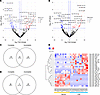 Figure 1
Differential gene expression analysis between serum and CSF. (A and B) Volcano plots for identified DEGs between CSF and serum in complete injury (A) and incomplete injury (B). Blue dots represent read counts with fold changes ≤ –1.5 and P value < 0.05. Red dots show read counts with fold changes ≥ 1.5 and P value < 0.05. (C and D) The Venn diagram displays the common upregulated DEGs in CSF (C) and serum (D). (E) Heatmap shows 15 genes significantly enriched in CSF and 9 genes in serum.
Proteins in CSF as biomarkers for SCI. In this study, we used a multiplex immunoassay to measure the abundance of 18 neuronal proteins and 30 cytokines in CSF exosomes (see Methods for details). To assess the clinical utility of these markers, we developed predictive models using receiver operating characteristic (ROC) curve analysis. ROC analysis was conducted to evaluate the discriminatory power of the protein biomarkers in distinguishing between injury severity and recovery outcomes. An area under the curve (AUC) value approaching 1 indicates high sensitivity and specificity, underscoring the potential of these biomarkers for early diagnosis and ongoing monitoring of SCI progression. First, we identified signature proteins associated with injury severity (complete AIS A vs. incomplete AIS B/C SCI) and functional outcome (improvement vs. non-improvement) by performing ROC analysis on all detected proteins and cytokines. Based on these findings, we constructed 2 distinct predictive models using combined protein profiles for early diagnosis and monitoring SCI recovery.
A prediction model incorporating 5 proteins — amyloid-β1–42, KLK6, NCAM-1, IP-10, and IFN-α — demonstrated strong performance in distinguishing complete (AIS A) from incomplete (AIS B/C) patients, achieving an AUC of 0.854 (Figure 2A). These biomarkers reflect a broad spectrum of biological functions, including roles as inflammatory mediators, structural and cell adhesion molecules, and growth factors. For instance, among them, NCAM-1 is a member of the immunoglobulin superfamily and is predominantly expressed in neurons and glial cells (19). It plays critical roles in CNS development, synaptic plasticity, and cell-cell communication (19, 20). Streijger et al. reported significantly decreased NCAM-1 levels in the whole CSF of patients with complete injuries compared with those with incomplete injuries, suggesting a loss or dysfunction of NCAM-1–expressing cells in more severe injury states (21). Notably, the elevations of amyloid-β (Aβ) in CSF exosome can be observed in a very early stage of SCI, which may pave the way for the development of neurodegeneration at a later time. In addition, a separate prediction model using 4 different proteins — MIF, HGF, IL-1RA, and IL-10 — was developed to assess neurological improvement, defined as an AIS grade improvement of greater than 1 at ≥3 months after injury. This model achieved an AUC of 0.813 (Figure 2B). Patients who exhibited neurological recovery showed elevated levels of antiinflammatory and neuroprotective cytokines, particularly IL-1RA and IL-10. These cytokines are known to suppress proinflammatory mediator production, regulate immune cell activation, and support tissue repair and regeneration (22, 23). In preclinical SCI models, administration of IL-1RA or IL-10 has led to reduced lesion volumes and improved motor outcomes, further underscoring their therapeutic potential (22). Together, these biomarker panels offer predictive insight into injury severity and recovery potential, and also highlight molecular pathways that may be targeted to improve outcomes following SCI.
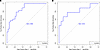 Figure 2
Proteomic profiling of CSF exosomes after SCI. (A and B) ROC curves illustrate the performance of combination proteins in distinguishing patient outcomes: 5 proteins (amyloid-β1–42 + KLK6 + NCAM-1 + IFN-α + IP-10) for differentiating between complete and incomplete injuries (AUC = 0.854) (A) and 4 proteins (MIF + HGF + IL-1RA + IL-10) for distinguishing between improvement and non-improvement in the severe group (AUC = 0.813) (B).
Construction of weighted co-expression networks and identification of modules associated with injury severity and recovery. CSF-derived exosomal miRNAs are more likely to reflect CNS-specific processes, making them valuable candidates for biomarkers to assess SCI severity and predict recovery outcomes. In order to investigate exosome miRNAs associated with injury severity, we first addressed potential serum contamination by normalizing all miRNA reads using the transcripts per million method and performing sample clustering to distinguish CSF from serum profiles. Some samples were excluded as a result of contamination with blood. Samples with low read counts (<5) and poor clustering performance were excluded. After quality control, 38 high-quality CSF samples were selected for downstream analysis.
Further, to identify coordinated gene expression networks associated with SCI severity and recovery, we applied weighted gene co-expression network analysis (WGCNA) to miRNAs with read counts ≥5 in at least 3 filtered CSF samples. This approach reduced transcriptome-wide expression variability and yielded 450 miRNAs for further analysis. Before network construction, samples were clustered and visualized via heatmap to assess the relationship between clinical traits and sample groupings (Figure 3A). For network construction, pairwise miRNA expression correlations were computed, and the resulting similarity matrix was transformed into an adjacency matrix to reflect connection strength between miRNAs. A soft-thresholding power of β = 7 was chosen to approximate scale-free network topology (scale-free R2 = 0.90; Figure 3, B and C). Hierarchical agglomerative clustering using 1-Topological Overlap Matrix as the distance metric was then performed to identify modules of co-expressed miRNAs, using the dynamic tree cut algorithm with a minimum module size of 10. This process identified 17 distinct co-expression modules (Figure 3, D and E).
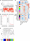 Figure 3
Identification and correlation of WGCNA modules across all time points. (A) Sample dendrogram and trait heatmap, displaying 5 traits: AIS, AIS improvement, motor improvement, BASIC, and sex. (B) Scale-free topology fit index (y axis) across different soft-thresholding powers (β) (x axis). (C) Mean connectivity (degree, y axis) analysis for various soft-thresholding powers (x axis). (D) Gene clustering dendrogram based on topological overlap, with assigned module colors. (E) Module-trait relationships, where each row represents a module eigengene (ME) and each column a trait. Correlation values (top) and P values (bottom) are displayed in each cell, color-coded by correlation strength.
We then examined correlations between module eigengenes (MEs) and clinical traits, including injury severity measures (complete vs. incomplete status, assessed by AIS grade and Brain and Spinal Injury Center [BASIC] score), recovery-related outcomes (AIS improvement and motor function improvement), and sex. At a significance threshold of P < 0.05, the MEblue module showed a significant negative correlation with BASIC score (r = –0.39; P = 0.02), suggesting that its constituent miRNAs are associated with more severe injury. On the other hand, the brown (r = 0.38; P = 0.02) and turquoise (r = 0.40; P = 0.01) modules were positively correlated with motor improvement. In contrast, the cyan (r = –0.35; P = 0.03), midnight blue (r = –0.39; P = 0.02), and purple (r = –0.34; P = 0.03) modules were negatively correlated with AIS improvement.
To investigate the functional roles of these modules, hub miRNAs were identified within each module by |module membership| > 0.8 and |gene significance| > 0.2 (Supplemental Table 2), and their target genes were determined by intersecting results from 2 validated miRNA-target databases: TarBase v9.0 (24) and miRTarBase v9.0 (25). Only targets present in both databases were retained for downstream analysis. Functional enrichment analysis of the miRNA target genes in each module was performed using the clusterProfiler package. As shown in Figure 4, A and B, the turquoise, brown, and blue modules were significantly enriched in pathways related to unfolded protein binding, stem cell differentiation, gliogenesis, and axon development, etc. In contrast, the midnight blue, cyan, and purple modules were enriched in immune response, neuronal apoptosis, toxin transport, and type I interferon signaling (Figure 4, C and D, and Supplemental Figure 2, A–D).
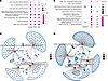 Figure 4
Gene set enrichment and the network of functional categories and their associated genes in WGCNA modules across all time points. (A and B) Dot plots of Gene Ontology enrichment analysis for turquoise, brown, and blue modules (A) and midnight blue, cyan, and purple modules (B). (C and D) The network in each module visualization highlights enriched functional categories and their associated genes. Functional enrichment analysis for the brown (C) and turquoise (D) modules was performed based on the target genes of hub miRNAs.
Differential expression of exosomal miRNAs in complete versus incomplete SCI groups. To identify miRNA signatures based on injury severity (complete AIS A vs. incomplete AIS B/C), we performed differential expression analysis using DESeq2 to identify significantly altered genes from days 1, 3, and 5 (Figure 5, A and B). This analysis identified 18 significantly differentially expressed genes (DEGs), including 7 miRNAs upregulated and 11 miRNAs downregulated in the complete injury group (Figure 5, A and B).
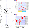 Figure 5
Differential gene expression analysis between complete and incomplete injuries in CSF. (A and B) Volcano plot (A) and heatmap (B) of DEGs among all patients with complete and incomplete injuries. (C and D) Volcano plot (C) and heatmap (D) of DEGs among patients (from day 1) with complete and incomplete injuries. Blue dots indicate DEGs with fold changes ≤ –1.5 and P value < 0.05, while red dots represent DEGs with fold changes ≥ 1.5 and P value < 0.05.
We additionally analyzed expression patterns in the acute phase of SCI at different time points both to better understand this secondary injury cascade and to potentially improve clinical decision-making. Evaluating DEGs on day 1 is clinically relevant, as SCI patients present to the hospital within 24 hours of injury for rapid evaluation and treatment. Therefore, the diagnostic utility of significant day 1 DEGs can aid in decision-making, especially if a patient has an unreliable neurological exam. Figure 5, C and D, displays 19 significant DEGs, including 6 genes upregulated in complete cases and 13 downregulated. Analysis of miRNA expression profiles revealed a severity-dependent pattern, with AIS A patients showing reduced expression of several miRNAs. On day 1 after injury, AIS A patients exhibited significantly lower levels of hsa-miR-429, hsa-miR-182-5p, and hsa-miR-7-1-3p. miR-429 has been previously shown to regulate neuronal survival. In a rat SCI model, reduced miR-429 levels were associated with increased neuronal apoptosis through activation of PTEN, which inhibits the PI3K/Akt signaling pathway — a critical axis for promoting cell survival (26). Additionally, miR-429 has been shown to suppress Notch1 signaling, which is implicated in neural injury under hypoxic conditions (27), suggesting that its downregulation may exacerbate neurodegenerative processes following SCI. Similarly, miR-182 plays a protective role in SCI by targeting the IKKβ/NF-κB pathway. Inhibition of this proinflammatory cascade by miR-182 results in reduced levels of IL-1, IL-6, and TNF, thereby suppressing inflammation and apoptosis during secondary injury (28). In our cohort, the decreased expression of miR-182 in AIS A patients is consistent with increased inflammatory damage and more severe outcomes. miR-7, particularly miR-7a, has also been reported as neuroprotective. It downregulates NF-κB signaling while upregulating Bcl-2, an anti-apoptotic protein, promoting cell survival after SCI. In rat models, intrathecal administration of miR-7a led to increased expression of neurotrophic factors such as BDNF and NT-3, as well as enhanced axonal regeneration and structural repair at the injury site (29). Beyond these functions, miR-7 also targets mitochondrial pathways by regulating VDAC1 and promotes antioxidative defense by inhibiting Keap1, thereby activating the Nrf2 pathway and protecting against ROS-induced toxicity (30). Together, these data highlight the dynamic and injury severity–dependent regulation of neuroprotective and inflammatory miRNAs in acute SCI, supporting their potential roles as both biomarkers and therapeutic targets.
Differential expression of exosomal miRNAs in AIS A improvement versus non-improvement groups. Given the variability in patient recovery outcomes, we aimed to predict recovery results based on early miRNA characteristics. Using clinical data on neurological exams recorded at 3 months after injury, we classified samples into improvement (>1 AIS grade) and non-improvement groups and identified significant DEGs between the two groups (Supplemental Figure 2, E and F). This analysis revealed only 7 significant DEGs, with 4 upregulated and 3 downregulated in the improvement group. For example, miR-449a suppresses glioma cell proliferation by downregulating PKCα expression (31) and also facilitates neurological recovery and promotes neuronal differentiation (32, 33).
Further evaluation revealed that most patients with incomplete injuries (AIS B/C) demonstrated improvement at 3 months, whereas only about half of those with complete injuries (AIS A) showed recovery. Given this strong association between initial injury severity and recovery potential, predictive modeling across the full cohort would largely reflect baseline severity differences rather than true biological signals of recovery (34). Therefore, to identify miRNAs specifically associated with neurological improvement independent of injury severity, we focused the recovery prediction analysis on patients with complete SCI, whose recovery outcomes are more variable, so our findings would be more clinically relevant. This approach allowed us to identify miRNAs potentially associated with differential recovery trajectories within the complete injury group. In day 1 samples, we identified 23 significant DEGs, with 11 upregulated in the AIS A improvement group (Supplemental Figure 3). We found an increase in certain genes associated with inflammation inhibition, including hsa-miR-375-3p (35), in the improvement groups (Supplemental Figure 3, A and B). Additionally, the AIS A improvement group exhibited enrichment in axon development, mesenchymal cell differentiation, and glutamatergic synapses (Supplemental Figure 3C), whereas the non-improvement groups were enriched in inflammatory response, autophagy, neuro-apoptotic processes, and fibroblast proliferation (Supplemental Figure 3D).
Additionally, we analyzed marker genes from days 3 and 5 and a combination of all days 1, 3, and 5 (Supplemental Figure 4). For days 1/3/5, we identified 30 significant DEGs, with 9 upregulated and 21 downregulated in the AIS A improvement group (Supplemental Figure 4, A and B). On days 3/5, we identified 42 significant DEGs, including 5 upregulated and 37 downregulated in the AIS A improvement group (Supplemental Figure 4, C and D). In AIS A patients who demonstrated neurological improvement, differential expression of specific miRNAs appeared to contribute to reduced neuroinflammation, inhibition of apoptosis, and enhanced neuroregeneration (e.g., hsa-miR-708-5p, hsa-miR-362-5p, hsa-miR-195-5p). For instance, miR-195 functions as a negative regulator of HIF-1α, a transcription factor that promotes neuroprotection under ischemic conditions by upregulating survival-related genes such as VEGF and Bcl-2. This regulation supports neuronal survival and facilitates functional recovery (36). Although miR-195 suppresses HIF-1α, its precise role may depend on timing and cellular context, potentially balancing protective and detrimental responses after injury. Another example is miR-362, which has been shown to inhibit PAX2 expression. In a rat SCI model, overexpression of miR-362 led to downregulation of the ERK/MEK and p38 MAPK signaling pathways — both of which are associated with inflammation and cell death. This suppression resulted in reduced neuroinflammation and apoptosis, ultimately leading to improved motor outcomes (37). miR-708-5p may suppress inflammation in rheumatoid arthritis by inhibiting the Wnt3a/β-catenin pathway (38). It is also significantly downregulated in glioma, and its restoration reduces glioma cell proliferation and invasion (39). These findings suggest that recovery-associated miRNAs modulate key signaling pathways involved in inflammation, apoptosis, and regeneration, highlighting their potential role in facilitating long-term recovery and serving as therapeutic targets in SCI.
Astrocyte gap closure assay and miRNA mimic treatment to investigate the role of CSF exosome miRNAs in astrocyte activation. CSF exosomes originate from multiple CNS cell types, including astrocytes, microglia, and neurons. In the context of SCI, exosomes released into CSF may reflect the cellular and molecular events occurring within the CNS and facilitate long-distance intercellular communication. Emerging evidence suggests that exosomes contribute to secondary injury by delivering cell type–specific molecular cargo, such as miRNAs. Following SCI, astrocytes rapidly become reactive and undergo significant morphological and functional changes, impacting neuronal recovery (40, 41). To investigate the effects of CSF exosomal miRNAs on astrocyte behavior, we selected 10 miRNAs that were differentially expressed in the improvement group versus the non-improvement group and examined their effects using a gap closure assay after scratch to assess astrocyte proliferation and migration. Treatment of human astrocytes with mimics of hsa-miR-449a, hsa-miR-375-3p, hsa-miR-30c-5p, hsa-miR-6873, and hsa-miR-708-5p, as well as inhibitors of hsa-miR-7111-3p and hsa-miR-195-5p, resulted in a significant reduction in the rate of gap closure in comparison with the control group (Figure 6 and Supplemental Figure 5). These findings indicate that modulation of these miRNAs — either by mimic or inhibitor — suppresses astrocyte proliferation and migration.
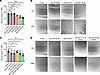 Figure 6
Results of astrocyte gap closure assay. Results of scratch assay at 36 hours after miRNA mimic/inhibitor treatments. (A and C) Results of gap closure measurements reveal the miRNAs that significantly inhibit astrocyte proliferation and migration in filling the gaps. Asterisks indicate significance based on unadjusted P values. Data are represented as mean ± SD; n = 4; 1-tailed independent t test. *P < 0.05, **P < 0.01, ***P < 0.001. Multiple-testing–adjusted P values (Benjamini-Hochberg, false discovery rate correction) are shown in the Supporting Data Values. In A, all results remained significant after correction. In C, miR-195-5p inhibitor retained significance after correction (adjusted P = 0.0085), and adjusted P = 0.0616 for miR-30c-5p, miR-6873, and miR-7111-3p inhibitors. (B and D) Images of scratch area from groups at 0- and 36-hour time points. Scale bars: 200 μm.
miRNA-mediated regulation of astrocyte secretory factors shapes neurite growth and synaptogenesis. Based on the observed association between specific miRNA expression patterns and patient recovery outcomes, we further examined whether astrocyte-derived factors regulated by these miRNAs could influence neuronal growth and connectivity. To this end, astrocytes were scratched and transfected with selected miRNA mimics or inhibitors and subsequently cocultured with SH-SY5Y neurons in a compartmentalized system. Neurite extension and synaptogenesis were then assessed (Figure 7). Our results demonstrated that in astrocyte-neuron coculture transfected with hsa-miR-16-2-3p, hsa-miR-449a, hsa-miR-375-3p, hsa-miR-30c-5p, hsa-miR-6873-3p, and hsa-miR-362-5p mimics, as well as hsa-miR-195-5p and hsa-miR-7111-3p inhibitors, neurite outgrowth was significantly enhanced in comparison with scramble control. Synapse formation, as quantified by synapsin-1 puncta per neuron, was also significantly increased following astrocyte transfection with hsa-miR-16-2-3p, hsa-miR-449a, hsa-miR-375-3p, and hsa-miR-30c-5p mimics, or hsa-miR-195-5p and hsa-miR-7111-3p inhibitors. In contrast, treatment with hsa-miR-20a-5p or hsa-miR-708-5p reduced synapse density despite their modest or neutral effects on neurite elongation. Importantly, 6 miRNAs — hsa-miR-16-2-3p, hsa-miR-449a, hsa-miR-375-3p, hsa-miR-30c-5p, hsa-miR-195-5p inhibitor, and hsa-miR-7111-3p inhibitor — displayed dual functions, simultaneously enhancing neurite extension and synapse formation. These results suggest that a subset of miRNAs exerts regulatory effects on astrocyte secretory programs that directly shape neuronal connectivity.
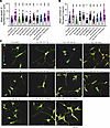 Figure 7
Effects of miRNA-mediated regulation on human neurite growth and synaptogenesis in a compartmentalized astrocyte-neuron coculture model. (A and B) Quantitative analysis of neurite length and synapsin-1 (Syn1) puncta per neuron in compartmentalized cocultures of injured astrocytes transfected with selected hsa-miRNA mimics or inhibitors. For neurite length and Syn1 puncta analysis, at least 20 randomly selected images per treatment group were acquired at ×40 magnification to measure the mean neurite length per neuron and the mean number of synaptic puncta per neuron. Asterisks indicate significance based on unadjusted P values (Benjamini-Hochberg). Multiple-testing–adjusted P values are shown in the Supporting Data Values. All comparisons significant by raw P value remained significant after correction. Data are represented as mean ± SD. *P < 0.05, **P < 0.01, ***P < 0.001 (1-tailed independent t test). (C) Representative immunofluorescence images of SH-SY5Y neurons stained for TUJ1 (green), Syn1 (red), and DAPI (blue). Scale bar: 100 μm.
-
Discussion
Recent research underscores the importance of identifying CSF and serum biomarkers that provide insight into SCI severity and can aid in predicting neurological recovery. Such biomarkers are essential for establishing objective indicators that are not captured by standard ASIA examinations or MRI. Despite growing interest, no prior studies have characterized CSF exosomal protein or miRNA biomarkers in the acute phase of SCI that are predictive of long-term outcomes. In this study, we comprehensively characterized the exosomal miRNAs and proteins in CSF and serum from SCI patients.
CSF more accurately reflects pathological changes in CNS than serum. Exosomes offer further advantages due to their cargo stability and ability to carry cell-specific signals. They can be engineered to deliver therapeutic miRNAs (42, 43) and have shown potential in SCI treatment (44–46). For example, Li et al. demonstrated that CSF-derived exosomes promoted vascular regeneration and improved motor function after SCI (47). Thus, we performed protein analysis on CSF exosomes and identified marker panels for distinguishing complete from incomplete SCI patients and improved from non-improved patients using ROC analysis. We found 15 CSF-specific and 9 serum-specific exosomal miRNAs that were consistently expressed regardless of injury severity. Using WGCNA, we identified 3 positively and 3 negatively correlated miRNA modules associated with SCI severity and recovery. Gene set enrichment analysis revealed that these modules were involved in specific biological processes. By combining clinical improvement data collected at 3 months after injury among AIS A complete SCI patients, we identified differentially expressed miRNAs between improvement and non-improvement cases. Functional tests using an astrocyte gap closure assay and astrocyte-neuron coculture demonstrated that a number of miRNAs enriched in the improvement group decreased astrocyte proliferation and migration, and promoted neurite extension and synapse formation. Given that previous knowledge of SCI pathophysiology is mostly rooted in experimental animal models, our results will help tie together preclinical data to better describe the human pathophysiology of the injury process, and highlight promising exosomal miRNA and protein biomarker candidates for improving early diagnosis, predicting long-term outcomes, and guiding therapeutic discovery in SCI.
Our findings demonstrate that protein and cytokine levels in CSF-derived exosomes are strongly associated with SCI severity and neurological recovery. Using a panel of 5 proteins, we developed a predictive model that classified injury severity with an accuracy of 0.854. A separate model, based on 4 proteins, predicted neurological improvement — defined as a gain of at least 1 AIS grade at or beyond 3 months after injury — with an accuracy of 0.813. Similarly to our approach, previous studies have sought to correlate protein biomarkers with clinical and imaging features in SCI and other neurological conditions (48). For example, Kwon et al. constructed a 6-protein model (IL-6, IL-8, MCP-1, tau, GFAP, and S100β) that predicted 6-month recovery with 83% accuracy (6), outperforming the prognostic utility of baseline ASIA grade (Concordance Index = 0.773). In addition, Skinnider et al. reported dynamic protein changes in acute SCI using total CSF protein profiling. They identified protein biomarkers associated with injury severity and recovery (49), providing valuable information. While these studies focused on whole CSF, our work specifically examines CSF-derived exosomes. CSF is a complex fluid that bathes the CNS and contains a mixture of proteins, nucleic acids, metabolites, and cellular debris released from various cell types, while exosomes are small EVs actively secreted by cells and selectively packaged with specific proteins, lipids, and RNAs, including miRNAs, reflecting the physiological or pathological state of their cells of origin. Because of this selective packaging, the molecular profile of CSF exosomes can differ substantially from that of whole CSF. This distinction is particularly important in disease contexts such as SCI, where exosomes may carry more precise information and low-abundance biomarkers that are diluted or masked in whole CSF. Therefore, analyzing CSF-derived exosomes can provide unique insights into cellular responses and disease mechanisms that are not always apparent from examining whole CSF alone.
Emerging evidence indicates that individuals with SCI are at elevated risk for developing Alzheimer’s disease (AD) compared with non-SCI populations (50, 51). Aβ is a well-established biomarker in AD, and our data suggest a link between Aβ42 levels and SCI severity. In particular, we found that Aβ42 levels in CSF-derived exosomes were lower in patients with severe injuries. Given that CSF plays a critical role in clearing waste from the CNS, including Aβ, this observation aligns with reports showing higher Aβ levels in healthy individuals relative to those with AD. Consistently, in our cohort, Aβ42 levels were higher in patients with incomplete injuries compared with those with complete injuries, suggesting that more effective Aβ clearance during the acute phase of SCI may support better long-term functional recovery. These findings underscore the importance of further investigating the role of Aβ in the transition from acute SCI to chronic neurodegeneration. A deeper understanding of the mechanisms could inform the development of exosome-based therapeutics targeting both SCI-related pathology and dementia risk.
miRNAs contained within CSF-derived exosomes reflect dynamic molecular changes within the CNS, making them promising biomarkers for distinguishing between complete and incomplete SCI. To identify miRNAs associated with injury severity and neurological recovery, we performed WGCNA, which revealed several modules correlated with key clinical outcomes. Functional inference was conducted by analysis of the target protein-coding genes of hub miRNAs within each module. Notably, the turquoise and brown modules were significantly associated with motor recovery. Target gene analysis of miRNAs in these modules revealed strong enrichment for pathways implicated in SCI. For example, prior studies have demonstrated that activation of the unfolded protein response can mitigate tissue damage and enhance locomotor recovery following SCI (52). Several hub miRNAs within these recovery-associated modules also appear to play roles in neuronal function. Among them, miR-449a, identified as a hub gene in the turquoise module, was enriched in patients who exhibited neurological improvement (Supplemental Figure 2F). Consistent with our findings, previous research using cerebral ischemia models showed that miR-449a upregulation promoted neurological recovery via activation of the PI3K/Akt signaling pathway (33). In contrast, the midnight blue, cyan, and purple modules were primarily associated with immune response pathways, which are generally considered detrimental to SCI recovery. These findings highlight the functional heterogeneity of miRNA modules and suggest that distinct regulatory networks contribute to either recovery or persistent dysfunction following SCI.
We observed that most patients with incomplete SCI showed neurological improvement within 3 months, whereas approximately half of the patients with complete injuries also demonstrated measurable recovery. To further investigate the molecular basis of these outcomes, we analyzed CSF exosomes collected from complete SCI cases at days 1, 3, and 5 after injury. This analysis identified miRNAs with significantly altered expression in patients who showed neurological improvement compared with those who did not. Functional studies indicated that these improvement-associated miRNAs exert important effects on astrocyte biology. Specifically, miRNAs can influence astrocyte proliferation and migration and, through astrocyte-derived secretory factors, modulate neurite outgrowth and synaptogenesis. We separately tested mimics for 8 miRNAs positively correlated with patient improvement. When astrocytes were treated with mimics of 5 of these miRNAs, cell proliferation and migration were inhibited, suggesting a regulatory role in astrocyte activation. Conversely, we inhibited 2 miRNAs that were negatively associated with patient improvement and also observed decreased astrocyte proliferation and migration. Of the 10 tested, 8 miRNAs demonstrated functional roles in promoting neurite extension, and 8 miRNAs in synapse formation in astrocyte-neuron coculture. Combining the results from both astrocyte gap closure assays and astrocyte-neuron coculture, we found that mimics of hsa-miR-375 and hsa-miR-30c-5p suppressed astrocyte proliferation and migration, and enhanced neurite growth and synaptogenesis. Also, inhibitors of hsa-miR-7111-3p and hsa-miR-195-5p produced similar effects, suggesting that these miRNAs may contribute to balancing glial activity and promote neuronal regeneration. Mechanistically, miR-375-3p has been reported to regulate neurogenesis and motor neuron development through modulation of PAX6 and CCND2, and to suppress p53-mediated apoptosis (53). However, the roles of many of the other miRNAs identified in our study remain poorly characterized, with limited prior research available regarding their impact on neuronal development or recovery. For example, hsa-miR-7111-3p was specifically enriched in non-improvement cases and, to our knowledge, has not been previously reported in the SCI literature. Consistent with these findings, qPCR validation demonstrated that hsa-miR-7111-3p expression was significantly elevated in non-improvement cases, at day 1 and days 1/3/5 after injury (Figure 8, A and C), supporting its potential as a robust biomarker predictive of SCI recovery outcomes. Although the comparison at days 3/5 alone did not reach statistical significance (Figure 8B), the expression trend remained consistent with the sequencing results. Together, these findings suggest that improvement-associated miRNAs may contribute to neurological recovery after SCI by regulating astrocyte activation and the release of astrocyte-derived factors that influence neuronal regeneration. By fine-tuning astrocyte secretory activity, these miRNAs may facilitate neuronal repair and functional recovery after SCI, highlighting their potential as therapeutic targets for enhancing repair mechanisms following SCI.
 Figure 8
qPCR analysis of hsa-miR-7111-3p expression in patient CSF exosomes. hsa-miR-7111-3p expression level was higher in the non-improvement patient group compared with the improvement patient group. (A) Day 1 after injury (improvement patients, n = 4; non-improvement patients, n = 4). (B) Day 3/5 after injury represents pooled samples from days 3 and 5 (improvement patients, n = 4; non-improvement patients, n = 5). (C) Day 1/3/5 after injury represents pooled samples from days 1, 3, and 5 (improvement patients, n = 5; non-improvement patients, n = 5). Results are expressed as mean ± SEM for each group. *P < 0.05, **P < 0.01 (1-tailed independent t test).
Despite its strengths, our study has limitations. Attrition is a common challenge in SCI research owing to patient mobility limitations and follow-up barriers. However, our cohort had the benefit of clinical follow-up through 3 months, which is the time with the most rapid rate of motor recovery (54). Although CSF was collected on days 1, 3, and 5, some samples were excluded because of insufficient volume or contamination with blood. This is due to the inherent nature of SCI, with traumatic bleeding into the CSF occasionally causing issues with drainage through the lumbar catheter. CSF was obtained only during the initial hospitalization and not in the clinic at follow-up, as obtaining CSF at follow-up is not the standard of care. Lumbar puncture has risks such as bleeding, infection, CSF leak, and nerve injury; therefore, it is not medically/ethically appropriate to obtain in these patients without any diagnostic or therapeutic benefit. For statistical analysis, to control type I error, multiple-testing correction was applied using the Benjamini-Hochberg false discovery rate method. The adjusted P values demonstrated that the majority of findings remained significant after correction; for example, 27 of 30 comparisons in the Supporting Data Values file (corresponding to Figure 6, A and C, and Figure 7, A and B) were consistent between raw and adjusted analyses. Therefore, adjusted P value analyses support our overall conclusions. Future studies with larger sample sizes will further enhance the rigor of the study. Finally, the experiments were primarily designed to investigate the effects of miRNAs on astrocytes, and astrocyte-neuron interactions. Although the effects on microglia are outside the scope of the current study, this could be an interesting area for future study.
In conclusion, our study presents an extensive investigation of the human exosome in the CSF and serum of patients with acute SCI. We identified miRNA and protein biomarkers in CSF-derived exosomes that could potentially improve early diagnosis and predict outcome and treatment of SCI. These findings provide a foundation for future mechanistic investigations into targeted therapies and personalized medicine approaches for SCI recovery.


