
Research ArticleCell biologyImmunology
Open Access |  10.1172/jci.insight.206701
CXCR4 coordinates adhesion, migration, and development of human NK cells
Shira E. Eisman,1,2 Francesca E. Grossberg,1,2 Batya S. Koenigsberg,1,2 David H. McDermott,3 Frédérique van den Haak,1 Luis A. Pedroza,1 Everardo Hegewisch-Solloa,1,2 Philip M. Murphy,3 and Emily M. Mace1,2
1Department of Pediatrics, Vagelos College of Physicians and Surgeons, and
2Columbia Stem Cell Initiative, Columbia University Irving Medical Center, New York, New York, USA.
3Laboratory of Molecular Immunology, National Institute of Allergy and Infectious Diseases, Bethesda, Maryland, USA.
Address correspondence to: Emily M. Mace, Columbia University Irving Medical Center, 630 W. 168th Street, New York, New York 10032, USA. Phone: 212.305.7836; Email: em3375@cumc.columbia.edu. LAP’s present address is: Department of Pediatrics, Children’s Hospital of Philadelphia, Philadelphia, Pennsylvania, USA.
Find articles by Eisman, S. in: PubMed | Google Scholar
1Department of Pediatrics, Vagelos College of Physicians and Surgeons, and
2Columbia Stem Cell Initiative, Columbia University Irving Medical Center, New York, New York, USA.
3Laboratory of Molecular Immunology, National Institute of Allergy and Infectious Diseases, Bethesda, Maryland, USA.
Address correspondence to: Emily M. Mace, Columbia University Irving Medical Center, 630 W. 168th Street, New York, New York 10032, USA. Phone: 212.305.7836; Email: em3375@cumc.columbia.edu. LAP’s present address is: Department of Pediatrics, Children’s Hospital of Philadelphia, Philadelphia, Pennsylvania, USA.
Find articles by Grossberg, F. in: PubMed | Google Scholar
1Department of Pediatrics, Vagelos College of Physicians and Surgeons, and
2Columbia Stem Cell Initiative, Columbia University Irving Medical Center, New York, New York, USA.
3Laboratory of Molecular Immunology, National Institute of Allergy and Infectious Diseases, Bethesda, Maryland, USA.
Address correspondence to: Emily M. Mace, Columbia University Irving Medical Center, 630 W. 168th Street, New York, New York 10032, USA. Phone: 212.305.7836; Email: em3375@cumc.columbia.edu. LAP’s present address is: Department of Pediatrics, Children’s Hospital of Philadelphia, Philadelphia, Pennsylvania, USA.
Find articles by Koenigsberg, B. in: PubMed | Google Scholar
1Department of Pediatrics, Vagelos College of Physicians and Surgeons, and
2Columbia Stem Cell Initiative, Columbia University Irving Medical Center, New York, New York, USA.
3Laboratory of Molecular Immunology, National Institute of Allergy and Infectious Diseases, Bethesda, Maryland, USA.
Address correspondence to: Emily M. Mace, Columbia University Irving Medical Center, 630 W. 168th Street, New York, New York 10032, USA. Phone: 212.305.7836; Email: em3375@cumc.columbia.edu. LAP’s present address is: Department of Pediatrics, Children’s Hospital of Philadelphia, Philadelphia, Pennsylvania, USA.
Find articles by
McDermott, D.
in:
PubMed
|
Google Scholar
|

1Department of Pediatrics, Vagelos College of Physicians and Surgeons, and
2Columbia Stem Cell Initiative, Columbia University Irving Medical Center, New York, New York, USA.
3Laboratory of Molecular Immunology, National Institute of Allergy and Infectious Diseases, Bethesda, Maryland, USA.
Address correspondence to: Emily M. Mace, Columbia University Irving Medical Center, 630 W. 168th Street, New York, New York 10032, USA. Phone: 212.305.7836; Email: em3375@cumc.columbia.edu. LAP’s present address is: Department of Pediatrics, Children’s Hospital of Philadelphia, Philadelphia, Pennsylvania, USA.
Find articles by van den Haak, F. in: PubMed | Google Scholar
1Department of Pediatrics, Vagelos College of Physicians and Surgeons, and
2Columbia Stem Cell Initiative, Columbia University Irving Medical Center, New York, New York, USA.
3Laboratory of Molecular Immunology, National Institute of Allergy and Infectious Diseases, Bethesda, Maryland, USA.
Address correspondence to: Emily M. Mace, Columbia University Irving Medical Center, 630 W. 168th Street, New York, New York 10032, USA. Phone: 212.305.7836; Email: em3375@cumc.columbia.edu. LAP’s present address is: Department of Pediatrics, Children’s Hospital of Philadelphia, Philadelphia, Pennsylvania, USA.
Find articles by Pedroza, L. in: PubMed | Google Scholar
1Department of Pediatrics, Vagelos College of Physicians and Surgeons, and
2Columbia Stem Cell Initiative, Columbia University Irving Medical Center, New York, New York, USA.
3Laboratory of Molecular Immunology, National Institute of Allergy and Infectious Diseases, Bethesda, Maryland, USA.
Address correspondence to: Emily M. Mace, Columbia University Irving Medical Center, 630 W. 168th Street, New York, New York 10032, USA. Phone: 212.305.7836; Email: em3375@cumc.columbia.edu. LAP’s present address is: Department of Pediatrics, Children’s Hospital of Philadelphia, Philadelphia, Pennsylvania, USA.
Find articles by
Hegewisch-Solloa, E.
in:
PubMed
|
Google Scholar
|
1Department of Pediatrics, Vagelos College of Physicians and Surgeons, and
2Columbia Stem Cell Initiative, Columbia University Irving Medical Center, New York, New York, USA.
3Laboratory of Molecular Immunology, National Institute of Allergy and Infectious Diseases, Bethesda, Maryland, USA.
Address correspondence to: Emily M. Mace, Columbia University Irving Medical Center, 630 W. 168th Street, New York, New York 10032, USA. Phone: 212.305.7836; Email: em3375@cumc.columbia.edu. LAP’s present address is: Department of Pediatrics, Children’s Hospital of Philadelphia, Philadelphia, Pennsylvania, USA.
Find articles by Murphy, P. in: PubMed | Google Scholar
1Department of Pediatrics, Vagelos College of Physicians and Surgeons, and
2Columbia Stem Cell Initiative, Columbia University Irving Medical Center, New York, New York, USA.
3Laboratory of Molecular Immunology, National Institute of Allergy and Infectious Diseases, Bethesda, Maryland, USA.
Address correspondence to: Emily M. Mace, Columbia University Irving Medical Center, 630 W. 168th Street, New York, New York 10032, USA. Phone: 212.305.7836; Email: em3375@cumc.columbia.edu. LAP’s present address is: Department of Pediatrics, Children’s Hospital of Philadelphia, Philadelphia, Pennsylvania, USA.
Find articles by
Mace, E.
in:
PubMed
|
Google Scholar
|
Published August 4, 2026 - More info
JCI Insight. 2026;11(17):e206701. https://doi.org/10.1172/jci.insight.206701.
© 2026 Eisman et al. This work is licensed under the Creative Commons Attribution 4.0 International License. To view a copy of this license, visit http://creativecommons.org/licenses/by/4.0/.
Received: March 31, 2026; Accepted: July 20, 2026

-
Results
CXCR4 is expressed on immature and mature human NK cells. To investigate whether CXCR4/CXCL12 signaling might play a role in human NK cell development, we first assessed CXCR4 expression across NK cell developmental stages by examining CXCR4 transcript levels in circulating lymphoid progenitors and NK cell subsets using data from the Human Immune Health Atlas (36). CXCR4 was detected in the innate lymphoid cell (ILC) progenitors detectable in PBMCs in this dataset, CD56bright NK cells, CD56dim NK cells, and proliferating NK cell subsets, with the highest mean expression in CD56dim NK cells (Figure 1A).
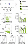 Figure 1
CXCR4 is expressed throughout human NK cell development. (A) CXCR4 transcript in innate lymphoid cell progenitors (ILCPs) and NK subsets from the Human Immune Health Atlas (66). Dot size indicates frequency of CXCR4+ cells; color represents mean expression. (B) NK cell development: CD34+ hematopoietic stem and progenitor cells (HSPCs), CD117+CD94– NK precursors (NKPs), CD94+CD16– NK cells, and CD94+CD16+ NK cells. (C) Extracellular CXCR4 in NK developmental intermediates from tonsil. Gray histogram represents fluorescence minus one (FMO) control. (D) Pooled data from 6 donors corresponding to flow cytometry data in C. n = 6; Friedman test with Dunn’s multiple-comparison correction (paired by donor). Only comparisons with P < 0.05 are displayed. (E) Extracellular CXCR4 mean fluorescence intensity (MFI) in NK developmental intermediates from peripheral blood. (F) Pooled data from 9 donors corresponding to flow cytometry data in E. n = 9; Friedman test with Dunn’s multiple-comparison correction (paired by donor). Only comparisons with P < 0.05 are displayed. (G) Representative histograms of cell surface CXCR4 MFI in primary NK cells (left) and NK92 cell lines (right) after 1-hour treatment with vehicle (solid lines) or 10 μM AMD3100 (dashed lines). Representative of more than 10 biological replicates. (H) Validation of CXCR4 knockout (KO) in NK92 cell line. CXCR4 on WT (solid line) and CRISPR/Cas9 CXCR4-KO (dashed line) cells. Representative of 4 replicates.
We next assessed CXCR4 surface expression by flow cytometry on NK cell subsets and NK progenitors from tonsil, a site of human NK cell maturation (Figure 1B, table of antibodies in Supplemental Table 1, and gating strategy in Supplemental Figure 1A; supplemental material available online with this article; https://doi.org/10.1172/jci.insight.206701DS1). As predicted by its association with early hematopoietic differentiation (37, 38), we found significantly higher CXCR4 expression in CD34+ hematopoietic stem and progenitor cells (HSPCs) from tonsil when compared with mature CD16– NK cells (Figure 1, C and D). Additionally, CD16+ NK cells from tonsil showed a significant increase in CXCR4 mean fluorescence intensity (MFI) relative to CD16– NK cells (Figure 1, C and D), confirming that the higher expression of CXCR4 transcript in CD56dim NK cells (Figure 1A) was also associated with higher protein expression. This higher protein expression seemed to be associated with NK cell developmental subsets, as CD16– and CD16+ subsets of CD56dim NK cells did not have significant differences in CXCR4 MFI (Supplemental Figure 1B). These data were supported by our analysis of scRNA-seq data from human bone marrow (39) showing similar levels of CXCR4 transcript in mature (CD56+) NK cell common lymphoid progenitors and other lineage-primed CD34+ subsets (Supplemental Figure 1C).
We then performed the same analysis using peripheral blood. Although CD34+ HSPCs are rare in peripheral blood, a similar trend toward higher expression in CD34+ HSPCs was observed relative to CD16– NK cells (Figure 1, E and F). Circulating NK/ILC precursors (Lin–CD117+CD94–) in peripheral blood (40) showed comparable CXCR4 expression to HSPCs. As in tonsil, CD16+ NK cells displayed higher CXCR4 MFI than CD16– NK cell subsets (Figure 1, E and F). Together, these flow cytometry data demonstrated that CXCR4 is expressed at all stages of human NK cell development in peripheral blood and tonsil, with differences in expression associated with changes in NK cell maturation and location.
To establish experimental models for CXCR4 perturbation, we used pharmacologic blockade with a CXCR4 antagonist (AMD3100) on primary NK cells and generated a CRISPR/Cas9–mediated CXCR4 knockout (KO) in the NK92 NK cell line. As expected, AMD3100 treatment of primary NK cells and NK92 cells reduced CXCR4 detection by flow cytometry as a result of competitive binding with the CXCR4-specific antibody clone 12G5 (Figure 1G) (41, 42). Targeted knockout of CXCR4 using CRISPR/Cas9 resulted in a single population with approximately 50% decreased CXCR4 protein expression by flow cytometry (Figure 1H). Neither pharmacologic blockade nor genetic deletion affected NK cell viability or proliferation (Supplemental Figure 2A). In Transwell assays, both AMD3100 and CXCR4 knockout significantly impaired directed migration toward CXCL12, confirming that both approaches impacted the canonical function of CXCR4 in chemotaxis (Supplemental Figure 2B). Decreased chemotaxis in the CXCR4-KO cells was moderate but could be further reduced in the presence of AMD3100 (Supplemental Figure 2B). In contrast, NK cell cytotoxic function in a standard 4-hour killing assay against K562 target cells was only modestly reduced by CXCR4 knockout or AMD3100 (Supplemental Figure 2C). Together, these results demonstrate that CXCR4 is expressed throughout NK cell development and in NK cell lines, and that it is not required for NK cell survival or cytotoxic function.
CXCR4 ligand CXCL12 is found in NK cell developmental niches. CD34+ progenitors in peripheral blood that give rise to NK cells are thought to enter the tonsil through high endothelial venules (HEVs) within the interfollicular domain, then migrate to the parafollicular domain, where they undergo further maturation (3). As CXCL12 is the primary ligand for CXCR4, we sought to define its spatial localization in tonsil using cyclic immunofluorescence (CycIF) on formalin-fixed, paraffin-embedded sections from routine pediatric tonsillectomies (43).
Observing CXCL12 expression in CycIF images, we found that CXCL12 protein was distributed throughout the tonsil but enriched in inter- and parafollicular stromal regions surrounding follicles and in HEVs (Figure 2A). Quantification of multiple regions of interest from samples from multiple donors confirmed higher CXCL12 intensity in inter- and parafollicular regions relative to B cell follicles (Figure 2B). Using our multiplexed images, we defined CD56+CD3− NK cells and used Ki67 to visualize proliferating cells (Figure 2C; additional channels shown in Supplemental Figure 3). High-magnification images showed that NK cells were often adjacent to CXCL12+ stromal cells; and many, but not all, of these were Ki67+ (Figure 2C). Further analysis revealed that CD34+ progenitors were also found adjacent to CXCL12+ stroma (Supplemental Figure 2B). These data were supported by recent studies using multiplexed immunofluorescence that showed that proliferative NK cell progenitors were significantly enriched proximal to CXCL12+ cells (43).
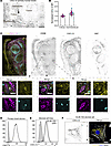 Figure 2
CXCL12 is found in sites that support human NK cell development. Cyclic immunofluorescence was performed on pediatric human tonsil. (A) Representative immunofluorescence staining of CXCL12 in primary human tonsil tissue with follicles and interfollicular and parafollicular regions annotated. Scale bar: 200 μm. (B) Quantification of normalized CXCL12 MFI comparing follicular versus para- and interfollicular regions; each point represents a region of interest from 3 donors, 8–12 regions per donor. Large symbols represent mean of values from individual donors. (C) Cyclic immunofluorescence staining of pediatric tonsil showing CD56+ NK cells (yellow), CXCL12 (magenta), and Ki67 (cyan). Grayscale images show individual channels CD56, CXCL12, and Ki67. Bottom panels show higher-magnification views of NK cell–rich para- and interfollicular stromal regions with CD56+ NK cells adjacent to CXCL12+ stroma. Scale bars: 25 μm. (D) Flow cytometric analysis of primary tonsil stromal cells stained for intracellular CXCL12 (white) compared with fluorescence minus one (FMO) control (gray). (E) CXCL12 expression by stromal cell lines. Flow cytometry comparing intracellular CXCL12 expression on EL08.1D2 (solid line) versus OP9 (dashed line) stromal cells with FMO control (gray). (F) Confocal imaging of EL08.1D2 stromal cells showing CXCL12 (yellow), DAPI (blue), and actin (white). Scale bar: 5 μm.
To further demonstrate that tonsil stromal cells are a source of CXCL12, we isolated CD45− primary tonsil stromal cells from routine tonsillectomies. Flow cytometry demonstrated that primary tonsil stroma cells uniformly expressed CXCL12 that was detected following fixation and permeabilization (Figure 2D). We also found that cell lines that support NK cell development, including OP9 and EL08.1D2, similarly expressed CXCL12 (Figure 2E). We confirmed this finding with confocal microscopy of EL08.1D2, which showed intracellular and cell surface expression of CXCL12 (Figure 2F). These results are consistent with CXCL12 being produced intracellularly for secretion and subsequent binding to the extracellular matrix (44, 45). Together, these data demonstrate that CXCL12 is produced by stromal cells in the inter- and parafollicular zones, forming chemokine-rich microenvironments that likely guide NK progenitors and other CXCR4+ cells in the tonsil (43).
CXCR4 promotes integrin-mediated interactions between NK cells and stroma. Since NK cell development depends on direct contact with stromal cells and NK cells undergo surveillance-like migration on stromal monolayers in vitro (12, 13), we asked whether CXCR4/CXCL12 contributes to these migratory behaviors. CD56+ NK cells and CD34+ progenitors from cord blood were pretreated with vehicle (water) or AMD3100 and plated on EL08.1D2 stromal monolayers. To assess these interactions in the context of spontaneous cell migration, cells were imaged every 2 minutes for 2–6 hours by live-cell confocal microscopy (Figure 3A). We used Cellpose (46) to segment the cells, btrack (47) to track the cells, and Cell Plasticity Analysis Tool (cellPLATO) (48) to quantify and plot cell motility and shape parameters (Figure 3B). This analysis focuses on spontaneous contact-mediated motility, in contrast to the directed chemotactic migration measured in Transwell assays (Supplemental Figure 2B).
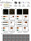 Figure 3
CXCR4-CXCL12 interaction promotes integrin-mediated arrest on stromal cells. Primary CD56+ NK cells and CD34+ progenitors were plated on EL08.1D2 stromal monolayers or integrin ligand surfaces and imaged by confocal microscopy every 2 minutes for 2–6 hours. (A) Schematic of NK cell migration assay. (B) Overview of the cellPLATO analysis pipeline. Time-lapse images were segmented using Cellpose, tracked using btrack, and processed through the cellPLATO workflow. (C) Migration of primary NK cells treated with 10 μM AMD3100 or vehicle on stroma. Top: Representative segmentation masks and cell trajectories. Scale bar: 100 μm. Color gradient signifies time. Plots of differences show mean speed, arrest coefficient, and aspect ratio. Each point represents a single cell. Effect size was calculated by bootstrap resampling. (D) Migration of CD34+ progenitor cells treated with 10 μM AMD3100 or vehicle on stroma. Top: Representative segmented images and cell tracks of migrating CD34+ cells. Scale bar: 100 μm. Color gradient signifies time. Migration and shape parameters for CD34+ cells, including mean speed, arrest coefficient, and aspect ratio, are shown. (E) Time-lapse sequence of CD34+ HSPC with segmentation shown in red interacting with a stromal cell. Asterisk shows the point of contact. Time is shown as hh:mm. See also Supplemental Video 2 and Supplemental Table 2.
We first analyzed the migration of primary NK cells (Figure 3C). Individual cell trajectories over time were visibly longer and less confined in AMD3100-treated cultures (Figure 3C and Supplemental Videos 1 and 2), suggesting changes in migratory behavior resulting from CXCR4 inhibition. We quantified cell migration parameters, including cell speed and arrest coefficient (the frequency of time a cell is immotile as defined by moving under 1 μm/min). To visualize these differences, we used plots of differences (49) to display the effect size distribution of more than 8,000 cells between conditions. In the vehicle-treated condition, NK cells exhibited characteristic non-directed migration on stromal monolayers, with heterogeneous motility patterns and intermittent arrest (Supplemental Video 1). Overall, AMD3100 treatment increased motility and reduced arrest on stroma (Figure 3C and Supplemental Video 2). Specifically, AMD3100-treated cells had significantly higher average speeds (1.233 μm/min vs. 0.986 μm/min for vehicle) with a reduction in arrest coefficient (0.374 vs. 0.483 for vehicle). These data reflect the AMD3100-treated NK cells becoming more motile and less adherent to the stromal surface. Additionally, shape analysis demonstrated rounder, less elongated cells under AMD3100 treatment (aspect ratio of 1.312 vs. 1.319 for vehicle). Similar trends were observed in additional biological replicates (Supplemental Figure 4 and Supplemental Table 2).
Next, given the expression of CXCR4 on CD34+ HSPCs (Figure 1), we asked whether AMD3100 treatment similarly affects CD34+ progenitor cell migration and engagement with stroma (Figure 3D and Supplemental Videos 3 and 4). Although these cells were overall less motile than mature NK cells (13, 50), AMD3100 similarly increased cell speed and modestly reduced arrest coefficient, with rounder and less polarized cells. These data indicate that CXCR4 contributes to progenitor-stroma interactions as well. Consistent with these population-level differences, time-lapse imaging showed cells in the CD34+ vehicle-treated condition that remained stably anchored to individual stromal cells for more than 1 hour, with ongoing protrusive activity but little translocation (Figure 3E and Supplemental Video 5), a phenotype that has been previously described for HSPCs in contact with bone marrow stroma (16) and was reduced upon AMD3100 treatment (Supplemental Video 6).
To confirm that these observations were not unique to primary cells or pharmacologic blocking of CXCR4, we next analyzed migration of CXCR4-wild-type (WT) and CXCR4-KO NK92 cell lines. Like primary NK cells treated with AMD3100, CXCR4 knockout increased cell speed while decreasing arrest coefficient (Supplemental Figure 4B, Supplemental Videos 7 and 8, and Supplemental Table 2).
Since NK cell migration and adhesion are known to be mediated by integrin-ligand interactions (51), we next assessed the migration of primary NK cells with and without AMD3100 on integrin ligands ICAM-1 or VCAM-1 (Supplemental Videos 9–12). In both cases, AMD3100 treatment similarly increased motility and reduced arrest, suggesting that CXCR4 signaling directly reinforces integrin-dependent contact with both stromal cells and integrin ligands (Supplemental Figure 4C and Supplemental Table 2). Altogether, these findings indicate that CXCR4 contributes to integrin-dependent adhesion and motility.
CXCR4 promotes integrin activation and NK cell adhesion. To directly test whether CXCR4 modulates NK cell adhesion via integrin activation, we measured adhesion to ICAM-1. Pharmacologic inhibition of CXCR4 significantly reduced the adhesion of primary NK cells to ICAM-1–coated surfaces, and similar reductions were observed in CXCR4-KO NK92 cells, indicating that CXCR4 activity enhances ICAM-1 engagement (Figure 4, A and B). Despite the decrease in adhesion, surface expression of CD18 was unchanged (Figure 4C), indicating that reduced adhesion is not due to changes in integrin surface abundance. We also found no obvious differences in cell morphology or actin architecture after inhibition of CXCR4 function (Figure 4D).
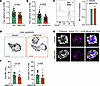 Figure 4
CXCR4 signaling promotes adhesion and integrin activation. (A) Cell adhesion assays of primary CD56+ NK cells on ICAM-1 following vehicle or 10 μM AMD3100 treatment. n = 4 donors; mean of each vehicle-treated donor is normalized to 1. (B) Adhesion of NK92 WT or CXCR4-KO cells to ICAM-1–coated plates. n = 3 independent experiments; large symbols represent the mean of each technical replicate. Statistical analysis by 2-tailed 1-sample t test compared with a theoretical mean of 1. (C) Surface CD18 expression in primary NK cells after treatment with vehicle (water) or AMD3100 displayed as representative histograms (left) and normalized MFI (right). Gray, fluorescence minus one (FMO) control. n = 3 independent experiments (mean ± SD). No statistical significance by 2-tailed Wilcoxon’s matched-pairs signed-rank test. (D) Representative confocal imaging of actin in NK92 cells treated with 10 μM of AMD3100 or vehicle and incubated on ICAM-1, then fixed and immunostained with phalloidin. Scale bars: 5 μm. Data are representative of 3 independent experiments/donors. (E) Primary CD56+ NK cells were treated with 10 μM of AMD3100 or vehicle and incubated on ICAM-1 (LFA-1 ligand) before fixation and immunostaining with anti–activated LFA-1 (clone m24), phalloidin, and DAPI. Scale bars: 5 μm; maximum projection shown. (F) Quantification of m24 MFI and percentage area from one representative donor. Each dot represents a single cell from one donor. Horizontal bars indicate mean ± SD. Statistical comparison by 2-tailed Mann-Whitney test across single-cell measurements within this donor. Data are representative of 3 independent experiments/donors (see also Supplemental Figure 5).
We therefore examined integrin activation on primary CD56+ NK cells using the conformation-sensitive antibody m24, which recognizes the high-affinity state of LFA-1 (52). CXCR4 inhibition with AMD3100 markedly reduced m24 intensity and cluster formation on the NK cell surface following ICAM-1 engagement (Figure 4E). Quantification of these observations confirmed that AMD3100 significantly decreased the size and intensity of m24 foci (Figure 4F). Two additional donors are included in Supplemental Figure 5. Together, these findings demonstrate that CXCR4 signaling promotes β2 integrin activation and ICAM-1–dependent adhesion. This establishes a mechanistic link between CXCR4 and the stabilization of NK cell–stroma interactions, positioning CXCR4 as an upstream regulator of the adhesion-migration balance in human NK cells.
CXCR4 and CXCL12 are required for effective NK cell differentiation from CD34+ cells. Because stromal contact is required for human NK cell maturation, we next asked whether disruption of CXCR4-dependent adhesion affects NK cell developmental progression. To test this, we performed NK cell differentiation from HSPCs in the presence or absence of AMD3100. CD34+ cord blood progenitors were isolated by FACS and differentiated on EL08.1D2 stromal monolayers with cytokines (8). Cultures were treated with 10 μM AMD3100 or vehicle (water) at weekly media exchanges (Figure 5A). At weekly harvests, we quantified total live CD45+ cells and NK developmental subsets by flow cytometry.
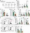 Figure 5
CXCR4-CXCL12 interactions are required for effective NK cell differentiation from CD34+ cells. (A) Schematic of NK cell in vitro differentiation protocol. Human CD34+ cells were isolated from cord blood using density centrifugation and fluorescence-activated cell sorting (FACS). Sorted CD34+ cells were plated on EL08.1D2 stromal cells with cytokines as indicated. Cultures were treated with 10 μM AMD3100 or vehicle at weekly media exchanges. (B) Fold increase in the absolute number of live CD45+ cells from AMD3100-treated (orange) and vehicle-treated (green) cultures. n = 4–10 donors. (C) Frequency of CD34+ cells gated on CD45+ cells at days 7 and 14. n = 7–10 donors. (D) Absolute number (×103) of NK cells at day 14. n = 5 donors. (E) Frequency of Lin–CD117+ NK/ILC progenitors gated on CD45+ live cells by week. n = 7–9 donors. For B–E, each dot represents an individual donor, and lines connect paired samples. Statistical comparisons between vehicle and AMD3100 conditions at each time point were performed using Wilcoxon’s matched-pairs signed-rank tests. (F) Representative flow plots from days 14 and 28 showing frequencies of CD56+ Lin– NK cells. Previously gated on CD45+ live cells. (G) Frequencies of CD94+ conventional NK cells at days 14, 21, and 28. n = 7–9 donors. (H) Absolute number (×105) of CD94+ conventional NK cells at day 28. n = 9 donors. (I) Frequencies of granzyme B+ (GzmB) and EOMES+ NK cells at day 28. n = 4–7 donors.
We first examined the fold increase of live CD45+ cells in each culture condition over time (Figure 5B). Vehicle- and AMD3100-treated cultures were not significantly different through day 14. However, by day 21, a clear divergence emerged whereby vehicle-treated cultures underwent a robust expansion and AMD3100-treated cultures failed to expand. This indicates that CXCR4 signaling becomes particularly important during later stages of in vitro NK cell differentiation when cells normally proliferate in response to stromal cues.
To test whether reduced output reflected impaired hematopoietic progenitor maintenance or differentiation, we measured CD34+ frequencies at days 7 and 14 and found no significant difference between conditions at day 7 (Figure 5C), suggesting that early differentiation is not stalled in the presence of AMD3100. As expected, the frequency of CD34+ cells decreased from about 30% to under 10% between weeks 1 and 2 in both groups, consistent with CD34 downregulation during progression through differentiation, and at day 14 we found significantly fewer CD34+ progenitors in the AMD3100-treated condition (Figure 5C) (10). This progression through differentiation was accompanied by the appearance of NK cells in these cultures, with a decrease in the absolute number of NK cells at 14 days noted in the AMD3100 condition (Figure 5D). Next, we looked at CD117+ Lin– NK/ILC precursors (40), a substantial population of cells in in vitro differentiation cultures (8, 9). AMD3100 cultures contained significantly fewer NK precursors (NKPs) than vehicle controls at multiple time points when measured by frequency (Figure 5E). This reduction in intermediate precursors suggests that CXCR4 signaling supports the progression and/or maintenance of developing NK cells beyond early progenitor stages. Representative flow cytometry plots illustrate the robust accumulation of CD56+ NK cells in vehicle-treated cultures compared with the reduced population in AMD3100-treated cultures (Figure 5F). The effect of AMD3100 on other, non-NK cell populations in these cultures cannot be ruled out; however, the decreased absolute number of NKPs and mature NK cells in our cultures precludes the different frequency of these cells being a result of altered frequencies of other subsets.
By day 28, AMD3100-treated cultures showed a clear reduction in CD94+CD56+ mature NK cells (Figure 5G). This observation was consistent with the failure of AMD3100 cultures to undergo the late expansion observed in vehicle-treated cultures. In addition to frequency, the absolute number of NK cells generated at 28 days was also significantly lower for AMD3100 cultures in comparison with vehicle (Figure 5H). NK cells generated in AMD3100 cultures also showed significantly reduced frequency of granzyme B+ cells at day 28, with a consistent reduction in EOMES+ cells across donors that did not reach statistical significance (paired Wilcoxon’s test) (Figure 5I). These markers reflect acquisition of effector functions and transcriptional maturation, indicating that CXCR4 blockade affects multiple markers of NK cell maturation and/or homeostasis. Whether there are effects on non-NK cells, particularly myeloid cells and other ILC subsets, that are generated in concert with conventional NK cells remains to be determined.
In summary, AMD3100 treatment during in vitro differentiation limits the generation and maturation of NK cells from CD34+ progenitors by reducing NK/ILC precursors and committed NK output and decreasing cytotoxic and transcriptional maturation markers. These findings suggest that CXCR4/CXCL12 signaling provides critical, stage-specific developmental cues, likely through niche positioning and stromal contact interactions, that support efficient human NK cell development.
Plerixafor treatment of WHIM syndrome patients alters NK cell motility, mirroring in vitro AMD3100 effects. Because our in vitro data suggest that CXCR4 blockade alters NK cell phenotypes, we asked whether these features are mirrored ex vivo in samples from WHIM patients with gain-of-function CXCR4 mutations undergoing treatment (28). To characterize NK cell phenotype in WHIM syndrome, we analyzed peripheral blood from healthy donors (HDs) and WHIM patients by flow cytometry. All WHIM samples were collected after 8 months of treatment in a 12-month course of filgrastim (G-CSF) or the CXCR4 antagonist plerixafor (AMD3100) (Figure 6A). Filgrastim mobilizes CXCR4+ cells by altering the CXCL12 in the stromal niche, while plerixafor directly blocks CXCR4-CXCL12 interactions (28). Since paired samples were available, we compared phenotypes during filgrastim versus plerixafor treatment from the same individual. Pretreatment samples were not available.
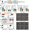 Figure 6
Plerixafor treatment of WHIM syndrome patients alters NK cell phenotype and motility. (A) WHIM samples were obtained after 8 months of treatment following either filgrastim or plerixafor administration as previously described (29). (B) NK cell frequency and CD16+ and CD57+ subsets in paired samples from the same individual undergoing filgrastim (Filg) or plerixafor (Plerix) treatments. n = 6 donors; mean ± SD; Wilcoxon’s matched-pairs signed-rank tests. (C) Surface CXCR4 and CD62L expression on NK cells from paired samples from the same individual undergoing filgrastim or plerixafor treatments. n = 6; mean ± SD; Mann-Whitney test. (D) NK cells were isolated from peripheral blood of WHIM syndrome individuals undergoing plerixafor or filgrastim treatment. Cells were imaged on EL08.1D2 stromal cells for 4 hours at 2-minute intervals. Motility and shape parameters comparing NK cells of treated WHIM patients. Data were paired between post-filgrastim and post-plerixafor samples from 12 WHIM donors. Point plots display pooled single-cell metrics with effect size distributions. (E) Representative time-lapse imaging of post-plerixafor and post-filgrastim treated NK cells (white arrows) on stromal cells.
WHIM syndrome patients undergoing treatment with filgrastim or plerixafor collectively displayed a significantly increased percentage of NK cells among live CD45+ cells from peripheral blood compared with HDs, with skewing toward the less mature CD16– population (Supplemental Figure 6). Surface CXCR4 expression was modestly lower on NK cells from treated WHIM patients compared with HDs (Supplemental Figure 6). Given that CXCR4 gain-of-function mutations in WHIM are classically associated with impaired receptor internalization and heightened CXCL12 responsiveness, this is surprising. However, this reduction in detection likely reflects the residual binding of plerixafor to CXCR4, which is known to compete for antibody binding with detection antibody. We also found that CD62L expression was significantly reduced in WHIM patient NK cells (Supplemental Figure 6), suggesting additional mechanisms of altered maturation and/or homing of NK cells in individuals with WHIM syndrome undergoing treatment.
To analyze treatment-specific effects, we compared paired samples during filgrastim or plerixafor treatment from individual WHIM donors. The frequency of CD56+ NK cells among CD45+ leukocytes was lower after plerixafor treatment compared with filgrastim (Figure 6B). This is expected, as more lymphocyte populations are released into circulation in plerixafor-treated versus filgrastim-treated patients (28). Moreover, CXCR4 surface levels were lower in plerixafor-treated NK cells relative to filgrastim-treated samples (Figure 6C), consistent with plerixafor occupancy blocking the 12G5 antibody epitope. CD62L expression also trended lower under plerixafor treatment, though this difference did not reach significance.
To assess how treatment influences NK cell migratory behavior, CD56+ NK cells were sorted from patient samples and plated on EL08.1D2 stromal monolayers. NK cells from filgrastim- versus plerixafor-treated WHIM patients displayed striking differences in morphology and motility (Figure 6, D and E, and Supplemental Table 2). NK cells from filgrastim-treated patients showed more elongated and polarized morphologies, whereas NK cells from plerixafor-treated patients were markedly rounded. Additionally, NK cells from plerixafor-treated patients exhibited lower aspect ratio, reduced elongation, and increased rounding (Figure 6D), indicating impaired polarization. They also showed significantly lower arrest coefficient, suggesting reduced stable substrate engagement. Instantaneous speeds were modestly higher in NK cells from plerixafor-treated patients, consistent with a non-adherent, exploratory mode of movement. Examples from time-lapse imaging confirmed these differences, as filgrastim-treated NK cells formed persistent leading edges and stable contacts with stroma, whereas plerixafor-treated NK cells remained spherical, transiently contacting the surface but failing to establish durable adhesions (Figure 6E). This phenotype closely mirrors the reduced adhesiveness and polarization we observed with AMD3100 (plerixafor) treatment in vitro, reinforcing that CXCR4 blockade directly disrupts adhesion and cytoskeletal organization.
Taken together, these findings indicate that pharmacologic CXCR4 antagonism disrupts polarization and stable substrate engagement of NK cells, potentially impairing their ability to navigate tissues or form long-lived immune synapses. While plerixafor is clinically effective for leukocyte mobilization and WHIM management, our results suggest that transient impairment of NK cell adhesion and polarity may represent a short-term tradeoff of CXCR4 blockade, with potential implications for NK surveillance and effector functions during treatment.


